Bio lecture 20
 Call Kai
 Learn
 Practice Test
 Spaced Repetition
 Match
 Flashcards
 Knowt Play
1/35

There's no tags or description
Looks like no tags are added yet.
36 Terms
How do cells organize into tissue? What is the tissue formed of?
They secrete proteins that help form the ECM. Cells plus the ECM make the tissue
What are the 4 major type of proteins?
Epithelial, Connective, muscle, and nervous
What ECM is found in epithelial tissue?
Only the Basil Lamina. This is a sheet like meshwork of ECM. The epi cell is composed of mostly cells.
What ECM is found under the basil lamina?
The connective tissue. This region is sparse in cells and has a lot of ECM
Why is the ECM not abundant in cells?
Cell-to-cell junctions and the cytoskeleton provide strength and support. Not as much ECM is needed.
How does having lots of ECM in connective tissue help it?
The ECM will help bear the mechanical load and tension in the tissues

Where can the basil lamina be located?
In muscle cells, it supports the cell. In epithelial cells, it is located on the basal surface. In both cases, it is between the cell and the connective tissue.

What happens to cell organization in cancer cells?
Normally the basil lamina will connect to the cell and the basil lamina. cancer cells are able to break through the lamina and connective tissue and enter the bloodstream. This allows the cancer cells to metastasize.
What are integrans?
TM proteins on the surface of the cell. They cluster band form adhesive structures that help with cell to ECM contact.

What domains are present in the integrin protein, and where are they located?
Consist of a alpha and beta subunit. These subunits have domains both on the inside and outside of the cell.
Are integrin proteins only used for cell to ECM contact?
No. They can also be used to bind other proteins on the surface of other cells.
Are integrins always active?
No. When they are inactive, the domains are bent over. When active, the cell unfolds and it is able to bind to proteins.
What are the three components of the ECM?
Collagen, Proteoglycans, and Multi-adhesive matrix proteins
In which ECM types are the 3 components present?
They are present in both the basal lamina and connective tissue. The amount differs in these types.
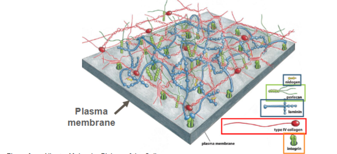
How is the basal lamina organized?
Cross link between collagen, proteoglycans, and MAMP. Integrins will bind these ECM components to the cell.
What is the role of collagen?
It provides structural integrity and mechanical strength
What are the 2 types of collagen ECM?
Sheet Forming collagen (collagen 4) and Fibrillar collagen
Where is collagen 4 found?
Found in the basal lamina
How do the collagen-4 subunits form? And how do subunits form into a network?
3 collagen-4 molecules come together and form a triple helix subunit. Globular domains are present at the N- and C-terminal ends of these subunits. Several subunits will then interact, N to N and C to C. These form a 2D sheet-like network.

What type of collagen does connective tissue have?
Contains collagen fibers rather than sheet like fibers

What are fibrillar collagens?
Collagen forms in the connective tissue. They will assemble into triple-stranded molecules. Many of these triple-stranded molecules will interact along their lengths and assemble into the fibers found in connective tissue.
What is the difference in how the collagen 4 subunit interact vs how fibrillar collagens interact?
Collagen-4 subunits interact end to end
fibrillar collagens interact side to side along their lengths
How are ECM components made/what pathway?
They are made up through an internal cell secretory pathway. Despite this, the ECM components do not end up inside the cell

Why don’t collagen fibers form inside the cell before they are secreted, and how are they activated?
When procollagen is formed on the inside of the cell, it contains extra domains at its end. These prevent the collagen from forming into fibers. Once the procollagen is secreted outside of the cell, a protease will come and chop these domains. It goes from procollagen to a collagen
What are proteoglycans?
Proteins that have attached sugar molecules and provide cushioning
How are proteoglycans formed?
Proteoglycans get a sugar molecule attached to them as they migrate to the golgi. Specifically a GAG sugar will attach to it.
Does a GAG protein non-cov or cov bind to the proteoglycan?
Covalent binding
What is the function of a proteoglycan?
The proteoglycan when attached with GAG occupies a lot of space because the GAG is very extended. This allows it to act as a space filler and give the cell gell like cushioning.
What are a multi adhesive matrix proteins?
These are proteins that cross link to each other and create an ECM meshwork. The proteins are also bind to integrins!
What are two examples of MAMP proteins?
Laminin and Fibronectin
What is laminin?
Found in the basil lamina. It is cross shaped and contains many domains with globular domains. These serve as binding site that allow binding to many proteins
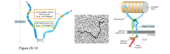
What is fibronectin?
A MAMP that bind to fibrillar collagens in connective tissue OR sheet forming collagens in the basil lamina. It also contains a domain that binds to integrins, helping collagens link to the ECM.
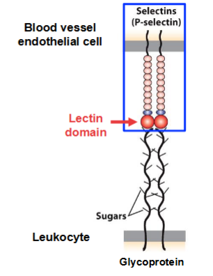
What proteins mediate weak adhesions?
Glycoproteins with attached sugar molecules are on the surface of the leukocyte (WBCs).
P-selectin. A protein that contains a lectin domain that binds a sugar molecule on the glycoprotein. This is found on the surface of cells lining the blood vessels.
Both are weak bindings that allow the cell to roll
What is the difference between a glycoprotein and a proteoglycan?
Glycoproteins are proteins with attached sugar residues and Proteoglycans are a specific type of glycoprotein that
contain GAGs
How does the cell interaction change from weak to strong?
When leuk reaches the site of infection, there is a signal to switch to tight adhesion. This signal will activate the iCAM integran inside the leuk (go from being folded to unfolded). The integrin will then tightly bind to the blood vessel cells.
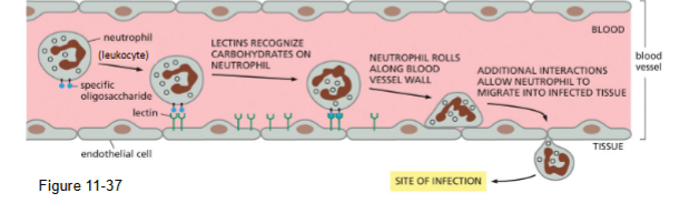
Describe the whole process of cell binding during extraversion
1) Inflammatory signals trigger the WBCs to start rolling to the site of infection
2) Selectins on the blood vessel cells will bind to glycoproteins on the leukocytes
3)These are weak interactions. As blood flows, the cells roll forward
4) Once the cell reaches the site of infection iCAM integrin will activate and bind to the blood vessel cell.
5) Leuk stops rolling and can crawl between the cell into the infected tissue.