AE 1.9 - Prosthetic Valve, Valve Repair, Hypertension

0.0(0)
Studied by 0 people
call kaiCall Kai
learnLearn
examPractice Test
spaced repetitionSpaced Repetition
heart puzzleMatch
flashcardsFlashcards
GameKnowt Play
Card Sorting

1/52

encourage image

There's no tags or description

Looks like no tags are added yet.

53 Terms

1
New cards

Major categories of prosthetic valves (4)

Mechanical

  • bileaflet tilting disk

  • single tilting disk

  • ball cage


Bioprosthetic

  • percutaneous (TAVR, mitraclip)

  • human

  • animal

    • stented porcine (typically no leaks or clots)

    • stentless porcine (AV only, large flow area, no real regurg)

    • bovine (regurg or stenosis common from leaflet thickening)


Homograft

  • from cadaver

  • usually only for aortic valve

  • stentless

  • low stenosis rate

  • low thrombosis rate

  • failure secondary (degeneration) to increasing regurgitation


Autograft (Ross Procedure)

  • take own pulmonic valve & use it to replace the aortic valve

  • replace the PV with a homograft

  • coronary arteries reimplanted

  • pediatrics w/congenital AS

  • failure due to:

    • dilatation of autograft root leading to progression of aortic regurgitation

    • pulmonary homograft degeneration


<p><strong>Mechanical</strong></p><ul><li><p>bileaflet tilting disk</p></li><li><p>single tilting disk</p></li><li><p>ball cage</p></li></ul><p></p><p><strong>Bioprosthetic</strong></p><ul><li><p>percutaneous (TAVR, mitraclip)</p></li><li><p>human</p></li><li><p>animal</p><ul><li><p>stented porcine (typically no leaks or clots) </p></li><li><p>stentless porcine (AV only, large flow area, no real regurg)</p></li><li><p>bovine (regurg or stenosis common from leaflet thickening)</p></li></ul></li></ul><p></p><p><strong>Homograft</strong></p><ul><li><p>from cadaver</p></li><li><p>usually only for aortic valve</p></li><li><p>stentless</p></li><li><p>low stenosis rate</p></li></ul><ul><li><p>low thrombosis rate</p></li></ul><ul><li><p>failure secondary (degeneration) to increasing regurgitation</p></li></ul><p></p><p><strong>Autograft (Ross Procedure)</strong></p><ul><li><p>take own pulmonic valve &amp; use it to replace the aortic valve</p></li><li><p>replace the PV with a homograft</p></li><li><p>coronary arteries reimplanted</p></li><li><p>pediatrics w/congenital AS</p></li><li><p><span style="background-color: transparent;">failure due to:</span></p><ul><li><p><span style="background-color: transparent;">dilatation of autograft root leading to progression of aortic regurgitation</span></p></li><li><p><span style="background-color: transparent;">pulmonary homograft degeneration</span></p></li></ul></li></ul><p></p>
2
New cards

Basic design characteristics of prosthetic valves

  • all prosthetic valves have reduced orifice size and are inherently stenotic

  • need to know type of valve & size

  • inflow profile & regurgitation characteristics


3
New cards

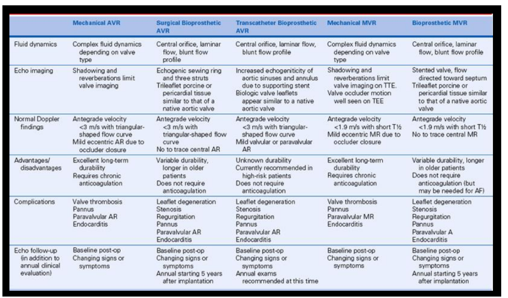
Advantages & disadvantages of each prosthetic valve type

Mechanical

  • high risk of clots / have to be on blood thinners

  • clicking noise

  • tissue overgrowth/pannus risk

  • last very long / doesn’t degenerate


Bioprosthetic

  • don’t last as long / degenerate over time

  • endocarditis risk

  • no need for blood thinners / low clot risk

  • no clicking sound

  • no inherent regurg


<p><strong>Mechanical</strong></p><ul><li><p>high risk of clots / have to be on blood thinners</p></li><li><p>clicking noise</p></li><li><p>tissue overgrowth/pannus risk</p></li><li><p>last very long / doesn’t degenerate</p></li></ul><p></p><p><strong>Bioprosthetic</strong></p><ul><li><p>don’t last as long / degenerate over time</p></li><li><p>endocarditis risk</p></li><li><p>no need for blood thinners / low clot risk</p></li><li><p>no clicking sound</p></li><li><p>no inherent regurg</p></li></ul><p></p>
4
New cards

Mechanical valves

  • More durable than tissue valves (some last decades)

  • Made of carbon, polymeric silicone, or titanium

  • May have overgrowth of a tissue around the valve perimeter called a pannus

  • Typically more suitable for younger male patients or females who don’t intend to become pregnant

    • Will need blood thinners for life (Coumadin)

    • Problems with increased thrombogenicity

  • Can hear the “clicking” sound outside of the body

  • Physiological trivial regurgitation is normal


  • ball & cage (rare)

  • bileaflet tilting disk

  • single tilting disk


<ul><li><p><span style="background-color: transparent;">More durable than tissue valves (some last decades)</span></p></li><li><p><span style="background-color: transparent;">Made of carbon, polymeric silicone, or titanium</span></p></li><li><p><span style="background-color: transparent;">May have overgrowth of a tissue around the valve perimeter called a <strong>pannus</strong></span></p></li><li><p><span style="background-color: transparent;">Typically more suitable for younger male patients or females who don’t intend to become pregnant</span></p><ul><li><p><span style="background-color: transparent;"><strong>Will need blood thinners for life (Coumadin)</strong></span></p></li><li><p><span style="background-color: transparent;">Problems with increased thrombogenicity</span></p></li></ul></li><li><p><span style="background-color: transparent;">Can hear the “clicking” sound outside of the body</span></p></li><li><p><span style="background-color: transparent;">Physiological <strong>trivial regurgitation</strong> is normal</span></p></li></ul><p></p><ul><li><p>ball &amp; cage (rare)</p></li><li><p>bileaflet tilting disk </p></li><li><p>single tilting disk </p></li></ul><p></p>
5
New cards

Expected flow characteristics of mechanical valves

ball & cage

  • non-central flow

  • lateral flow around the ball occluder

  • stenotic

  • closing volume


bileaflet tilting disk

  • 3 orifices/jets when open (1 small central, 2 larger lateral)

  • high velocities in central one


  • leakage volume / washing jets

    • built-in transprosthetic regurgitation aims to prevent thrombus formation at potential sites of stasis 

    • common in bileaflet valve 


single tilting disk

  • 2 orifices/jets (1 major, 1 minor)

  • leaking & closing volume regurgitation common around central strut & between disc & sewing ring


<p><strong>ball &amp; cage </strong></p><ul><li><p><span style="background-color: transparent;">non-central flow </span></p></li><li><p><span style="background-color: transparent;">lateral flow around the ball occluder</span></p></li><li><p><span style="background-color: transparent;">stenotic </span></p></li><li><p><span style="background-color: transparent;">closing volume </span></p></li></ul><p></p><p><strong>bileaflet tilting disk </strong></p><ul><li><p>3 orifices/jets when open (1 small central, 2 larger lateral) </p></li><li><p>high velocities in central one </p></li></ul><p></p><ul><li><p>leakage volume / washing jets </p><ul><li><p><span style="background-color: transparent;">built-in transprosthetic regurgitation aims to prevent thrombus formation at potential sites of stasis&nbsp;</span></p></li><li><p><span style="background-color: transparent;">common in bileaflet valve&nbsp;</span></p></li></ul></li></ul><p></p><p><strong>single tilting disk </strong></p><ul><li><p>2 orifices/jets (1 major, 1 minor) </p></li><li><p><span style="background-color: transparent;">leaking &amp; closing volume regurgitation common around central strut &amp; between disc &amp; sewing ring</span></p></li></ul><p></p>
6
New cards

Expected flow characteristics of tissue valves

homograft

  • similar flow characteristics as native valve

  • central flow

  • trivial/no regurgitation

  • low stenosis rate


autograft

  • native valve flow characteristics for both valves

  • central flow

  • trivial/no regurgitation


stentless valves

  • large flow area

  • trivial/no regurgitation


7
New cards

Expected normal regurgitation patterns of prosthetic valves

bioprosthetic valves

  • trivial/no regurg


percutaneous valves (transcatheter)

  • paravalvular leaks in AV


mechanical valves

  • washing jets / built-in regurg


ball & cage and single tilting disk

  • closing volume: blood displaced when the occluder shuts

  • leaking common around central strut & between disk and ring


<p><strong>bioprosthetic valves</strong></p><ul><li><p>trivial/no regurg</p></li></ul><p></p><p><strong>percutaneous valves (</strong>transcatheter)</p><ul><li><p>paravalvular leaks in AV</p></li></ul><p></p><p><strong>mechanical valves</strong></p><ul><li><p>washing jets / built-in regurg</p></li></ul><p></p><p><strong>ball &amp; cage and single tilting disk</strong></p><ul><li><p>closing volume: blood displaced when the occluder shuts</p></li><li><p>leaking common around central strut &amp; between disk and ring</p></li></ul><p></p>
8
New cards

Which prosthetic valves require lifelong anticoagulation

  • mechanical prosthetic valves

  • high risk of thrombosis on valve


<ul><li><p>mechanical prosthetic valves </p></li></ul><ul><li><p>high risk of thrombosis on valve </p></li></ul><p></p>
9
New cards

Indications for valve replacement vs repair

replacement

  • valve itself is severely damaged

  • can no longer function normally

  • advanced stenosis, regurgitation, endocarditis

  • usually the aortic valve is replaced


repair

  • when cause is secondary to valve

    • annular dilation

    • chordal issue

  • leaflets/cusps are not severely damaged or calcified

    • small perforation

  • usually the mitral & tricuspid valves are repaired


10
New cards

Advantages of valve repair compared with valve replacement

  • repair is always the best option when available

  • lasts very long

    • much more durable / less chance of degeneration

  • less complications with own native valve

    • less chance of infection

    • no need for blood thinners


11
New cards

Components of a comprehensive prosthetic valve evaluation

  • on & off axis views

  • Valve function

  • Chamber measurements

  • Systolic/diastolic function

  • Color Doppler

  • Spectral Doppler

    • CW peak/mean gradients

  • Valve appearance

  • Seating of valve within the native annulus

  • Opening and closing motion of the leaflets

  • Valve bed/appearance of sewing ring stability

    • “rocking motion” = dehiscence

  • bioprosthetic leaflet thickness


12
New cards

Importance of knowing valve type & size

  • so you know what to look for on echo

  • normal hemodynamic values are based on this:

    • unique normal flow profiles

    • unique regurgitation patterns

    • unique normal hemodynamic values


13
New cards

Expected 2D appearance of prosthetic valves

post-op

  • IVS paradoxical motion


mechanical

  • very echogenic

  • shadowing below valve

  • microbubbles / degassing


bioprosthetic

  • sometimes look like native valve

  • 3 struts


14
New cards

Causes of acoustic shadowing & reverberation artifact

  • Sewing rings & mechanical valves are strong echo reflectors – results in acoustic shadows & reverberations

    • Blocks imaging & Doppler abnormalities in shadow region


<ul><li><p><span style="background-color: transparent;">Sewing rings &amp; mechanical valves are strong echo reflectors – results in acoustic shadows &amp; reverberations</span></p><ul><li><p><span style="background-color: transparent;">Blocks imaging &amp; Doppler abnormalities in shadow region</span></p></li></ul></li></ul><p></p>
15
New cards

Purpose of baseline post-operative echocardiography

(4-6 weeks post-op)

  • Full comprehensive echo

  • Primary goal is to acquire hemodynamic data when valve is working normally

  • Can serve as a “normal reference”

  • Advantages:

    • recognizes PPM – prosthesis patient mismatch

    • detects prosthetic valve dysfunction

    • identifying infective endocarditis


16
New cards

Importance of comparing serial studies with baseline examinations

So many numbers to remember! (each valve has its own unique ones)

  • This is why a high quality baseline echo is so important!!!

  • Use the baseline as the fingerprint (new normal) and compare all serial echoes to this

  • can see a change from “normal” values

  • Comparison with post-operative studies is essential in cases of high gradients


17
New cards

Prosthesis-patient mismatch (PPM)

  • Occurs when the effective orifice area (IEOA) of the prosthetic valve is too small in relation to a patient’s body size

  • Resulting in high residual postoperative pressure gradients across the prosthesis

  • More common in aortic valves


<ul><li><p><span style="background-color: transparent;">Occurs when the effective orifice area (IEOA) of the prosthetic valve is too small in relation to a patient’s body size</span></p></li><li><p><span style="background-color: transparent;">Resulting in high residual postoperative pressure gradients across the prosthesis</span></p></li><li><p><span style="background-color: transparent;">More common in aortic valves</span></p></li></ul><p></p>
18
New cards

Prosthetic valve thrombosis

  • Formation of blood clot around or over prosthetic valve

  • Observed with bioprosthetic and mechanical valves

  • More common in mechanical valves with inadequate antithrombotic therapy (INR ≤ 2.5)

  • Can lead to obstruction &/or regurgitation


<ul><li><p><span style="background-color: transparent;">Formation of blood clot around or over prosthetic valve</span></p></li><li><p><span style="background-color: transparent;">Observed with bioprosthetic and mechanical valves</span></p></li><li><p><span style="background-color: transparent;">More common in mechanical valves with inadequate antithrombotic therapy (INR ≤ 2.5)</span></p></li><li><p><span style="background-color: transparent;">Can lead to obstruction &amp;/or regurgitation</span></p></li></ul><p></p>
19
New cards

Pannus formation

growth of abnormal tissue around a prosthetic heart valve
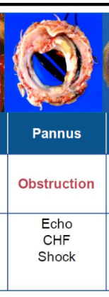
<p><span style="background-color: transparent;">growth of abnormal tissue around a prosthetic heart valve</span></p>
20
New cards

Prosthetic valve obstruction

TAVR = HALT (hypo-attenuated leaflet thickening) 

<p><span style="background-color: transparent;">TAVR = HALT (hypo-attenuated leaflet thickening)&nbsp;</span></p>
21
New cards

Prosthetic valve endocarditis

  • Infection of the valve leaflets or between the sewing ring and native annulus

  • More likely in bioprosthetic valves

  • Timing of the infection reflects different mechanisms

  • May create regurgitation and/or stenosis

  • May also cause other complications like leaflet perforation, abscesses, dehiscence, etc

  • TEE often required


<ul><li><p><span style="background-color: transparent;">Infection of the valve leaflets or between the sewing ring and native annulus</span></p></li><li><p><span style="background-color: transparent;">More likely in bioprosthetic valves</span></p></li><li><p><span style="background-color: transparent;">Timing of the infection reflects different mechanisms</span></p></li><li><p><span style="background-color: transparent;">May create regurgitation and/or stenosis</span></p></li><li><p><span style="background-color: transparent;">May also cause other complications like leaflet perforation, abscesses, dehiscence, etc</span></p></li><li><p><span style="background-color: transparent;">TEE often required</span></p></li></ul><p></p>
22
New cards

Paravalvular leak vs. transvalvular regurgitation

paravalvular

  • a leak caused by a space left between natural heart tissue and the valve replacement from a previous transcatheter aortic or mitral valve replacement

  • Color flow Doppler: arises outside the sewing ring, between the sewing ring and the native annulus


transvalvular

  • Occurs within the sewing ring

  • Most mechanical valves have physiologic regur

    • May be multiple jets - usually short, narrow and nonturbulent

  • Most common cause of mechanical is thrombosis or pannus formation

  • Most common cause of bioprosthetic is leaflet degeneration


<p><strong>paravalvular </strong></p><ul><li><p><span style="background-color: transparent;">a leak caused by a space left between natural heart tissue and the valve replacement from a previous transcatheter aortic or mitral valve replacement</span></p></li><li><p><span style="background-color: transparent;">Color flow Doppler: arises outside the sewing ring, between the sewing ring and the native annulus</span></p></li></ul><p></p><p><strong>transvalvular </strong></p><ul><li><p><span style="background-color: transparent;">Occurs within the sewing ring</span></p></li><li><p><span style="background-color: transparent;">Most mechanical valves have physiologic regur</span></p><ul><li><p><span style="background-color: transparent;">May be multiple jets - usually short, narrow and nonturbulent</span></p></li></ul></li><li><p><span style="background-color: transparent;">Most common cause of <strong>mechanical </strong>is thrombosis or pannus formation</span></p></li><li><p><span style="background-color: transparent;">Most common cause of <strong>bioprosthetic </strong>is leaflet degeneration</span></p></li></ul><p></p>
23
New cards

Valve dehiscence

serious prosthetic valve complication characterized by “rocking” of valve at an angle far in excess of its normal excursion

24
New cards

Typical echocardiographic findings associated with each complication

PPM

  • ↑ pressure gradients

  • EOA & DVI normal

  • ↓ IEOA

  • No change ΔEOA/DVI (from baseline)


Thrombosis

  • obstruction/regurg

  • echodensity on the occluder or leaflets

  • restricted motion


Pannus

  • tissue on valve


Obstruction/Stenosis

  • ↑ Pressure gradients

  • ↓ EOA & DVI (AVR)

  • ↓ EOA & ↑ DVI (MVR)

  • ↑ PHT (MVR, TVR)


Endocarditis

  • regurgitation / stenosis

  • leaflet perforation, abscesses, dehiscence


Dehiscence

  • rocking motion

  • paravalvular leak


<p><strong>PPM</strong></p><ul><li><p><span style="background-color: transparent;">↑ pressure gradients</span></p></li><li><p><span style="background-color: transparent;">EOA &amp; DVI normal</span></p></li><li><p><span style="background-color: transparent;">↓ IEOA</span></p></li><li><p><span style="background-color: transparent;">No change ΔEOA/DVI (from baseline)</span></p></li></ul><p></p><p><strong>Thrombosis </strong></p><ul><li><p>obstruction/regurg</p></li><li><p>echodensity on the occluder or leaflets</p></li><li><p>restricted motion</p></li></ul><p></p><p><strong>Pannus </strong></p><ul><li><p>tissue on valve </p></li></ul><p></p><p><span style="background-color: transparent;"><strong>Obstruction/Stenosis</strong></span></p><ul><li><p><span style="background-color: transparent;">↑ Pressure gradients</span></p></li><li><p><span style="background-color: transparent;">↓ EOA &amp; DVI (AVR)</span></p></li><li><p><span style="background-color: transparent;">↓ EOA &amp; ↑ DVI (MVR)</span></p></li><li><p><span style="background-color: transparent;">↑ PHT (MVR, TVR)</span></p></li></ul><p></p><p><strong>Endocarditis </strong></p><ul><li><p><span style="background-color: transparent;">regurgitation / stenosis</span></p></li><li><p><span style="background-color: transparent;">leaflet perforation, abscesses, dehiscence</span></p></li></ul><p></p><p><strong>Dehiscence </strong></p><ul><li><p>rocking motion </p></li><li><p>paravalvular leak </p></li></ul><p></p>
25
New cards

Mitral valve repair techniques

  • Annuloplasty rings

    • used to reshape and strengthen the natural valve annulus

    • a ring is placed around a stretched-out valve

      • the ring shrinks the valve down so the leaflets can close better


  • Gore-Tex

    • chordal replacement


  • Leaflet resection

    • removing the damaged part of the leaflet and then sews the remaining edges together


26
New cards

Common post-repair complications

  • Recurrent MV regurgitation

  • Newly developed septal contact / SAM

    • systolic anterior motion of anterior MV leaflet

  • Pericardial effusion/constriction


27
New cards

Tricuspid Annuloplasty

  • implantation of an undersized ring (rigid, semirigid, flexible) or a flexible band

  • has importance among patients with functional TR

  • annuloplasty rings or bands do not cover the whole tricuspid annulus, but remain open at the septal annulus to avoid injury to the conduction system


<ul><li><p><span style="background-color: transparent;">implantation of an undersized ring (rigid, semirigid, flexible) or a flexible band</span></p></li><li><p><span style="background-color: transparent;">has importance among patients with functional TR</span></p></li><li><p><span style="background-color: transparent;">annuloplasty rings or bands do not cover the whole tricuspid annulus, but remain open at the septal annulus to avoid injury to the conduction system</span></p></li></ul><p></p>
28
New cards

Valve-sparing aortic root repair

  • Feasible in some instances:

    • leaflets are not deformed, scarred, or calcified


  • Indications for AV repair include

    • 1. bicuspid valve with prolapsed of a single leaflet

    • 2. tricuspid valve with prolapsed of one leaflet

    • 3. normal aortic leaflets with dilated sinotubular junction

    • 4. Limited cusp destruction caused by infective endocarditis may also be amenable to repair in select instances. However, such repair should preferably be done at centers with specialized expertise


29
New cards

Definition & classification of systemic hypertension

  • Begins at 130/80

  • Should be based on the mean of 2+ readings on two separate visits


  • 1. Essential

    • Known as primary or idiopathic

    • No identifiable cause

    • Approx 90-95% of patients will have essential systemic HTN

  • 2. Secondary

    • Has identifiable mechanism

      • Arterial renal disease, aortic coarctation, endocrine diseases, drugs, food ingestion


<ul><li><p><span style="background-color: transparent;">Begins at 130/80</span></p></li><li><p><span style="background-color: transparent;">Should be based on the mean of 2+ readings on two separate visits</span></p></li></ul><p></p><ul><li><p><span style="background-color: transparent;">1. <strong>Essential</strong></span></p><ul><li><p><span style="background-color: transparent;">Known as primary or idiopathic</span></p></li><li><p><span style="background-color: transparent;">No identifiable cause</span></p></li><li><p><span style="background-color: transparent;">Approx 90-95% of patients will have essential systemic HTN</span></p></li></ul></li><li><p><span style="background-color: transparent;">2. <strong>Secondary</strong></span></p><ul><li><p><span style="background-color: transparent;">Has identifiable mechanism</span></p><ul><li><p><span style="background-color: transparent;">Arterial renal disease, aortic coarctation, endocrine diseases, drugs, food ingestion</span></p></li></ul></li></ul></li></ul><p></p>
30
New cards

Modifiable & non-modifiable risk factors (systemic hypertension)

Modifiable risk factors

  • Smoking 

  • Type 2 Diabetes

  • Obesity (esp abdominal) 

  • Hypercholesteremia

  • Inactivity 

  • Unhealthy diet

  • Sodium intake 

  • Alcohol consumption

  • Emotional stress


Non-Modifiable risk factors

  • Chronic Kidney Disease 

  • Family History

  • Increased age

  • Male

  • Race (higher in African Americans)

  • Low socioeconomic status

  • Obstructive Sleep Apnea


31
New cards

Common complications (systemic hypertension)

  • Atherosclerotic complications

  • Left ventricular hypertrophy

  • Heart failure associated symptoms

  • TIA/stroke

  • Hypertensive encephalopathy, intracerebral hemorrhage, dementia

  • Renal disease (nephrosclerosis, renal failure, glomerulosclerosis)

  • Hypertensive retinopathy, optic neuropathy

  • Aortic aneurysm (ascending aorta, abdominal aorta)

  • Aortic dissection


32
New cards

Basic treatment approaches (systemic hypertension)

  • Behavior modification

    • Weight loss, healthy diet, reduce alcohol consumption, exercise, tobacco cessation, stress reduction

  • Pharmacologic

    • Diuretics, Beta blockers, ACE inhibitor, Calcium channel blocker, vasodilator


33
New cards

Role of echocardiography in patients with systemic hypertension

  • Evaluate secondary complications

    • LVH/increased mass

    • LV systolic & diastolic function

    • LAE

  • Exclude any causes


34
New cards

Left ventricular mass

  • LV mass is the weight of the LV

  • Typically measured using wall and LV dimensions


35
New cards

Relative wall thickness (RWT)

  • A ratio that indicates the relationship between the wall thickness and cavity size of the left ventricle

  • RWT = 2 x PW / LVIDd 


36
New cards

LV Geometric Patterns (Left Ventricular Remodeling)

Concentric hypertrophy

  • thicker walls

  • normal cavity

  • increased mass

  • higher ratio between wall and cavity


Eccentric hypertrophy

  • normal thickness walls

  • increased cavity size

  • increased mass

  • lower ratio between wall and cavity


Concentric remodeling

  • thicker walls

  • normal or smaller sized cavity

  • normal mass

  • higher ratio between wall and cavity


<p><strong>Concentric hypertrophy</strong></p><ul><li><p>thicker walls </p></li><li><p>normal cavity </p></li><li><p>increased mass </p></li><li><p>higher ratio between wall and cavity </p></li></ul><p></p><p><strong>Eccentric hypertrophy</strong></p><ul><li><p>normal thickness walls </p></li><li><p>increased cavity size </p></li><li><p>increased mass </p></li><li><p>lower ratio between wall and cavity</p></li></ul><p></p><p><strong>Concentric remodeling</strong></p><ul><li><p>thicker walls </p></li><li><p>normal or smaller sized cavity </p></li><li><p>normal mass </p></li><li><p>higher ratio between wall and cavity </p></li></ul><p></p>
37
New cards

Echocardiographic findings associated with chronic systemic hypertension

EKG

  • thickened LV wall leads to prolonged depolarization (increased R wave peak time)

  • delayed repolarization (ST and T-wave abnormalities) in the lateral leads


Echo

  • LVH

  • LAE

  • Ao root dilation

  • diastolic dysfunction


38
New cards

Increased Wall Thickness vs LVH

  • Increased Wall Thickness

    • Thick walls can happen for several reasons, some of which are not due to true muscle hypertrophy (cell growth)

    • Processes can cause an increase in wall thickness which is not due to hypertrophy of the myocardium

      • Infiltrative disease, tumors, transplant rejection

    • Typically result in an increased mass

  • LVH

    • From pressure overload 

    • From increased afterload 


39
New cards

Definition & classification of pulmonary hypertension

  • A clinical condition characterized by elevated pulmonary vascular pressure

    • Plaque, fibrosis, and narrowing

    • Increased pulmonary vascular resistance (afterload) 

  • Over time sustained increased pressure in the arterial bed leads to increased right ventricular RV) afterload and eventually RV failure

  • Can occur at any age

  • Cause: idiopathic (no real reason) or secondary (has a reason) 


40
New cards

5 groups & their underlying causes (pulm htn)

1) Pulmonary Arterial Hypertension (PAH)

  • Arteries in the lungs become narrowed, thickened or stiff

  • Right heart must work harder to push blood through these narrowed arteries

  • Extra stress can cause the heart to lose its ability to pump enough blood through the lungs to meet the needs of the rest of the body


2) PHTN due to left heart disease

  • Arteries and lungs are not as thick or stiff as Group 1

  • Caused by:

    • Systolic or diastolic dysfunction

    • Left sided valve disease (MR backs up into pulm vns → lungs → right side) 

  • “backup” of blood raises pressure in the lungs


3) PHTN due to lung disease

  • Arteries in the lungs tighten so that blood can only go to areas of the lungs that are receiving the most air and oxygen

    • This tightening leads to high blood pressure throughout the lungs

  • Restrictive lung disease in which the lungs have a tough time expanding when one inhales (ILD, pulm fibrosis)

  • Lung airways narrow and make it harder to exhale (COPD/Emphysema)

  • Sleep apnea


4) PHTN due to chronic blood clots in the lungs

  • CTEPH – Chronic Thromboembolic Pulmonary Hypertension

    • Body is unable to dissolve blood clots in the lungs

    • Leads to scar tissue in the blood vessels of the lungs

    • Makes right side work harder


5) PHTN due to unknown causes

  • Not really understood

  • Associated conditions include sarcoidosis, sickle cell anemia, certain metabolic disorders


<p>1) <span style="background-color: transparent;"><strong>Pulmonary Arterial Hypertension (PAH)</strong></span></p><ul><li><p><span style="background-color: transparent;">Arteries in the lungs become narrowed, thickened or stiff</span></p></li><li><p><span style="background-color: transparent;">Right heart must work harder to push blood through these narrowed arteries</span></p></li><li><p><span style="background-color: transparent;">Extra stress can cause the heart to lose its ability to pump enough blood through the lungs to meet the needs of the rest of the body</span></p></li></ul><p></p><p>2) <span style="background-color: transparent;"><strong>PHTN due to left heart disease</strong></span></p><ul><li><p><span style="background-color: transparent;">Arteries and lungs are not as thick or stiff as Group 1</span></p></li><li><p><span style="background-color: transparent;">Caused by:</span></p><ul><li><p><span style="background-color: transparent;">Systolic or diastolic dysfunction</span></p></li><li><p><span style="background-color: transparent;">Left sided valve disease (MR backs up into pulm vns → lungs → right side)&nbsp;</span></p></li></ul></li><li><p><span style="background-color: transparent;">“backup” of blood raises pressure in the lungs</span></p></li></ul><p></p><p>3) <span style="background-color: transparent;"><strong>PHTN due to lung disease</strong></span></p><ul><li><p><span style="background-color: transparent;">Arteries in the lungs tighten so that blood can only go to areas of the lungs that are receiving the most air and oxygen</span></p><ul><li><p><span style="background-color: transparent;">This tightening leads to high blood pressure throughout the lungs</span></p></li></ul></li><li><p><span style="background-color: transparent;">Restrictive lung disease in which the lungs have a tough time expanding when one inhales (ILD, pulm fibrosis)</span></p></li><li><p><span style="background-color: transparent;">Lung airways narrow and make it harder to exhale (COPD/Emphysema)</span></p></li><li><p><span style="background-color: transparent;">Sleep apnea</span></p></li></ul><p></p><p>4) <span style="background-color: transparent;"><strong>PHTN due to chronic blood clots in the lungs</strong></span></p><ul><li><p><span style="background-color: transparent;">CTEPH – Chronic Thromboembolic Pulmonary Hypertension</span></p><ul><li><p><span style="background-color: transparent;">Body is unable to dissolve blood clots in the lungs</span></p></li><li><p><span style="background-color: transparent;">Leads to scar tissue in the blood vessels of the lungs</span></p></li><li><p><span style="background-color: transparent;">Makes right side work harder</span></p></li></ul></li></ul><p></p><p>5) <span style="background-color: transparent;"><strong>PHTN due to unknown causes</strong></span></p><ul><li><p><span style="background-color: transparent;">Not really understood</span></p></li><li><p><span style="background-color: transparent;">Associated conditions include sarcoidosis, sickle cell anemia, certain metabolic disorders</span></p></li></ul><p></p>
41
New cards

PAH

  • Pulmonary Arterial Hypertension

  • Idiopathic PAH occurs without a clear cause

  • Heritable PAH is linked to genes that are inherited from family members

  • PAH developed in association with:

    • congenital heart disease, liver disease, HIV and connective tissue diseases such as scleroderma and lupus

    • past or present drug use, such as methamphetamine or certain diet pills

  • While there are treatment options for PAH, there is no known cure


<ul><li><p>Pulmonary Arterial Hypertension</p></li><li><p><span style="background-color: transparent;"><strong>Idiopathic PAH</strong> occurs without a clear cause</span></p></li><li><p><span style="background-color: transparent;"><strong>Heritable PAH</strong> is linked to genes that are inherited from family members</span></p></li><li><p><span style="background-color: transparent;">PAH developed in association with:</span></p><ul><li><p><span style="background-color: transparent;">congenital heart disease, liver disease, HIV and connective tissue diseases such as scleroderma and lupus</span></p></li><li><p><span style="background-color: transparent;">past or present drug use, such as methamphetamine or certain diet pills</span></p></li></ul></li><li><p><span style="background-color: transparent;">While there are treatment options for PAH, there is no known cure</span></p></li></ul><p></p>
42
New cards

Gold standard for diagnosis (pulm htn)

right heart catherization

Dicrotic notch = SL valve opens

Mean PA pressure > 20 mmHg

<p>right heart catherization</p><p><span style="background-color: transparent;">Dicrotic notch = SL valve opens</span></p><p><span style="background-color: transparent;"><u>Mean PA pressure</u> &gt; <u>20 mmHg</u></span></p>
43
New cards

Role of echocardiography in evaluation (pulm htn)

  • Identify potential cause of PHTN

  • Estimate PA pressures

  • Estimate RAP

  • Assess RV structure, thickness, and function

  • Observe for shunts or other hemodynamic flow abnormalities

  • Assess PA

  • RV

    • On-axis: measurements & size

    • Off-axis: function & doppler 


m-mode

  • Mid-systolic notching (flying W) of pulmonary valve cusps

    • Flying “W” 

  • Reduced RV TAPSE


<ul><li><p><span style="background-color: transparent;">Identify potential cause of PHTN</span></p></li><li><p><span style="background-color: transparent;">Estimate PA pressures</span></p></li><li><p><span style="background-color: transparent;">Estimate RAP</span></p></li><li><p><span style="background-color: transparent;">Assess RV structure, thickness, and function</span></p></li><li><p><span style="background-color: transparent;">Observe for shunts or other hemodynamic flow abnormalities</span></p></li><li><p><span style="background-color: transparent;">Assess PA</span></p></li><li><p><span style="background-color: transparent;">RV</span></p><ul><li><p><span style="background-color: transparent;"><u>On-axis</u>: measurements &amp; size</span></p></li><li><p><span style="background-color: transparent;"><u>Off-axis</u>: function &amp; doppler&nbsp;</span></p></li></ul></li></ul><p></p><p>m-mode</p><ul><li><p><span style="background-color: transparent;">Mid-systolic notching (flying W) of pulmonary valve cusps</span></p><ul><li><p><span style="background-color: transparent;"><strong>Flying “W”&nbsp;</strong></span></p></li></ul></li><li><p><span style="background-color: transparent;">Reduced RV TAPSE</span></p></li></ul><p></p>
44
New cards

Right ventricular adaptation to chronic pulmonary hypertension

hypertrophy

dilation


echo findings

  • RV dysfunction

  • TV annulus dilation

  • PA dilation

  • RA dilation (RAE)

  • D-shaped LV

  • Dilated IVC


45
New cards

Septal flattening in pressure overload versus volume overload

pressure

  • during systole


volume

  • during diastole


<p><strong>pressure </strong></p><ul><li><p>during systole </p></li></ul><p></p><p><strong>volume </strong></p><ul><li><p>during diastole </p></li></ul><p></p>
46
New cards

Assessment of RV systolic function

  • 2D RV fractional area change

    • >35%

  • TAPSE

    • >1.7 cm

  • Tissue Doppler s’

    • >9.5 cm/s

  • RV longitudinal strain

    • > -20%

  • RVEF%

    • >45%

  • 3D RV volumes

    • <90 mL/m2

  • RV free wall

    • < 5mm


47
New cards

Qualitative assessment of RV size

  • “Eyeball” for initial judgement

  • From a non-foreshortened A4C view (location of probe not too medial)

    • Normal: RV area or mid-cavity diameter should be less than 2/3 of LV

    • Moderate dilatation: RV same as LV, share the apex

    • Severe dilatation: RV exceeds LV, RV forms apex


<ul><li><p><span style="background-color: transparent;">“Eyeball” for initial judgement</span></p></li><li><p><span style="background-color: transparent;">From a non-foreshortened A4C view (location of probe not too medial)</span></p><ul><li><p><span style="background-color: transparent;">Normal: RV area or mid-cavity diameter should be less than 2/3 of LV</span></p></li><li><p><span style="background-color: transparent;">Moderate dilatation: RV same as LV, share the apex</span></p></li><li><p><span style="background-color: transparent;">Severe dilatation: RV exceeds LV, RV forms apex</span></p></li></ul></li></ul><p></p>
48
New cards

Estimation of right ventricular systolic pressure (RVSP)

  • TR velocity

    • RVSP = PASP (as long as no PS)

  • Evaluate RAP with IVC

  • Hepatic vein flow


49
New cards

Relationship between RVSP & pulmonary artery systolic pressure

RVSP = PASP (as long as no PS)

50
New cards

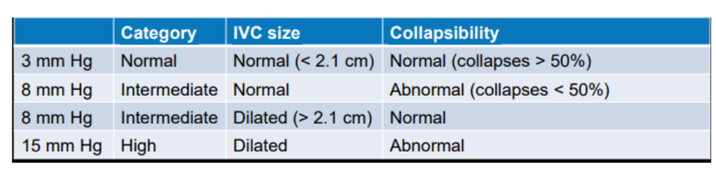
Use of tricuspid regurgitation velocity & estimated right atrial pressure

RVSP = Tricuspid Regurgitation Pressure Gradient + RA pressure


TR = 4V2

RAP = IVC size + collapsibility

51
New cards

Evaluation of the inferior vena cava (IVC)

  • sniff test

  • size of IVC (is it dilated)

  • does diameter decreases > 50%


<ul><li><p>sniff test </p></li></ul><ul><li><p>size of IVC (is it dilated) </p></li><li><p>does diameter decreases &gt; 50%</p></li></ul><p></p>
52
New cards

Hepatic vein Doppler

  • PW Doppler of hepatic veins

  • Looking for s & d  (forward flow) 

  • Sample Volume size of 3-4 mm

  • Placed at 1-2 cm into the hep veins using color guidance


<ul><li><p><span style="background-color: transparent;">PW Doppler of hepatic veins</span></p></li><li><p><span style="background-color: transparent;">Looking for s &amp; d&nbsp; (forward flow)&nbsp;</span></p></li><li><p><span style="background-color: transparent;">Sample Volume size of 3-4 mm</span></p></li><li><p><span style="background-color: transparent;">Placed at 1-2 cm into the hep veins using color guidance</span></p></li></ul><p></p>
53
New cards

Pulmonary acceleration time (PAT)

  • Time to get from baseline to peak of RVOT 

  • Short = increased pressure (Pulm Art Pres) 


<ul><li><p><span style="background-color: transparent;">Time to get from baseline to peak of RVOT&nbsp;</span></p></li><li><p><span style="background-color: transparent;">Short = increased pressure (Pulm Art Pres)&nbsp;</span></p></li></ul><p></p>