Session 7 - Pediatric Cardiac
 Call Kai
 Learn
Learn Practice Test
 Spaced Repetition
 Match
Match Flashcards
 Knowt Play
1/133

There's no tags or description
Looks like no tags are added yet.
Name | Mastery | Learn | Test | Matching | Spaced | Call with Kai | Chat |
|---|
No analytics yet
Send a link to your students to track their progress
134 Terms
what does congenital mean?
present at birth
why does fetal blood bypass the lungs in utero?
fetal lungs are fluid-filled and nonfunctional; oxygen comes from the placenta
what are the three fetal shunts?
Ductus venosus, Foramen ovale, ductus arteriosus (VOA)
what does the ductus venosus do?
connects the umbilical vein to the inferior vena cava, bypassing the liver

what does the foramen ovale do?
allows blood to flow from the right atrium —> left atrium, bypassing the lungs

what does the ductus arteriosus do?
connects the pulmonary artery to the descending aorta, sending blood away from the lungs into systemic circulation

when do the ductus venosus and ductus arteriosus begin closing?
shortly after birth
what can happen if a fetal shunt remains open after birth?
it creates an abnormal pathway for blood flow and may cause a congnital heart defect
why might a newborn with a severe heart defect need temporary oxygen/support when fetal structures close?
the defect may depend on an open fetal shunt to maintain adequate circulation until surgeru
CHD vs. acquired heart disease?
CHD: anatomical malformation present at birth.
Acquired: develops after birth from causes such as infection or inflammation
what is the major cardiac cause of death during the first year of life?
Congenital heart disease.
what is the most common congenital heart defect?
ventricular septal defect (VSD)
which chromosomal condition is commonly associated with congenital heart defects?
down syndrome
what two major clues may suggest cardiovascular disease in an infant?
heart murmur and diaphoresis, especially during feeding
what other findings may occur with pediatric cardiovascular problems?
poor feeding, activity intolerance, tachypnea, tachycardia, crackles, hepatomegaly, and cyanosis
🚨 NCLEX
can normal or pink skin color rule out critical cardiac distress?
no. never rely on skin color alone. a child may be critically ill without cyanosis
what produces a heart murmur?
blood passing through a defective valve, narrowed vessel, or abnormal cardiac structure
how are heart murmurs graded?
on a 1-6 scale. 1 is barely audible, 6 is audible without a stethoscope
which murmur grade is loud but has no palpable thrill?
grade 3
which murmur grade does a palpable thrill begin?
grade 4
which murmur is heard with the stethoscope partly off the chest?
grade 5
which murmur can be heart without a stethoscope
grade 6
🚨 NCLEX
which murmur grades are considered pathologic in these course notes?
grade 3 or higher
how long should the nurse auscultate a pediatric apical pulse?
one full minute
when is the best time to measure a child’s resting heart rate?
while the child is asleep or calm because crying, anxiety, and breath-holding alter the rate
are rhythm irregularities always abnormal in infants and young children?
no. rhythm irregularities may be common in this age group
which pulse site should be used in a child under age 2?
brachial pulse; radial pulses may be difficult and inaccurate
which pulse site is acceptable after age 2?
radial pulse
🚨 NCLEX
what do weaker leg pulses or lower leg BP than arm BP suggest?
coarctation of the aorta (CoA)
what do bounding pulses suggest?
patent ductus arteriosus (PDA) or aortic insufficiency
when is tachycardia especially concerning for cardiac disease?
when it occurs without fever, crying, or acute stress.
what do tachypnea and chest retractions suggest in a cardiac patient?
respiratory distress, often related to heart failure.
how should a child be prepared for cardiac testing?
match teaching to erikson/piaget stage, use simple nonthreatening language, minimize fear, and never lie about discomfort
what does a chest x-ray assess in cardiac disease?
heart size and shape, pulmonary vascular markings, and cardiomegaly
what does an ECG/EKG assess?
electrical activity of the heart
what does an echocardiogram assess?
cardiac anatomical structures
what information can cardiac catherization provide?
visualization of chambers/vessels, chamber oxygen saturations, intracardiac pressures, and pumping function
what findings may indicate a contrast-dye reaction?
increased temperature, urticaria, wheezing, dyspnea, or edema
what safety question is essential before cardiac catherization?
ask about allergies, especially prior contrast reactions
🚨 NCLEX
what are major pre-cardiac-catherization nursing actions?
assess vital signs and Hgb/Hct, document pedal pulses, keep NPO, and hold digoxin as ordered/protocol
🚨 NCLEX
what are major post-cardaic-catherization nursing actions?
vital signs every 15 minutes for the first hour, apical pulse for one full minute, assess peda pulses, and maintain strict bed rest
when can oral fuids be given after cardiac catherization?
only when the child is fully awake and alert/oriented enough to swallow safely
what is the priority fluid assessment for pediatric cardiac patients?
strict intake and output. fluid overload can rapidly worsen pulmonary congestion
why are cardiac infants at risk for failure to thrive?
they use extra calories to breathe, suck, and maintain metabolic balance
what feeding schedule reduces exhaustion in cardiac infants?
small, frequent feedings; limit nipple feeding to 20-30 minutes
why use a soft nipple?
it reduces the energy required to suck.
what formula concentration may be used for extra calories?
24 calories/oz
what nutrient/electrolyte should be monitored in cardiac feeding plans?
sodium
why is adequate weight gain important before open-heart surgery?
the child needs sufficient growth and nutritional reserve to tolerate surgery and recovery
why does blood normally move left to right through an abnormal opening?
left-sided heart pressure is normally higher than right-sided pressure
describe left-to-right shunting
oxygenated blood moves left side → right side → lungs again, increasing pulmonary blood flow and reducing effective systemic output
🚨 NCLEX
why is a child with a left-to-right shunt usually not cyanotic?
the blood reaching systemic circulation is still oxygenated, oxygenated blood is being recirculated to the lungs
what are common left-to-right shunt manifestations?
recurrent respiratory infections, tachycardia, cardiomegaly, dyspnea, and pulmonary edema
why can left-to-right shunts cause pulmonary edema and dyspnea?
excess blood is sent to the lungs under high pressure
what is a VSD?
a hole in the ventricular septum; oxygenated blood flows LV → RV and returns to the lungs
why can a small VSD produce a loud murmur?
blood moves forcefully thourgh the small opening.
memory: smaller hole, louder flow
how many small versus medium/large VSDs present?
small VSDs may be asymptomatic with a loud murmur; larger VSDs may cause pressure problems and heart failure.
what may happen if a significant VSD is untreated?
pulmonary vessels thicken, pulmonary pressure rises, and pulmonary vascular disease/pulmonary hypertension develops
what care is associated with a symptomatic VSD?
treat heart failure with diuretics and digoxin, provide high-calorie nutrition, and follow prescribed dental-prophylaxis guidance.
what is an ASD?
a hole in the atrial septum; oxygenated blood flows LA → RA and returns to the lungs
what finding and test are associated with ASD?
murmur over the pulmonary valve area; diagnosed with echocardiography
what happens to heart size after ASD closure?
it may return toward normal over approximately 4-6 months
what is PDA?
failure of the ductus arteriosus to close after birth, allowing aortic blood to flow back into the pulmonary artery
what classic pulse finding occurs with PDA?
bounding pulses
how is PDA diagnosed?
chest x-ray and echocardiogram
which medications promote PDA closure?
indomethacin and ibuprofen lysine; they inhibit prostagandin effects and promote constriction/closure
what is the treatment sequence if medication does not close a PDA
cardiac catherization with a closure device/coil, then surgical ligation if catheter closure is unsuccessful or unsuitable
what causes a right-to-left shunt?
right-sided pressure exceeds left-sided pressure, often due to high pulmonary resistance or restricted pulmonary outflow
describe right-to-left shunting
deoxygenated blood moves right side → left side → systemic circulation
what chain occurs with chronic hypoxemia?
hypoxemia → polycythemia (increased RBC production) → increased blood viscocity → increased cardiac workload
what are common right-to-left shunt manifestations?
cyanosis, cubbing, irritability, and polycythemia
why is thrombus formation a complication of right-to-left shunts?
polycythemia makes blood thick and circulation sluggish
why can a right-to-left shunt cause brain abscess or stroke?
blood bypasses the filtering action of the lungs, allowing bacteria or microemboli to reach the brain
what are the four defects of Tetralogy of Fallot?
pulmonary stenosis, right ventricular hypertrophy, VSD, and overriding aorta
memory trick: PROVe = Pulmonary stenosis, RVH, Overriding aorta, VSD
what is TOF known for in children older than 1 year?
it is a common cardiac malformation responsible for cyanosis
🚨 NCLEX
why may supplemental oxygen not fully correct cyanosis in TOF?
the right-to-left shunt allows blood to bypass ventilated lung tissue
what is a “Tet spell”?
a sudden episode of severe cyanosis, dyspnea, and agitation caused by reduced pulmonary blood flow
what may trigger a Tet spell?
crying, feeding, or agitation causing spasm/increased obstruction of the pulmonary outflow tract.
🚨 NCLEX
what is the immediate action during a Tet spell?
place the child in the knee-chest position
why are prostaglandins used in severe TOF before repair?
to keep the PDA open and support pulmonary blood flow until surgery
what is coarctation of the aorta?
congenital narrowing of the aorta that obstructs blood flow from the heart

why does CoA produce different arm and leg blood pressures?
pressure is high before the narrowing and low distal to it: arm BP > leg BP
🚨 NCLEX
what pulse pattern occurs in CoA ?
bounding upper-extremity pulses, and diminished lower-extremity pulses; egs may feel cool
why may heart failure begin when the PDA closes in a newborn with CoA?
the PDA had provided an alternate pathway for lower-body blood flow; closure exposes the severe obstruction
why is prostaglandin E1 given for critical CoA?
to keep the PDA open and improve systemic blood flow past the narrowing until repair.
what renal complication is monitored during/after CoA repair?
kidney injury from reduced renal blood flow while the aorta is clamped.
what common postoperative problem follows CoA repair?
hypertension, which may require antihypertensives
what is aortic stenosis?
narrowing at the aortic valve/outflow that obstructs blood leaving the left ventricle
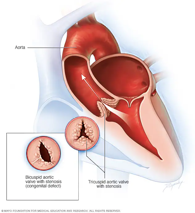
how does mild vs. moderate/severe aortic stenosis present?
mild: often asymptomatic or only a murmur
moderate/severe: dizziness, syncope, or chest pain with exertion
how may aortic stenosis be treated?
cardiac catheter-based intervention or surgical valvotomy, depending on severity and anatomy
what is transposition of the great arteries (TGA)?
the pulmonary artery exits the LV and the aorta exits the RV, creating aprallel rather than normally connected circulations.

why is an additional opening necessary for survival in TGA?
an atrial opening, PDA, or VSD allows oxygenated and deoxygenated blood to mix
what is the typical early manifestation of TGA?
moderate-to-severe cyanosis immediately after birth
🚨 NCLEX
why is continuous prostaglanding E1 used in TGA?
to keep the PDA open and permit blood mixing until catheter-based or surgical correction
what is the usual cause of heart failure in children under age 1?
congenital heart defects
what may heart failure after age 1 suggest when no congenital defect exists?
acquired heart disease
what findings suggest systemic venous congestion
weight fian, hepatomegaly, edema, and jugular venous distention
what findings suggest pulmonary venous congestion?
tachypnea, dyspnea, cough, and wheezing
what findings reflect compensation for poor cardiac output?
tachycardia, cardiomegaly, diaphoresis, fatigue, and failure to thrive
what findings suggest pulmonary venous congestion?
tachypnea, dyspnea, cough, and wheezing