BFA - Ch.6 Chemical Exam of Urine
 Call Kai
 Learn
Learn Practice Test
 Spaced Repetition
 Match
Match Flashcards
 Knowt Play
1/69

There's no tags or description
Looks like no tags are added yet.
Name | Mastery | Learn | Test | Matching | Spaced | Call with Kai | Chat |
|---|
No analytics yet
Send a link to your students to track their progress
70 Terms
what is used to perform routine chemical tests on urine
reagent strips
consist of chemical-impregnated absorbent pads on a plastic strip
test performed for pH, protein, glucose, ketones, blood, bilirubin, urobilinogen, nitrite, leukocyte esterase, and sp. grav
single and multitest strips available

what are the names 2 major types of reagent strips that are sold
Multistix and Chemstrip
some variations occur btwn the strips w/ regard to sensitivity, specificity, and interfering substances
users should be familiar w/ the product limitations
reagent strips are also used w. automated instruments
how is a reagent strip read
color comparison charts are supplied by the manufacturer
several degrees of color are shown to provide semiquantitative readings
neg, trace, 1+, 2+, 3+, and 4+
estimates of mg/dL are also provided for many test areas
reagent strip technqiue
dip strip briefly into well mixed specimen at rm temp
remove excess urine by touching edge of strip to container as strip is withdrawn
blot edge of strip on absorbent pad
wait specified amt of time
leukocyte esterase is the longest at 2 min
compare color rxn to manufacturer’s chart under good lighting
hold strip horizontally when comparing colors
Improper technique errors for reagent strips
not mixing sample - RBCs and WBCs sink to the bottom of an unmixed specimen
too cold/warm - enzyme reactions on strip are based on rm temp. readings
long dip time - reagents will leach off a strip remaining in urine too long - dip briefly
urine amt - excess urine on the strip will cause runover of reagents among the pads
quality control of reagent strips
all negative controls should = negative
pos. control readings should agree w/ published control values by ± 1 color block
be aware of manufacturer stated limitations and interfering substances
relate chemical readings to each other and physical and microscopic readings
liquid and tablet backup tests are available for some rxns
must also have + and - controls performed whenever these tests are required:
Protein: Sulfosalicylic Acid (SSA) Precipitation test
Galactose: Clinitest
Ketones: Acetest
Bilirubin: Ictotest
what tests require positive and negative controls
Protein: Sulfosalicylic Acid (SSA) Precipitation test
Galactose: Clinitest
Ketones: Acetest
Bilirubin: Ictotest
Urine pH - reagent strips
test pad measures acidity and alkalinity
pH sensitive dyes
kidneys are major regulator of acid-base balance
first morning specimen normally acidic (5-6)
postprandial specimen more alkaline
Ref range: 4.5-8
Normal fresh urine can’t reach pH 9
indicated old bacteria contaminated urine
too acidic or alkaline may form crystals and stones that can block passageways
what can affect urine pH?
diet and medication regulation
meat = acidic pH
vegetables = alkaline pH
medications for urinary tract infection → maintain an acid pH
Clinical significance of urine pH
respiratory or metabolic acidosis/ketosis
breathing problems, obesity, airway blockage
respiratory or metabolic alkalosis
hyperventilation, fever, sepsis, ventilators
defects in renal tubular secretion and reabsorption of acids and bases
renal tubular acidosis
renal calculi formation
treatment of urinary tract infections
precipitation/identification of crystals
protein in urine
most indicative of renal disease
proteinuria seen in early renal disease
Normal= <10 mg/dL or 100 mg/ 24 hr
low molecular weight serum proteins are filtered, while many are reabsorbed
albumin is primary protein of concern
clinical significance of proteinuria (values and causes)
30 mg/dL or 300 mg/24 hours
Variety of causes (prerenal, renal, post renal)
prerenal proteinuria
conditions affecting the plasma, not the kidney
transient causes due to reabsorption overflow
hemoglobin - intravascular hemolysis
myoglobin - muscle injury
infections/inflammation
Rarely seen on reagent strip b/c strip detects albumin
Prerenal proteinuria - Bence Jones Protein (BJP)
Multiple myeloma
elevated immunoglobulin light chains (monoclonal globulin protein)
body makes too many Abs, and they spill into the urine
requires confirmation by serum and urine immunoelectrophoresis
may not show on dipstick
Renal - Glomerular Proteinuria
damage to glomerular membrane which can impair selective filtration
allows for serum protein (albumin) to pass thru membrane and into the urine
Nephrotic syndrome - foamy urine
Inflammation
Nephritic syndrome - includes blood along w/ protein
Increased pressure on the filtration mechanism
hypertension
strenuous exercise
dehydration
preeclampsia
nephrotic syndrome typical features
onset: insidious
edema: ++++
bp: normal
jugular venous pressure: normal/low
proteinuria: ++++
Hematuria: May/may not occur
RBC casts: absent
serum albumin: low
nephritic syndrome typical features
onset: abrupt
edema: ++
bp: raised
jugular venous pressure: raised
proteinuria: ++
Hematuria: +++
RBC casts: present
serum albumin: normal/slightly reduced
renal - glomerular proteinuria causes
glomerulonephritis
glomerulosclerosis
Diabetes mellitus
sickl disease transplants and infectious diseases
cancers
drugs
increased pressure on the filtration mechanism (hypertension, strenuous exercise, dehydration, preeclampsia in pregnancy)
Renal - Tubular proteinuria
normal - small proteins filtered then reabsorbed
tubular damage affects reabsorptive ability
these proteins can no longer be absorbed
acute tubular dysfunction (toxic substances, heavy metals, viral infections, systemic diseases, hemolytic disorders, pyelonephritis
amount of protein is less than 4 g/day
difference in affects to the amount of albumin found in urine for glomerular damage in the kidney vs. tubular damge
glomerular - albumin
tubular - little to no albumin
microalbuminuria
small amounts of albumin
1st sign of diabetic nephropathy (could appear 5 years before seeing positive urine protein)
renal failure common in diabetics
early screening for albumin levels that are lower than seen on routine reagent strip
increased risk of cardiovascular disease
previously required 24 hr collection → reported mg/24 hr of albumin
random urine
postrenal proteinuria
protein added in the lower urinary and genitourinary tract
microbial infections causing inflammations and release of interstitial fluid protein
menstrual contamination or vaginal secretions
semen/ prostatic fluid
traumatic injury
principle of reagent strip reactions for protein
protein error of indicators
indicator dye release H+ if protein is present, causing a pH change and a change in color
pad maintained at pH 3.0 by a buffer
most sensitive to albumin → has more amino groups to accept H+ than other proteins
what is the color reaction that happens on the reagent strip for protein detection and what is it reported as
albumin + dye → albumin-dye complex + H+
color progresses thru green to blue
Report:
Neg, trace, 1+, 2+, 3+, 4+ OR
30, 100, 300, 2000 mg/dL
trace values are <30 mg/dL
what can interfere with protein detection on reagent strips
highly alkaline urine
produces a rise in pH and color change unrelated to protein
false positives
highly pigmented urine or high sp. grav
ammonium compounds, detergents or antiseptics
false-negatives for proteins other than albumin
Sulfosalicyclic Acid (SSA) Confirmation
screening or confirmation for protein in urine
precipitates proteins w/o heat
use centrifuged specimen → removed extraneous contamination
compare against standards for 1+, 2+, 3+, 4+
Interferences:
radiographic dye
antibiotics

clinical significance of glucose in urine
diabetes mellitus
renal threshold is 160-180 mg/dL
higher blood sugar= glycosuria
gestational diabetes
placental hormones block action of insulin
crosses placenta
hormonal disorders → insulin
renal glycosuria
glucose reagent strip rxn
glucose oxidase rxn specific for glucose
Glucose + O2 (glucose oxidase)→ gluconic acid + H2O2
H2O2 + chromogen (peroxidase)→ oxidized chromogen (colored) + H2O peroxidase
specific to glucose and no other sugar
reporting:
neg, trace, 1+, 2+, 3+, 4+
100-2000 mg/dL
what can interfere w/ glucose rxn on reagent strip
false positives
peroxide or oxidizing detergents
False negatives
enzymatic rxn interferences
ascorbic acid and strong reducing agents
high levels of ketons
high sp. grav
old specimens due to glycolysis - most common
clinitest procedure
not a specific test for glucose → tests for other reducing sugars (ex. galactose); confirmatory test
copper sulfate reacts w/ reducing substances to form cuprous oxide, which causes a color change
5 drops urine/ 10 drops diH2O
compare the color change to the color chart

ketones
three intermediate products of fat metabolism
acetone
acetoacetic acid
beta-hydroxybutyric acid
both acetone and beta-hydroxybutyric acid are produced from acetoacetic acid
appear in urine when fat is broken down to glucose for energy
what is the clinical significance of ketones in urine?
diabetes mellitus
inability to metabolize carbohydrate
ketonuria shows inadequate insulin
vomiting
creates loss of carbohydrates
starvation, malabsorption or dieting (fasting)
decrease in carbohydrate intake
reagent strip reaction for ketons
primarily measures acetoacetic acid
acetoacetic acid + Na nitroprusside → color change
Report:
neg, small (1+), moderate (2+), large (3+)
small (5,15), moderate (40), large (80, 160) mg/dL
what can interfere w/ ketone rxn on reagent strip
medications
abnormal urine color
only measures acetoacetate, not BHB/acetone
False-positives
improper timing
False-negative
old specimens
bacterial breakdown of acetoacetic acid
Acetest procedure
measures ketones
tablet form
sodium nitroprusside
provides a better color
slightly more sensitive
compare the tablet color w/ color chart
report as negative, small, moderate, or large
is the reagent strip or microscope more accurate for detecting blood?
the reagent strip
RBCs may lyse
what color urine does hematuria produce and what might it indicate?
intact RBCs→ causes a cloudy, red urine
causes:
renal calculi
glomerulonephritis
pyelonephritis
tumors
trauma
exposure to toxic chemicals
what color urine does hemoglobinuria produce and what might it indicate?
clear, red urine
causes:
transfusion rxns
hemolytic anemias
severe burns
infections/ malaria
strenuous exercise/ RBC trauma
brown recluse spider bites
hemosiderin
produces yellow-brown granules in sediment
reabsorption of filtered hemoglobin
myoglobinuria
myoglobin → heme containing protein w/i muscle tissue
produces clear, red/brown urine
caused by:
muscular trauma/ crush syndromes
prolonged coma
convulsions
muscle-wasting diseases
alcoholism/ overdose
drug abuse
cholesterol-lowering statin medications
clinical significance of hematuria
nephritis, trauma
clinical significance of hemoglobinuria
hemolysis
clinical significance of myoglobinuria
rhabdomyolysis, burns
reagent strip rxn for blood
lysing agent to lyse RBCs
Blood +peroxide lysing agent = hemoglobin
hemoglobin + indicator (bromphenol blue)= color change
intact RBCs show a speckled pattern
report:
trace, small (1+), moderate (2+), large (3+)
sensitivity to 5 RBCs/ µL
reaction interferences for blood on reagent strip
false positive
strong oxidizing agents
bacterial peroxidases
menstrual contamination
false-negative
high sp. grav
crenated cells
unmixed specimens
medications
ascorbic acid >25 mg/dL
bilirubin
a degradation product of hemoglobin
correlates w/ urobilinogen and serum bilirubin
clinical significance of bilirubin in urine
bile duct obstruction
bilirubin backs up into circulation
liver disease
bilirubin leaks back into circulation from damaged liver
bilirubin reagent strip rxn
bilirubin + diazo salt → azobilirubin
report: neg, small (1+), moderate (2+), large (3+)
sensitivity to 0.40 mg/dL
bilirubin reagent strip rxn interferences
false positive
urine pigments
false negative
old specimens
increased ascorbic acid and nitrite
exposure to light
Ictotest
confirmatory for bilirubin
diazo salt reaction
more sensitive: .05-.10 mg/dL
may be requested for early disease
report pos or neg

Bilirubin metabolism
Heme —heme oxygenase→ biliverdin —bilverdin reductase→ unconjugated bilirubin —UDPGT→ conjugated bilirubin —intestinal bacteria→ urobilinogen
urobiligen can travel from intestine to kidney → urinary urobilinogen
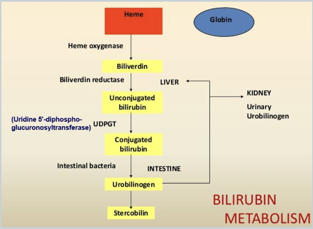
urobilinogen
bilirubin in intestine converted to _____ and sterocobilinogen
____ is reabsorbed into circulation
filtered by the glomerulus
always a small amount of ____ in the urine <1 mg/dL
clinical significance of urobilinogen in the body
early detection of liver disease
liver disorders, hepatitis, cirrhosis, or carcinoma
hemolytic disorders
excess bilirubin being converted to urobilinogen and increased urobilinogen recirculated to liver
negative bilirubin and strong positive urobilinogen
bile duct obstruction
no urobilinogen is seen in the urine
strip will give normal result
what does high urine bilirubin and normal urobilinogen indicate
bile duct obstruction
what can a ± urine bilirubin paired with a moderately high (++) urobilinogen indicate?
liver damage
what does negative urine bilirubin paired with strong positive (+++) urobilinogen indicate?
hemolytic disease
reagent strip reaction for urobilinogen
different principles for multistix and chemstrip
most common - Ehrlich reagent
urobilinogen + Ehrlich’s reagent —> colored complex
normal = 0.2-1 mg/dL, Abnormal= 2, 4, 8 mg/dL
rxn interference for urobilinogen - false positives for multistix
ehrlich reactive compounds
cold specimens
rxn interference for urobilinogen - false negatives for chemstrip
high nitrite interferences
rxn interference for urobilinogen -false results for both tests
due to urine colors
aged specimens'
timing of specimens
urobilinogen is highest after meals
clinical significance of nitrite (disease states and uses)
gram negative bacteria most common
UTI
Cystitis (bladder infection)
pyelonephritis (kidney infection)
evaluation of antibiotic therapy
monitoring of patients at high risk for urinary tract infection
nitrite reagent strip rxn
Nitrite + p-arsenilic acid → diazo compound
diazo compound + tetrahydrobenzaquinolinol → colored complex
tests ability of bacteria to reduce nitrate to nitrite
Greiss rxn
sensitive for 100,000 organisms/mL
Results: negative/positive
a true positive result should accompany a positive leukocyte esterase test
what can cause false negatives for nitrite test
other bacteria → skip nitrite stage to nitrogen gas
lack of urinary nitrate
presence of antibiotics
high conc. of ascorbic acid
high sp. grav
what can cause false positives for nitrite test
old specimens
bacterial multiplication
highly pigmented urine
pink edges or spotting on reagent strip is considered negative
Leukocyte esterase (LE)
detection of leukocytes (WBC) - “Pyuria”
not considered a quantitative test
perform microscopic evaluation is positive
detects presence of lysed leukocytes
release of leukocyte esterase
clinical significance of leukocyte esterase
UTI
acute inflammation
renal calculus
also seen w/ Trichomonas, Chlamydia, yeast, and interstitial nephritis
reagent strip rxn of LE
esterase + acid ester → acid + active alcohol
active alcohol + diazonium salt → color change
Report: trace, sm, mod, lg
15, 75, 125, 500 cells/ µL
false positive LE cause
strong oxidizing agents
highly pigmented urine
menstrual contamination
false negative LE causes
high conc. of protein, glucose, ascorbic acid, or antibiotics — reduces rxn sensitivity
leukocyte crenation from high sp. grav
prevents release of esterase
ascorbic acid
vitamin C
common interference w/ different urine tests
oxidizes certain compounds - effects hydrogen peroxide and diazo salt rxns
blood, bilirubin, glucose, and nitrite
low levels → false negatives
_____ pad on dipsticks