CHAPTER 16: PRINCIPLES OF MANAGEMENT & PREVENTION OF ODONTOGENIC INFECTIONS
 Call Kai
 Learn
Learn Practice Test
 Spaced Repetition
 Match
Match Flashcards
 Knowt Play
1/59

Earn XP
Description and Tags
ppt only / still need to get from book
Name | Mastery | Learn | Test | Matching | Spaced | Call with Kai | Chat |
|---|
No analytics yet
Send a link to your students to track their progress
60 Terms
odontogenic infections
infections that arise from teeth and is generally caused by bacteria
2 major origins:
periapical → from pulp necrosis (most common origin)
periodontal → from perio pocket
microbiology of odontogenic infections
polymicrobial
50% to 60% involves mixed bacteria
caused by normal oral bacterial flora (aerobic and anaerobic)
aerobic bacteria
gram-positive cocci → viridans-type Streptococci (45% occurrence)
anaerobic bacteria
gram-positive cocci:
Streptococcus
Peptostreptococcus
gram-negative rods:
Prevotella
Fusobacterium
Porphyromonas
pathophysiology of odontogenic infections
begins in periapical / periodontal area
↓
spreads in all directions favoring the path of least resistance
↓
gradually erodes cancellous bone toward facial or lingual cortices
↓
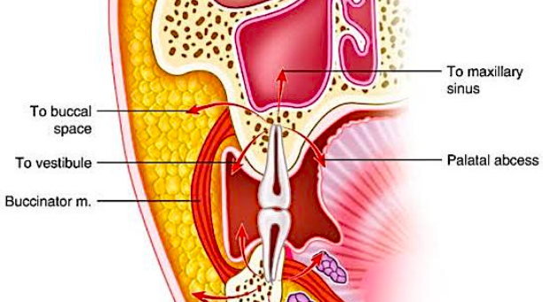
once through cortical bone, it spreads to fascial spaces (fascial spaces are not actual spaces but become created when invaded by infection)
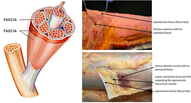
fascia
thin casing of connective tissue beneath the skin
these are “potential” spaces, only existing when invaded by bacteria
surrounds and holds every organ, blood vessel, bone, nerve fiber, muscle in place
the deep spaces of the head and neck are fascia-lined spaces containing loose areolar connective tissue
primary spaces
infratemporal
vestibular, buccal
canine / infraorbital
submandibular, submental, sublingual
secondary spaces
parapharyngeal
retropharyngeal
pterygomandibular
submasseteric / masseteric
superficial / deep temporal (masticator space)
prevertebral (deep cervical fascial spaces of the neck)
periorbital (masticator space) (deep cervical fascial spaces of the neck
location of the involved potential space is determined by:
the thickness of the bone overlying the apex of tooth
the relationship of the site of perforation of bone to muscle attachments of maxilla and mandible
thickness of the bone overlying the apex of tooth
periapical infections arising from mx molars generally erode through the thin buccal cortex
periapical infections arising from mn molars generally erode through the lingual cortex
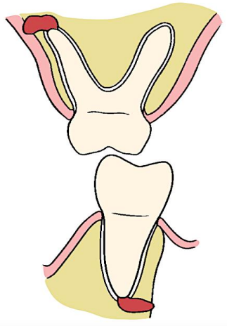

site of perforation of bone to muscle attachments of mx-mn
if bone erosion is below buccinator muscle attachment:
mx → vestibular space
mn → buccal space
if bone erosion is above buccinator muscle attachment:
mx → buccal space
mn → vestibular space

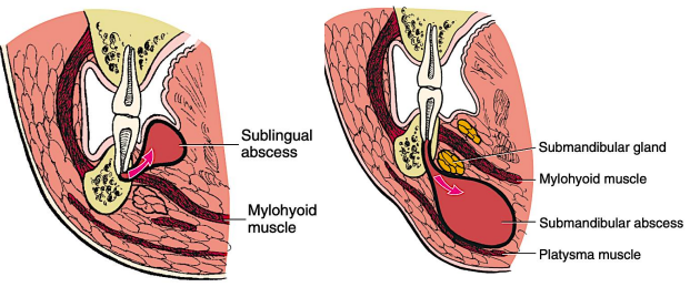
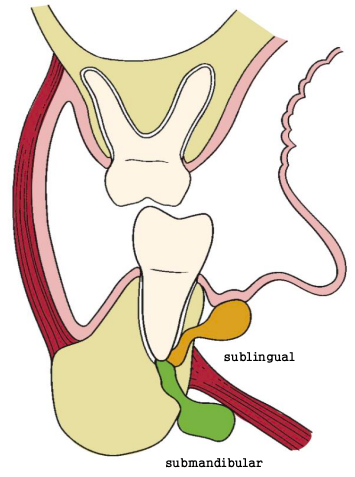
Lingual perforation
Lingual perforation superior to the mylohyoid attachment = sublingual space (shown in orange). Lingual perforation inferior to the mylohyoid attachment = submand
4 stages of odontogenic infections
edema or inoculation
cellulitis
abscess
resolution
resolution
spontaneously or surgically drainage of abscess destruction of bacteria by immune system-healing
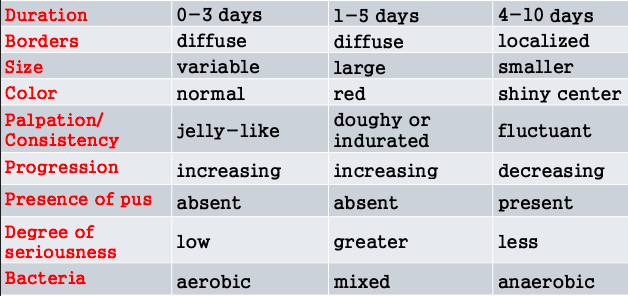
edema
0-3 days
diffuse borders
variable size
normal color
jelly-like consistency
increasing progression
absence of pus
low degree of seriousness
aerobic bacteria
cellulitis
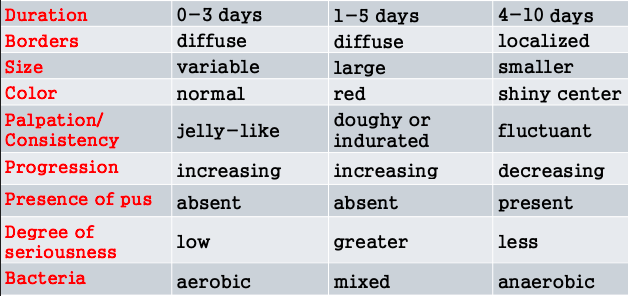
1-5 days
diffuse borders
larger size
red color
doughy or indurated consistency
increasing progression
absence of pus
greater degree of seriousness
mixed bacteria
abscess
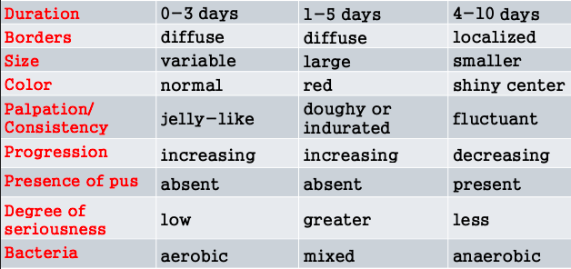
4-10 days
localized borders
smaller size
shiny center color
fluctuant consistency
decreasing progression
presence of pus
less degree of seriousness
anaerobic bacteria
principles of management of odontogenic infections
principle 1: determine severity of infection
principle 2: evaluate state of a px host defense mechanism
principle 3: determine whether px should be treated by general dentist or oral & maxillofacial surgeon
principle 4: treat infection surgically
principle 5: support px medically
principle 6: choose & prescribe appropriate antibiotics
principle 7: administer antibiotic properly
principle 8: evaluate the px frequently
principle 1: determine severity of infection
most odontogenic infections are mild and require only minor surgical therapy
severity can be determined by:
complete history taking
physical examination
complete history taking:
how long the infection has been present?
time of onset, progression of infection, rapidity of progression
information on patient’s symptoms
cardinal signs of inflammation
determine the general health of patient
malaise. fatigue, feverish, weak & sick patients
physical examination:
vital signs
pulse rate (increases with a rise in body temperature)
severe infection = >100 b/m
blood pressure
generally not altered by infection
pain and anxiety can cause an increase in BP
respiratory rate
normal: 12–20 b/m
mild-moderate infection: >20 b/m
temperature
normal: 36.5°C – 37.5°C (97.7°F – 99.5°F)
fever: more than 38°C (WHO)
[ergo, mild infection → vital signs normal, slight inc in temp
severe infection → vital signs abnormal, high inc in temp]
general appearance:
toxic appearance
suggests severe infection
elevated temperature, pulse, respiratory rate
head & neck examination
inspect cardinal signs of infection & inflammation
elevated temperature, pulse, respiratory rate
head & neck examination
assess tenderness, consistency of swelling
fleshy swelling → doughy swelling
firm or hard swelling → indurated swelling
fluctuant swelling → fluid-filled balloon (indicates liquid pus in the center)
others:
perform intra oral examination
radiographic examination
staging of infection
principle 2: evaluate state of a px host defense mechanism
estimate the patient's ability to defend against infection
can be reduced by:
medical conditions, such as:
uncontrolled diabetes mellitus
HIV, leukemia, malignancy,
congenital & acquired immunological disease
drugs, such as:
cancer chemotherapy
immunosuppressive drugs
citeria for referral to an OMFS
trismus
dehydration
need for GA
extra oral swelling
difficulty breathing
difficulty swallowing
temperature >101°F
failed previous treatment
compromised host defense
sever malaise / toxic appearance
criteria for immediate hospital emergency room admission
rapidly progressing infection
difficulty breathing (dyspnea)
temperature >101 °F (38.3 °C)
difficulty swallowing (dysphagia)
threat to airway or vital structures
trismus
results from involvement of the muscle of mastication by inflammatory process
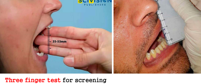
medial pterygoid muscle
the most commonly affected muscles during an IAN block
mild trismus
maximum interincisal opening between 30-40 mm
moderate trismus
maximum interincisal opening between 15-30mm
severe trismus
maximum interincisal opening is <15mm
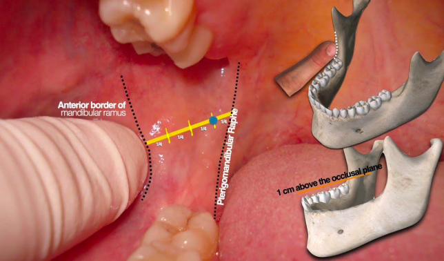
ways to anesthesize patients with trismus
vazirani akinosi
lingual nerve
inferior alveolar nerve
mylohyoid nerve (often)
mental and incisive nerves
gow-gates mandibular block
used if the patient has enough mouth opening
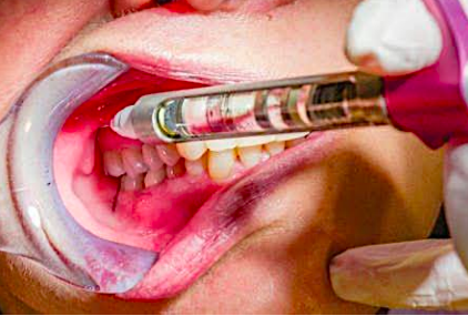
the principal managements of odontogenic infection
primary goal → remove the cause of infection
secondary goal → surgical drainage of pus
reasons for performing incision and drainage (I&D)
to decrease the load of bacteria and necrotic debris
to reduce hydrostatic pressure by decompressing tissues
to remove the accumulated pus and bacteria from the tissues
to prevent the spread of infection into deeper anatomic spaces
[ergo, decreases bacterial load and decreases pressure]
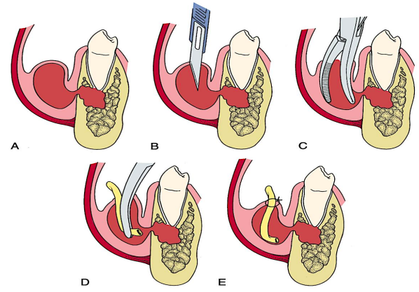
technique for I&D of a vestibular abscess or cellulitis
aka: Hilton’s Method
anesthesia
preferred → regional nerve block
alternative → infiltration
stab incision
at the site of maximum swelling
in mx: inferiorly for maximum gravity → dependent drainage
no more than 1cm in length
avoid frenum and mental nerve
insertion of hemostat or sinus forceps
if pus is not encountered, deepen the surgical site carefully with sinus forceps to avoid damaging vital structures
steps:
closed forceps are pushed through the deep fascia towards the pus collection
once the abscess cavity is entered, the forceps are opened parallel to vital structures
pus drains along the sides of the forceps
entire cavity is explored for additional loculi
placement of drain
a rubber drain is inserted into abscess cavity
external part is secured to the wound margin
drain is left for at least 2–5 days
purpose of drain:
allows continuous drainage of contents
maintains the opening for drainage
enables irrigation and debridement
commonly used drain:
¼ inch sterile penrose drain
alternative:
sterile glove or rubber dam
dressing
for extraoral incisions to protect the wound
principle 5: support patient medically
body's physiologic reserves are altered during infection
children:
susceptible to dehydration and high fever during infection
old patients:
less susceptible to fever, but in them dehydration occurs during infection
principle 6: choose & prescribe appropriate antibiotics
first, determine the NEED for antibiotic administration
the need can be determined from following 3 factors:
seriousness of infection
state of patient's host defenses
whether adequate surgical treatment can be achieved
[antibiotics do not speed up the wound healing and do not provide any benefit for non-bacterial conditions]
conditions that antibiotics are necessary
trismus & swelling
severe pericoronitis
temperature >101°F
deep fascial space infection
immunocompromised patient
osteomyelitis & lymphadenopathy
swelling extending beyond the alveolar process
acute onset infection with diffuse swelling & moderate to severe pain (cellulitis)
conditions that antibiotics are not necessary
toothache
severe pain
patient demand
mild pericoronitis
drained alveolar abscess
alveolar osteitis (dry socket)
minor, chronic, well-localized dentoalveolar, periapical, periodontal abscess
multiple extraction in patient who is NOT immunocompromised
empirical therapy
refers to treatment initiated before a definitive diagnosis is made
use of antibiotics on the assumption that it is the appropriate drug
recommended oral antibiotics
penicillin → first line (drug of choice)
penicillin G → original penicillin, IV route
penicillin V → oral route
amoxicillin
preferred due to better absorption
clindamycin
for penicillin allergy
cephalexin, clarithromycin, azithromycin
for clindamycin allergy
compliance decreases as dosage frequency increases
once daily → 80% compliance
twice daily → 69% compliance
indications for culture & sensitivity testing
recurrent infection
compromised host defense
rapidly progressive infection
previous multiple antibiotic therapy
nonresponsive infection (after >48hrs)
routine testing is NOT recommended but should be done in cases like nonresponsive infections!
principle 7: administer antibiotic properly
follow proper dose and at the proper dose interval
kills not only oral bacteria but also beneficial flora in the gut, skin
use cost effective antibiotics (do not always need to be expensive!)
choose drug which could be given once a day for not more than 4-5 days to get more compliance
avoid broad spectrum unless necessary, prefer narrow spectrum antibiotics (targets a limited range of bacteria)
plasma level of drug should be high enough to kill the bacteria but not so high to cause toxicity (peak plasma: 4-5x the minimal inhibitory concentration of bacteria)
use bactericidal antibiotic, if possible bc:
bactericidal
interferes with cell wall
bacteriostatic
interferes bacterial reproduction
use the antibiotic with lowest incidence of toxicity & side effects:
such as:
penicillin, clindamycin, macrolide, moxifloxacin
oral cephalosporins, tetracycline, metronidazole
narrow-spectrum antibiotics useful for treating simple odontogenic infections
penicillin
amoxicillin
clindamycin
metronidazole
[mnemonic: PACM]
broad-spectrum antibiotics useful for treating simple odontogenic infections
azithromycin
moxifloxacin
amoxi-clavulanic acid (for sinus infections)
[mnemonic: AMA]
penicillin
drug of choice
major side effect:
allergic reactions (hives, itching)
prophylactic dose:
2g ; 60mins before operation
clindamycin
advantages:
low incidence of toxicity & side effects
side effect:
pseudomembranous colitis caused by Clostridium difficile
macrolides
no longer used due to drug reactions
best alternative: azithromycin due to low toxicity
moxifloxacin (fluoroquinolone class)
advantages:
more effective than older fluoroquinolones
toxicity:
mental clouding
drug interactions
muscle weakness
oral cephalosporins (cephalexin & cefadroxil)
mild toxicity
cross-allergenicity with penicillin allergy
not recommended for odontogenic infections
tetracycline
mechanism: chelation to calcium
not used for odontogenic infections
used topically in high concentrations (periodontal pockets)
tooth discoloration (pregnant women; children under 12 year)
metronidazole
for anaerobic bacteria ONLY!
avoided in patients who are taking lithium
mild toxicity:
gastrointestinal disturbances
disulfiram-like reaction or antabuse effect
interacts with alcohol leading to:
severe abdominal cramping
violent vomiting
other side effects:
inhibits warfarin metabolism prolonging prothrombin time
principle 8: evaluate the patient frequently
if therapy is successful:
assess the I&D site to determine whether the drain can be removed
evaluate other key parameters:
trismus (should be improving)
swelling (should be reducing)
temperature (should be returning to normal)
patient’s subjective feelings (should report overall improvement)
if therapy is unsuccessful:
conduct a thorough examination to identify possible reasons for treatment failure such as:
inadequate drainage
persistent infection
antibiotic resistance
undiagnosed systemic conditions
prophylaxis of post-operative wound infection
antibiotics can reduce infection by up to 70% in OMFS
antibiotics DO NOT replace proper surgical technique
infection rate:
6–9% (clean-contaminated)
40% (dirty wounds)
prophylactic drug:
2x the usual therapeutic dose:
penicillin or amoxicillin → 2g (1hr before)
clindamycin → 600 mg (1hr before)
surgical features that need prophylaxis
complex surgeries
presence of foreign bodies
increase size of inoculum
immunocompromised patients
metastatic infection
refers to infections that occur in distant sites, not directly connected to the site of origin
most commonly occur via hematogenous route
sites most susceptible
heart valves = infective endocarditis
prosthetic joint replacements = prosthetic failure
prophylaxis against infective endocarditis
caused by bacteremia
an infection of the endocardial surface of the heart (heart valves)
non-antibiotic prophylactic measures:
oral hygiene
chlorhexidine gluconate rinses
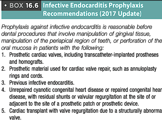
prophylaxis against prosthetic joint infection
for patients with recently placed prosthetic joints who are immunocompromised or have poor glycemic control
prophylaxis in px with other cardiovascular conditions
arterio-venous shunt (AV) shunts for dialysis
ventri-culo-atrial shunts (VA) shunts for hydrocephalus
antibiotic regimen for the prevention of infective endocarditis for adults
drug of choice
amoxicillin 2g 1hr prior the procedure (four 500mg tablets)
alternative if allergic to amox
clindamycin 600mg 1hr prior the procedure
antibiotic regimen for the prevention of infective endocarditis for children
drug of choice:
Amoxicillin 50mg/kg 1hr prior the procedure
alternative if allergic to amox:
Clindamycin 20mg/kg 1hr prior the procedure