1 - Retinal Anatomy and Physiology of the Healthy Retina
 Call Kai
 Learn
Learn Practice Test
 Spaced Repetition
 Match
Match Flashcards
 Knowt Play
1/38

Earn XP
Description and Tags
Block 1 (lecture 2 & 3 on ipad)
Name | Mastery | Learn | Test | Matching | Spaced | Call with Kai | Chat |
|---|
No analytics yet
Send a link to your students to track their progress
39 Terms
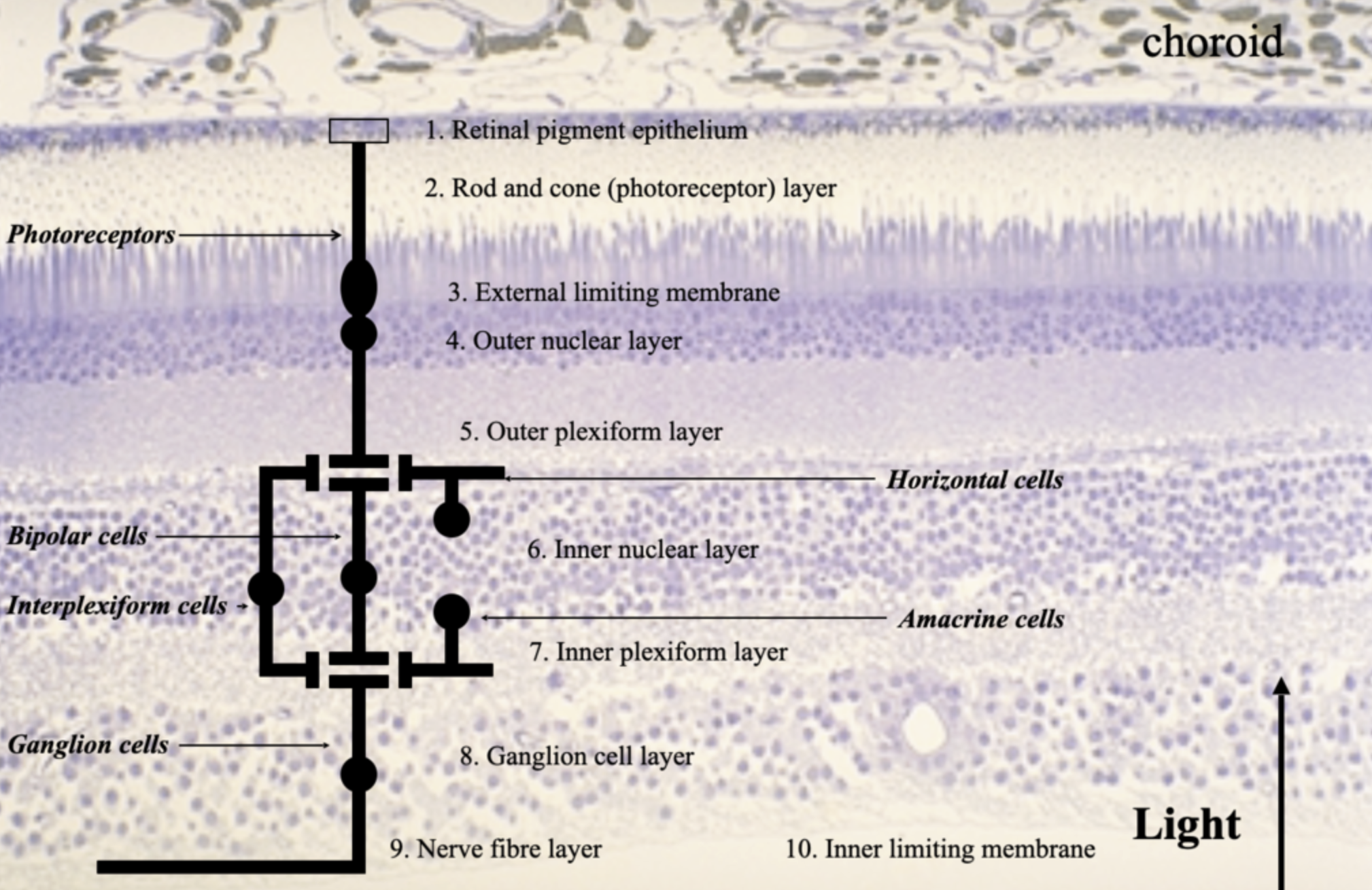
What are the main layers/cells of the retina from outer → inner retina?
RPE
Photoreceptor layer (rods [photopic vision] and cones [scotopic vision])
Outer limiting membrane
Outer nuclear layer
Outer plexiform layer
Inner nuclear layer
Inner plexiform layer
Ganglion cell layer
Retinal nerve fibre layer
Inner limiting membrane
![<ul><li><p>RPE</p></li><li><p>Photoreceptor layer (rods [photopic vision] and cones [scotopic vision])</p></li><li><p>Outer limiting membrane</p></li><li><p>Outer nuclear layer</p></li><li><p>Outer plexiform layer</p></li><li><p>Inner nuclear layer</p></li><li><p>Inner plexiform layer</p></li><li><p>Ganglion cell layer</p></li><li><p>Retinal nerve fibre layer</p></li><li><p>Inner limiting membrane</p></li></ul><p></p>](https://assets.knowt.com/user-attachments/48caa91c-7f36-4083-be8f-9e590786364a.png)
What are the main cells of the retina?
Photoreceptors - rods + cones
Bipolar cells
Horizontal cells
Amacrine cells
Retinal ganglion cells
Müller cells
RPE cells
What separates the choroid from the RPE?
Bruch’s membrane

What structures make up the outer retina, how is it supplied and whats the clinical relevance of these structures?
Structures
Choroid
Bruch’s membrane
RPE
Photoreceptors
Blood supply
Choroidal circulation
Also supplies foveal avascular region
Clinical relevance
Primary site of AMD pathogenesis
What is the choroid?
Its the vascular layer between sclera and Bruch’s membrane → supplied by long and short posterior ciliary arteries (originate from ophthalmic artery)
What is the anatomy choroid?
Haller layer = large vessels
Sattler layer = medium vessels
Choriocapillaris = fenestrated capillaries closest to Bruch’s

What is the function of the choroid?
Supplies outer retina with metabolites
Removes waste from outer retina
Why is the choroid containing the greatest density of vessels of the widest diameter important?
Higher uptake of O2 / nutrients due to increased metabolic demand
What are the age related changes of the choroid?
↓ choroidal thickness
↑ intercapillary spacing
↓ vessel number + diameter
↓ choroidal blood flow + volume
What are the layers of Bruch’s membrane?
RPE basal lamina
Inner collagenous layer
Elastic layer
Outer collagenous layer
Choriocapillaris basal lamina
Collagen + elastic fibres form a sieve-like structure

What are the functions of Bruch’s membrane?
Supports + anchors RPE
Semi-permeable barrier between RPE and choroid
Choroid → retina:
Nutrients
O₂
Retinoids
Retina/RPE → choroid:
Metabolic waste
How does Bruch’s membrane change with age?
↑ fibril cross-linking + density
Disrupted collagen/elastin turnover
Waste deposits in inner layers
↑ resistance to transport → ↑ waste
Lipid accumulation → ↓ water permeability
Thickens from ~2 µm at birth → 4–6 µm by 10th decade
What is the combined effect of ageing on Bruch’s membrane and the choroid?
↓ choroidal thickness
↓ capillary diameter + density
↑ Bruch’s membrane thickness
→ impaired O₂/metabolite delivery
→ impaired waste removal from outer retina
What is the structure of the RPE?
Monolayer of hexagonal cells
Basement membrane forms part of Bruch’s
Apical surface faces neural retina
Microvilli surround photoreceptor outer segments
No physical RPE–retina connections
What keeps the retina attached to the RPE?
Osmotic pressure
Fluid transport
Interphotoreceptor matrix
Vitreous
There are no physical connections between the RPE and retina
What are the main functions of the RPE?
Secretion → VEGF and PDF (maintains retinal integrity)
Phagocytosis → of outer segs of photoreceptors
Visual cycle → regenerates rhospsin
Glial-type support → blood retinal barrier in choroid
Epithelial transport → movement of O2 / H2O / nutrients

How does the RPE change with age?
Incomplete degradation of phagocytosed outer segments
Lipofuscin accumulates + extrudes into Bruch’s
↓ RPE cell density → ↑ demand on remaining cells
Dead cells add to RPE/Bruch’s waste
Lipofuscin:
Age 40: ~8% cytoplasmic space
Age 80: ~90%
What is the clinical significance of RPE lipofuscin?
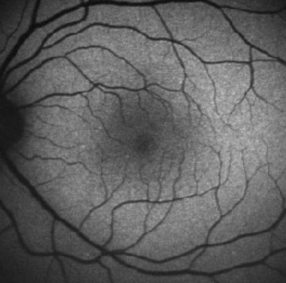
Bright on fundus autofluorescence
Peak density in parafovea
Toxic to RPE
Damages organelle membranes
↑ photo-oxidative damage
Can mechanically damage cell
What are the main structural features of photoreceptors?
Both rods + cones contain:
Outer segment
Inner segment
Cell body
Inner fibre
Synaptic terminal
Synaptic terminal:
Rod = spherule
Cone = pedicle
S-cones:
Longer inner segments
Smaller pedicles than L/M cones

How are rods and cones distributed across the retina?
Rods
~110–125 million
Maximum density ~18° from fovea
Cones
~6.3–6.8 million
Maximum density at fovea

How does the photoreceptor mosaic change with age?
Cones
No consistent age-related density change
Rods
~30% ↓ in central 28.5°
Loss begins in parafovea
Remaining rod outer segments expand to fill gaps
What are the functions and types of bipolar cells?
Function
Carry PR signals from OPL → amacrine cells + RGCs in IPL
Types
11 human types
1 rod-specific
Some involved in ON pathway others in OFF
Midget = few PR inputs
Diffuse = many PR inputs
What is the difference between ON and OFF bipolar pathways?
ON pathway
Excited by light on receptive-field centre
Synapses in inner IPL
OFF pathway
Inhibited by light on receptive-field centre
Synapses in outer IPL
What are the functions of horizontal and amacrine cells?
Horizontal cells
PR + bipolar synapses in OPL
Connected by gap junctions
Lateral communication
Feedback → PRs
Feedforward → bipolars
Amacrine cells
Bipolar + RGC synapses in IPL
Integrate/modulate RGC input
≥25 types
Compare midget and parasol retinal ganglion cells
Midget RGCs
~80%
Parvocellular pathway
Small receptive fields
High spatial-frequency sensitivity
Colour antagonism
Parasol RGCs
~10%
Magnocellular pathway
Larger receptive fields
Low spatial-frequency sensitivity
Strong response to transient/fast movement
What are bistratified, ON and OFF retinal ganglion cells?
Bistratified RGCs
Koniocellular pathway
Blue–yellow colour opponency
ON RGCs
Respond to light onset/light on dark
OFF RGCs
Respond to light offset/dark on light
How are fibres arranged in the RNFL?
Contains RGC axons + inner retinal vessels
Central fibres overlay peripheral fibres near ONH → thicker RNFL
Macular fibres → papillomacular bundle → temporal ONH
Temporal fibres → rgc axons arc below and above fovea to disc
Nasal fibres → rgc axons direct to disc
How is retinal blood flow divided between the choroidal and retinal circulations?
Choroid
65–85% of retinal blood flow
Via short + long posterior ciliary arteries
Supplies outer retina
Central retinal artery
~20–30%
Enters from ONH
Supplies inner retina
Cilioretinal artery:
Present in 15–20%
What capillary layers are supplied by the central retinal artery?
CRA → 4 main branches → 3 capillary layers:
Radial peripapillary capillaries → RNFL
Inner capillary layer → ganglion cell layer
Outer capillary layer → IPL to OPL
Drainage:
Capillaries → venules → central retinal vein
How is the macula defined?
Clinical
Central retina specialised for high-resolution VA
~6 mm diameter centred on fovea
~15–20° visual angle
Anatomical
Region where ganglion cell layer is >1 cell thick
What regions make up the macula?
Outer → inner:
Perifovea
Parafovea
Fovea
Foveola
Fovea
1.85 mm diameter
~5.5° visual field
Foveola
~1°20′ visual field
How are photoreceptors specialised at the foveola?
Adaptation
Maximum cone density: ~200,000/mm²
Foveal cones more rod-like due to high PR density
Consequence
Maximum visual resolution
Front: How are RGCs specialised in the macular region?
Adaptation
~50% of RGCs within 13° of foveola
Peak density ~35,100 cells/mm² at ~1 mm eccentricity
Consequence
Very high neural sampling density.
How does the cone:RGC ratio contribute to high foveal acuity?
Cone:RGC ratio lowest in macula
Foveal centre ≈ 1 cone : 2 RGCs
Each cone can contribute to ON + OFF pathways
→ low neural convergence
→ small receptive fields
→ high sampling density
→ high spatial acuity.
cones - highest = foveola, lowest = periphery
rods - highest = mid periphery, lowest = foveola (no rods present)

How does displacement of retinal layers specialise the foveola?
Adaptation
Cells proximal to PRs displaced laterally
Foveola = thinnest retina
Achieved by elongated cone axons → Henle fibre layer
Consequences
↓ light scatter before reaching PRs
Parafoveal GCL up to 6 layers → thickest retinal region.
How is the foveal blood supply specialised?
Adaptation
Fovea is vessel + capillary free
Macular vascular ring from superior temporal + inferotemporal arteries
Consequences
↓ light scatter from vessels
FAZ relies on choroidal circulation
What is macular pigment and where is it found?
Dietary xanthophyll carotenoids:
Lutein
Zeaxanthin
Found in:
Cone axons of Henle fibre layer
Interneurons of IPL
What are the functions of macular pigment?
Antioxidant → removes free radicals that cause oxidative damage to retina
Filters short-wavelength light
Improves image quality by removing wavelengths prone to chromatic aberrations
Blocks fundus autofluorescence → appears dark on FAF.
What features of the macula maximise central visual acuity?
Photoreceptors
Very high cone density
Neural
High RGC density
Low cone:RGC ratio
Low convergence
Small receptive fields
Structural
Inner retinal layers displaced from foveola
↓ light scatter
Vascular
Foveal avascular zone = minimise scatter
Macular pigment
Filters short wavelengths
Antioxidant
Improves image quality.