Toe, Foot, Ankle, Tib/fib Check off cards

0.0(0)
Studied by 0 people
call kaiCall Kai
learnLearn
examPractice Test
spaced repetitionSpaced Repetition
heart puzzleMatch
flashcardsFlashcards
GameKnowt Play
Card Sorting

1/12

encourage image

There's no tags or description

Looks like no tags are added yet.

13 Terms
1
New cards
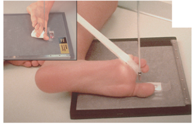
<p><strong>AP Toe</strong></p><p></p><p>Centered: ?</p><p>Collimation: ?</p><p>SID: ?</p><p>Technique: ?</p><p>Angle?</p><p>Anatomy included: ?</p>

AP Toe


Centered: ?

Collimation: ?

SID: ?

Technique: ?

Angle?

Anatomy included: ?

Centered: MTP Joint

Collimation: 10 x 12

SID: 40 - 45” SID

Technique: 2 @ 60

10 degree cephalic angle

Anatomy included: digit of interest and a minimum of the distal half of the metatarsal
<p>Centered: <span style="background-color: transparent;">MTP Joint</span></p><p>Collimation: <span style="background-color: transparent;">10 x 12</span></p><p>SID: <span style="background-color: transparent;">40 - 45” SID</span></p><p>Technique: <span style="background-color: transparent;">2 @ 60</span></p><p><span style="background-color: transparent;"><strong>10 degree cephalic angle</strong></span></p><p>Anatomy included: <span style="background-color: transparent;">digit of interest and a minimum of the distal half of the metatarsal</span></p>
2
New cards
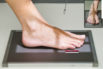
<p><strong>Oblique Toe</strong></p><p></p><p>Centered: ?</p><p>Collimation: ?</p><p>SID: ?</p><p>Technique: ?</p><p>Angle?</p><p>Medial or lateral roll for which digits? </p><p>Anatomy included: ?</p>

Oblique Toe


Centered: ?

Collimation: ?

SID: ?

Technique: ?

Angle?

Medial or lateral roll for which digits?

Anatomy included: ?

Centered: MTP Joint

Collimation: 10 x 12

SID: 40 - 45” SID

Technique: 2 @ 60

No Angle

Rotate leg & foot 30 degrees medially for the 1st - 3rd digits and laterally for the 4th & 5th digits

Anatomy included: digit of interest and a minimum of the distal half of the metatarsal

<p>Centered: <span style="background-color: transparent;">MTP Joint</span></p><p>Collimation: <span style="background-color: transparent;">10 x 12</span></p><p>SID: <span style="background-color: transparent;">40 - 45” SID</span></p><p>Technique: <span style="background-color: transparent;">2 @ 60</span></p><p><span style="background-color: transparent;">No Angle</span></p><p><span style="background-color: transparent;">Rotate leg &amp; foot <strong>30 degrees medially</strong> for the 1st - 3rd digits and laterally for the 4th &amp; 5th digits</span></p><p>Anatomy included: <span style="background-color: transparent;">digit of interest and a minimum of the distal half of the metatarsal</span></p>
3
New cards
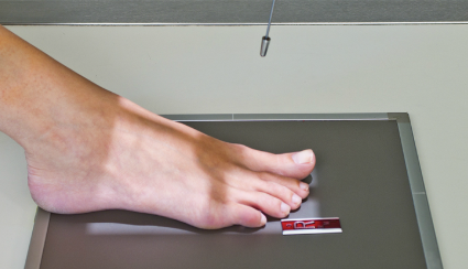
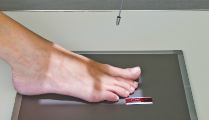
<p><strong>Lateral Toe</strong></p><p></p><p>Centered: ?</p><p>Collimation: ?</p><p>SID: ?</p><p>Technique: ?</p><p>Angle?</p><p>Medial or lateral roll for which digits? </p><p>Anatomy included: ?</p>

Lateral Toe


Centered: ?

Collimation: ?

SID: ?

Technique: ?

Angle?

Medial or lateral roll for which digits?

Anatomy included: ?

Centered: MTP Joint

Collimation: 10 x 12

SID: 40 - 45” SID

Technique: 2 @ 60

No Angle

Rotate affected leg medially for 1st - 3rd digits and laterally for 4th & 5th digits

Anatomy included: digit of interest and a minimum of the distal half of the metatarsal

<p>Centered: <span style="background-color: transparent;">MTP Joint</span></p><p>Collimation: <span style="background-color: transparent;">10 x 12</span></p><p>SID: <span style="background-color: transparent;">40 - 45” SID</span></p><p>Technique: <span style="background-color: transparent;">2 @ 60</span></p><p><span style="background-color: transparent;">No Angle</span></p><p><span style="background-color: transparent;">Rotate affected leg medially for 1st - 3rd digits and laterally for 4th &amp; 5th digits</span></p><p>Anatomy included: <span style="background-color: transparent;">digit of interest and a minimum of the distal half of the metatarsal</span></p>
4
New cards
<p><strong>AP Foot</strong></p><p></p><p>Centered: ?</p><p>Collimation: ?</p><p>SID: ?</p><p>Technique: ?</p><p>Angle?</p><p>Anatomy included: ?</p>

AP Foot


Centered: ?

Collimation: ?

SID: ?

Technique: ?

Angle?

Anatomy included: ?

Centered: Base of 3rd Metatarsal

Collimation: 10 x 12

SID: 40 - 45” SID

Technique: 2 @ 65

10 degree cephalic angle (CR perpendicular to metatarsals)

Anatomy included: entire foot up to lower ankle, Bases of 1st & 2nd metatarsals are separated, Bases of 2nd - 5th metatarsals overlap

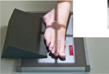
<p>Centered: <span style="background-color: transparent;">Base of 3rd Metatarsal</span></p><p>Collimation: <span style="background-color: transparent;">10 x 12</span></p><p>SID: <span style="background-color: transparent;">40 - 45” SID</span></p><p>Technique: <span style="background-color: transparent;">2 @ 65</span></p><p><span style="background-color: transparent;"><strong>10 degree cephalic angle</strong> (CR perpendicular to metatarsals)</span></p><p>Anatomy included: <span style="background-color: transparent;">entire foot up to lower ankle, Bases of 1st &amp; 2nd metatarsals are separated, Bases of 2nd - 5th metatarsals overlap</span></p>
5
New cards
<p><strong>Oblique Foot</strong></p><p></p><p>Centered: ?</p><p>Collimation: ?</p><p>SID: ?</p><p>Technique: ?</p><p>Angle?</p><p>Medial or lateral roll?</p><p>Anatomy included: ?</p>

Oblique Foot


Centered: ?

Collimation: ?

SID: ?

Technique: ?

Angle?

Medial or lateral roll?

Anatomy included: ?

Centered: Base of 3rd Metatarsal

Collimation: 10 x 12

SID: 40 - 45” SID

Technique: 2 @ 65

No Angle

Rotate foot 30 degrees medially

Anatomy included: entire foot up to lower ankle, 3rd - 5th metatarsals are free of superimposition, Tuberosity of 5th is seen in profile, Sinus Tarsi appears open
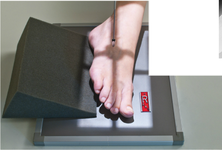

<p>Centered: <span style="background-color: transparent;">Base of 3rd Metatarsal</span></p><p>Collimation: <span style="background-color: transparent;">10 x 12</span></p><p>SID: <span style="background-color: transparent;">40 - 45” SID</span></p><p>Technique: <span style="background-color: transparent;">2 @ 65</span></p><p><span style="background-color: transparent;">No Angle</span></p><p><span style="background-color: transparent;">Rotate foot <strong>30 degrees</strong> medially</span></p><p><span style="background-color: transparent;">Anatomy included: entire foot up to lower ankle, 3rd - 5th metatarsals are free of superimposition, Tuberosity of 5th is seen in profile, Sinus Tarsi appears open</span></p><p></p>
6
New cards
<p><strong>Lateral Foot</strong></p><p></p><p>Centered: ?</p><p>Collimation: ?</p><p>SID: ?</p><p>Technique: ?</p><p>Angle?</p><p>Medial or lateral roll for which digits?</p><p>Anatomy included: ?</p>

Lateral Foot


Centered: ?

Collimation: ?

SID: ?

Technique: ?

Angle?

Medial or lateral roll for which digits?

Anatomy included: ?

Centered: CR @ Medial Cuneiform

Collimation: 10 x 12

SID: 40 - 45” SID

Technique: 4 @ 65

No Angle, Mediolateral projection

Roll so plantar surface is perpindicular to IR (laterally)

Anatomy included: entire foot up to lower ankle, Heads of metatarsals should be superimposed, 5th metatarsal seen in profile, distal fibula should be superimposed over posterior tibia

<p>Centered: <span style="background-color: transparent;">CR @ Medial Cuneiform</span></p><p>Collimation: <span style="background-color: transparent;">10 x 12</span></p><p>SID: <span style="background-color: transparent;">40 - 45” SID</span></p><p>Technique: <span style="background-color: transparent;">4 @ 65</span></p><p><span style="background-color: transparent;">No Angle, Mediolateral projection</span></p><p><span style="background-color: transparent;">Roll so plantar surface is perpindicular to IR (laterally)</span></p><p><span style="background-color: transparent;">Anatomy included: entire foot up to lower ankle, Heads of metatarsals should be superimposed, 5th metatarsal seen in profile, distal fibula should be superimposed over posterior tibia</span></p>
7
New cards
<p><span style="background-color: transparent;"><strong>Plantodorsal Calcaneus</strong></span></p><p></p><p>Centered: ?</p><p>Collimation: ?</p><p>SID: ?</p><p>Technique: ?</p><p>Angle?</p><p>Foot Flexed or Extended?</p><p>Anatomy included: ?</p>

Plantodorsal Calcaneus


Centered: ?

Collimation: ?

SID: ?

Technique: ?

Angle?

Foot Flexed or Extended?

Anatomy included: ?

Centered: Base of 3rd Metatarsal

Collimation: 10 x 12

SID: 40-45” SID

Technique: 5 @ 70

40 degree cephalic angle

Dorsiflex foot

Anatomy included: Entire foot from heel up to head of metatarsals, Sustentaculum Tali in profile
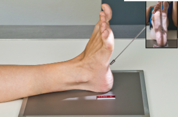
<p>Centered: <span style="background-color: transparent;">Base of 3rd Metatarsal</span></p><p>Collimation: <span style="background-color: transparent;">10 x 12</span></p><p>SID: <span style="background-color: transparent;">40-45” SID</span></p><p>Technique: <span style="background-color: transparent;">5 @ 70</span></p><p><span style="background-color: transparent;"><strong>40 degree cephalic angle</strong></span></p><p><span style="background-color: transparent;">Dorsiflex foot</span></p><p>Anatomy included: Entire foot from heel up to head of metatarsals, <span style="background-color: transparent;">Sustentaculum Tali in profile</span></p>
8
New cards
<p><span style="background-color: transparent;"><strong>Lateral Calcaneus</strong></span></p><p></p><p>Centered: ?</p><p>Collimation: ?</p><p>SID: ?</p><p>Technique: ?</p><p>Angle?</p><p>Foot Flexed or Extended?</p><p>Anatomy included: ?</p>

Lateral Calcaneus


Centered: ?

Collimation: ?

SID: ?

Technique: ?

Angle?

Foot Flexed or Extended?

Anatomy included: ?

Centered: Mid Os Calcis

Collimation: 10x 12

SID: 40-45” SID

Technique: 4 @ 65

No Angle, medio-lateral projection

Roll so plantar surface perpendicular to IR

Anatomy included: Lower ankle to bottom of foot vertically, and mid medial arch to back of heel horizontally. Include entire calcaneus, including joint spaces, Distal fibula centered over posterior half of tibia

<p>Centered: <span style="background-color: transparent;">Mid Os Calcis</span></p><p>Collimation: <span style="background-color: transparent;">10x 12</span></p><p>SID: <span style="background-color: transparent;">40-45” SID</span></p><p>Technique: <span style="background-color: transparent;">4 @ 65</span></p><p>No Angle, medio-lateral projection</p><p>Roll so <span style="background-color: transparent;">plantar surface perpendicular to IR</span></p><p>Anatomy included: Lower ankle to bottom of foot vertically, and mid medial arch to back of heel horizontally. <span style="background-color: transparent;">Include entire calcaneus, including joint spaces, Distal fibula centered over posterior half of tibia</span></p>
9
New cards
<p><span style="background-color: transparent;"><strong>AP Ankle</strong></span></p><p></p><p>Centered: ?</p><p>Collimation: ?</p><p>SID: ?</p><p>Technique: ?</p><p>Angle?</p><p>Foot Flexed or Extended?</p><p>Anatomy included: ?</p>

AP Ankle


Centered: ?

Collimation: ?

SID: ?

Technique: ?

Angle?

Foot Flexed or Extended?

Anatomy included: ?

Centered: Ankle Joint

Collimation: 10 x 12

SID: 40-45” SID

Technique: 6 @ 65

No Angle

Anatomy included: Dorsiflex if possible, do not force it. Include distal ⅓ of lower leg. Both malleoli should be visible, Lateral mortise joint should be closed, Superior and medial mortise joint should appear open
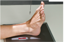
<p>Centered: <span style="background-color: transparent;">Ankle Joint</span></p><p>Collimation: <span style="background-color: transparent;">10 x 12</span></p><p>SID: <span style="background-color: transparent;">40-45” SID</span></p><p>Technique: <span style="background-color: transparent;">6 @ 65</span></p><p><span style="background-color: transparent;">No Angle</span></p><p>Anatomy included: <span style="background-color: transparent;">Dorsiflex if possible, do not force it. Include distal ⅓ of lower leg. Both malleoli should be visible, Lateral mortise joint should be closed, Superior and medial mortise joint should appear open</span></p>
10
New cards
<p><span style="background-color: transparent;"><strong>Mortise Ankle</strong></span></p><p></p><p>Centered: ?</p><p>Collimation: ?</p><p>SID: ?</p><p>Technique: ?</p><p>Angle?</p><p>Foot rotated medially or laterally?</p><p>Anatomy included: ?</p>

Mortise Ankle


Centered: ?

Collimation: ?

SID: ?

Technique: ?

Angle?

Foot rotated medially or laterally?

Anatomy included: ?

Centered: Midway between malleoli

Collimation: 10 x 12

SID: 40-45” SID

Technique: 6 @ 65

No Angle

Rotate entire leg & foot 15 degrees medially until intermalleolar line is parallel to IR

Anatomy included: Include distal ⅓ of lower leg, Both malleoli should be visible, Entire mortise joint should be open
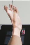
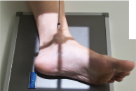
<p>Centered: <span style="background-color: transparent;">Midway between malleoli</span></p><p>Collimation: <span style="background-color: transparent;">10 x 12</span></p><p>SID: <span style="background-color: transparent;">40-45” SID</span></p><p>Technique: <span style="background-color: transparent;">6 @ 65</span></p><p>No Angle</p><p><span style="background-color: transparent;">Rotate entire leg &amp; foot 15 degrees medially until intermalleolar line is parallel to IR</span></p><p>Anatomy included: <span style="background-color: transparent;">Include distal ⅓ of lower leg, Both malleoli should be visible, Entire mortise joint should be open</span></p>
11
New cards
<p><strong>Lateral Ankle</strong></p><p></p><p>Centered: ?</p><p>Collimation: ?</p><p>SID: ?</p><p>Technique: ?</p><p>Angle?</p><p>Foot rotated medially or laterally?</p><p>Anatomy included: ?</p>

Lateral Ankle


Centered: ?

Collimation: ?

SID: ?

Technique: ?

Angle?

Foot rotated medially or laterally?

Anatomy included: ?

Centered: @ Medial Malleolus

Collimation: 10 x 12

SID: 40-45” SID

Technique: 5 @ 65

No Angle, mediolateral projection

Dorsiflex foot if possible, Rotate leg & foot 90 degrees so plantar surface is perpendicular to IR

Anatomy included: distal ⅓ of lower leg, Distal, posterior ½ of the tibia should cover the fibula, Tibiotalar joint should appear open

<p>Centered: <span style="background-color: transparent;">@ Medial Malleolus</span></p><p>Collimation: <span style="background-color: transparent;">10 x 12</span></p><p>SID: <span style="background-color: transparent;">40-45” SID</span></p><p>Technique: <span style="background-color: transparent;">5 @ 65</span></p><p>No Angle, mediolateral projection</p><p><span style="background-color: transparent;">Dorsiflex foot if possible, Rotate leg &amp; foot 90 degrees so plantar surface is perpendicular to IR</span></p><p><span style="background-color: transparent;">Anatomy included: distal ⅓ of lower leg, Distal, posterior ½ of the tibia should cover the fibula, Tibiotalar joint should appear open</span></p>
12
New cards
<p><strong>AP Tib/Fib</strong></p><p></p><p>Centered: ?</p><p>Collimation: ?</p><p>SID: ?</p><p>Technique: ?</p><p>Angle?</p><p>Foot positioned how?</p><p>Anatomy included: ?</p>

AP Tib/Fib


Centered: ?

Collimation: ?

SID: ?

Technique: ?

Angle?

Foot positioned how?

Anatomy included: ?

Centered: Mid Lower Leg

Collimation: 14 × 17

SID: 44-48”

Technique: 6 @ 70

No angle

Dorsiflex foot

Anatomy included: Knee joint down to and including malleoli

<p>Centered: Mid Lower Leg</p><p>Collimation: 14 × 17</p><p>SID: <strong>44-48”</strong></p><p>Technique: 6 @ 70</p><p>No angle</p><p><span style="background-color: transparent;">Dorsiflex foot</span></p><p>Anatomy included: Knee joint down to and including<span style="background-color: transparent;"> malleoli</span></p>
13
New cards
<p><strong>Lateral Tib/Fib</strong></p><p></p><p>Centered: ?</p><p>Collimation: ?</p><p>SID: ?</p><p>Technique: ?</p><p>Angle?</p><p>Foot positioned how?</p><p>Anatomy included: ?</p>

Lateral Tib/Fib


Centered: ?

Collimation: ?

SID: ?

Technique: ?

Angle?

Foot positioned how?

Anatomy included: ?

Centered: Mid Lower Leg

Collimation: 14 × 17

SID: 44-48”

Technique: 6 @ 70

No angle, mediolateral projection

Dorsiflex foot, roll laterally

Anatomy included: Knee joint down to and including malleoli, distal fibula needs cover posterior half of tibia

<p>Centered: Mid Lower Leg</p><p>Collimation: 14 × 17</p><p>SID: <strong>44-48”</strong></p><p>Technique: 6 @ 70</p><p>No angle, mediolateral projection</p><p><span style="background-color: transparent;">Dorsiflex foot, roll laterally</span></p><p>Anatomy included: Knee joint down to and including<span style="background-color: transparent;"> malleoli, distal fibula needs cover posterior half of tibia</span></p>