*Exam 2 practice AP 1*
 Call Kai
 Learn
Learn Practice Test
 Spaced Repetition
 Match
Match Flashcards
 Knowt Play
1/134

There's no tags or description
Looks like no tags are added yet.
Name | Mastery | Learn | Test | Matching | Spaced | Call with Kai | Chat |
|---|
No analytics yet
Send a link to your students to track their progress
135 Terms
A space within a bone filled with air and lined with mucus membrane is a:
Sinus
The hyoid belongs to which of the two main divisions of the skeleton?
Axial

Identify the indicated space within the long bone.
Medullary cavity

Identify the indicated type of bone within the long bone.
Compact bone

Identify the indicated space within compact bone.
Lacunae

Identify the indicated structure (layer) within compact bone.
Lamella

What type (shape) of bone is indicated?
Irregular

What type (shape) of bone is indicated?
Long
In adults, the inner (medullary) cavity of the shaft of long bones contains:
Yellow marrow
What is the name of the bone that forms the forehead?
Frontal

Identify the indicated bone.
Maxilla

Identify the indicated bone.
Temporal

Identify the indicated bone marking (projection).
Mastoid process

Identify the indicated bone marking (suture).
lambdoid suture


Identify the indicated bone marking (opening).
infraorbital foramen
THINK:
→ infra = below/beneath
→ orbital = eye / eye socket
→ foramen = hole


Identify the indicated bone marking (opening).
superior orbital fissure
THINK:
Superior = above
Orbital = eye
Fissure = a narrow slit or opening
“upper slit of the eye socket”


Identify the indicated bone.
sacrum

Identify the indicated bone.
atlas

What type of rib is indicated?
true rib

Identify the indicated sinus.
frontal

Identify the indicated bone marking (opening).
vertebral foramen
THINK:
vertebral = related to s vertebra, a bone of the spine
foramen = hole
“the opening in a vertebra”
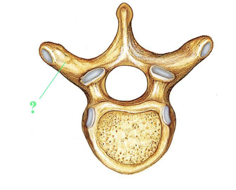
Identify the indicated bone marking (projection).
transverse process
THINK:
Transverse = across or sideways
Process = a projection sticking out from a bone
“A sideways projection of bone.”
Remember:
Transverse: to the sides
Spinous = toward the back
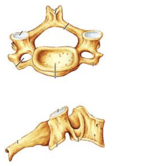
Identify the indicated type of vertebra.
cervical
7 → Cereal for breakfast at 7am
→ looks like jack-o-lantern
→ MUCH smaller than other vertebrae
→ DISTINGUISHING FEATURE:
transverse foramina (hole on each side
What structure on forms the medial bump on the "ankle"?
medial malleolus
What is the name of the lateral bone of the forearm?
radius (thumb side)

Identify the indicated bone.
femur

Identify the indicated bone.
tibia
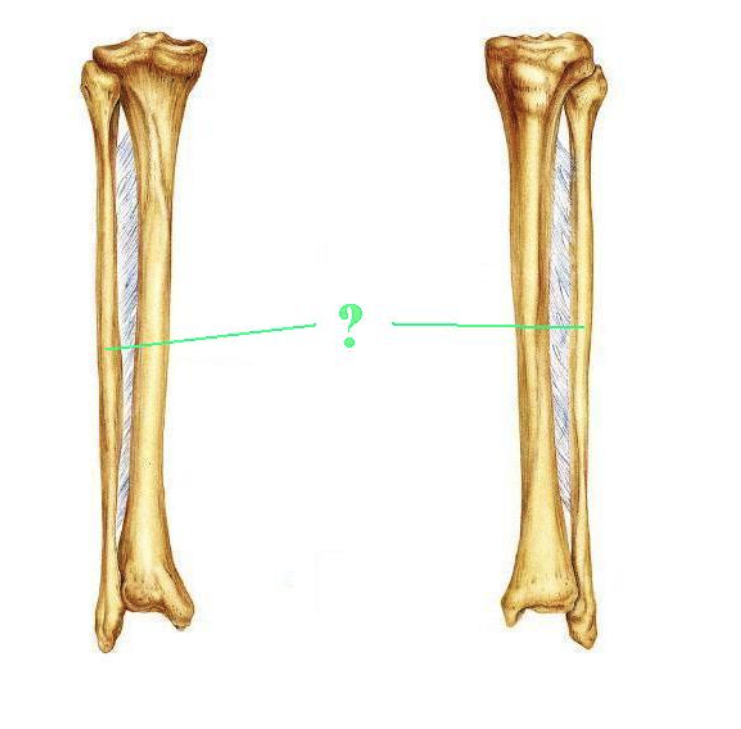
Identify the indicated bone.
fibula
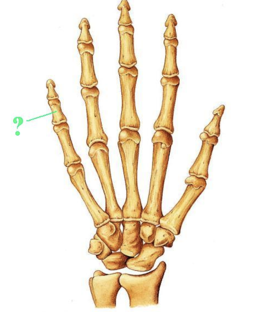
Identify the indicated bone.
middle phalanx

Identify the indicated bone.
Pisiform
THINK:
Some = Scaphoid
Lovers = Lunate
Try = Triquetrum
Positions = Pisiform
That = Trapezium
They = Trapezoid
Cannot = Capitate
Handle = Hamate
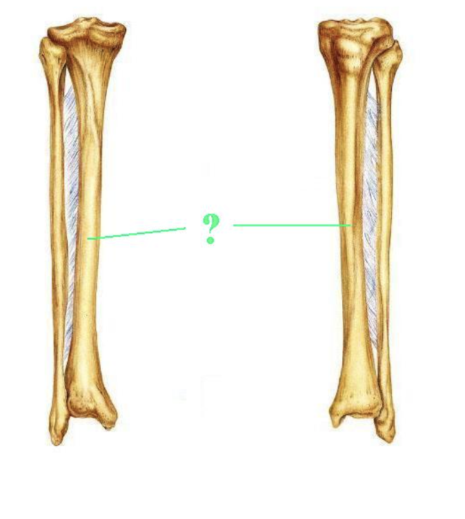
Is the indicated bone from the right of left side of the body?
left

Is the indicated bone from the right of left side of the body?
right

Identify the indicated bone marking (projection).
deltoid tuberosity
THINK:
Word | Meaning |
|---|---|
Deltoid | Triangle-shaped; the name of your shoulder muscle |
Tuberosity | A rough, raised area on a bone |
Humerus | Your upper arm bone |
“The rough area on the upper arm bone where the deltoid muscle attaches.”
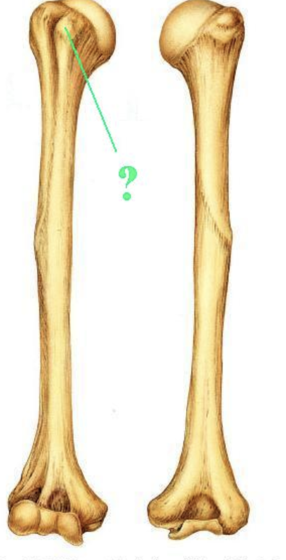
Identify the indicated bone marking (projection).
lesser tubercle
THINK:
Word | Meaning |
|---|---|
Lesser | Smaller |
Tubercle | A small, rounded bump on a bone |

Identify the indicated bone marking (surface)?
facet for the lateral condyle of femur
THINK:
Word | Meaning |
|---|---|
Facet | A smooth surface where bones meet |
Lateral | Toward the outer side |
Condyle | A rounded end of a bone that forms a joint |
Femur | Thigh bone |
Together: “The smooth surface that meets the outer rounded end of the thigh bone.”
Locate it: On the back of the patella (kneecap). The lateral facet is usually larger than the medial facet.
Function: Contacts the femur and helps the kneecap glide as you bend and straighten your knee.

Identify the indicated bone marking (projection)?
medial condyle
THINK:
Word | Meaning |
|---|---|
Medial | Toward the body’s midline |
Condyle | A rounded part of a bone that forms a joint |
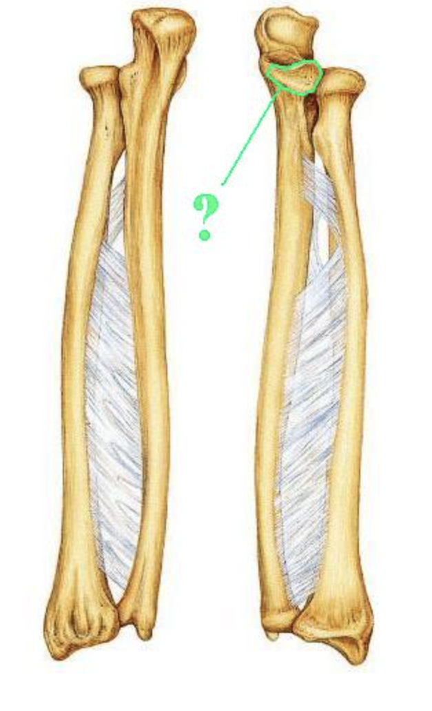
Identify the indicated bone marking (projection).
coronoid process
THINK:
Word | Meaning |
|---|---|
Coronoid | Crown-shaped |
Process | A projection sticking out from a bone |
Together: “A crown-shaped projection of bone.”
On the ulna: It’s the projection at the front of the upper ulna, forming the lower edge of the trochlear notch. It helps stabilize the elbow.

Identify the indicated bone marking (depression)?
acetabulum
“c-shaped”

Is this pelvis male or female?
female
How many carpal bones are present on each side?
8
Movement that results in an increase of the angle at a joint beyond 180 deg is called:
hyperextension

Identify the indicated body movement from A to B.
pronation

Identify the structure.
medial meniscus
THINK:
Word | Meaning |
|---|---|
Medial | Toward the body’s midline |
Meniscus | Crescent-shaped structure |
“The crescent-shaped cartilage pad on the inner side of the knee.”
Locate it: Between the medial condyles of the femur and tibia.
Function: Cushions the joint, distributes weight, and helps stabilize the knee.
Memory tip: Medial = inner; meniscus = cushion.

Identify the structure.
fibular colateral ligament
THINK:
Word | Meaning |
|---|---|
Fibular | Related to the fibula, the outer lower-leg bone |
Collateral | Located along the side |
Ligament | A strong band connecting bone to bone |
Together: “The ligament on the outer side of the knee that connects to the fibula.”
Locate it: Runs from the lateral epicondyle of the femur to the head of the fibula.
Function: Stabilizes the outer knee and resists forces that push the knee outward (varus stress).
Memory tip: Fibula = outer side → fibular collateral ligament = LCL.
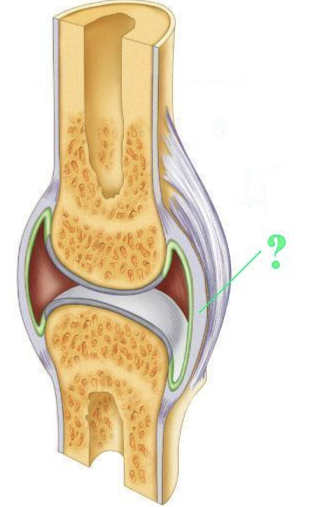
Identify the indicated layer of a synovial joint.
fibrous capsule
List two of the three structural types of articulations.
fibrous
cartilaginous
synovial
THINK:
Articulations = joints. Remember the 3 structural types with FCS.
Type | What connects the bones? | Example | Memory clue |
|---|---|---|---|
Fibrous | Dense fibrous connective tissue | Skull sutures | Fibers fasten bones together |
Cartilaginous | Cartilage | Pubic symphysis | Cartilage connects |
Synovial | A joint cavity containing synovial fluid | Knee, shoulder | Space + slippery fluid |
Structural = what the joint is made of.
Functional = how much the joint moves.
List two of the three functional types of articulations.
Structural = what the joint is made of.
Functional = how much the joint moves.
synarthroses
amphiarthroses
diarthroses
Functional types = how much a joint moves.
Remember “S–A–D: Still, A little, Does move freely.”
Functional type | Movement | Memory trick |
|---|---|---|
Synarthrosis | No movement | S = Still |
Amphiarthroses | Slight movement | A = A little |
Diarthroses | Free movement | D = Does move freely |
What structural type of articulation is a synchondrosis?
cartilaginous
What is the name of the type of synovial joint that has no rotational movement, but only gliding movement between flat or slightly curved bones?
plane
A site for muscle or ligament attachment that consists of a very large irregular process (found only on the femur) is a:
trochanter
The sternum belongs to which of the two main divisions of the skeleton?
axial

Identify the indicated structure within compact bone.
haversian canal or central canal

Identify the indicated space within compact bone.
lacunae

What type (shape) of bone is indicated?
sesamoid
Each osteon is composed of several layers of concentric material. Each layer is called a:
lamella
The cervical vertebra that articulates with the occipital condyles of the occipital bone is the:
atlas

Identify the indicated bone.
frontal

Identify the indicated bone.
temporal

Identify the indicated bone marking (depression).
anterior cranial fossa

Identify the indicated bone marking (suture).
squamosal suture
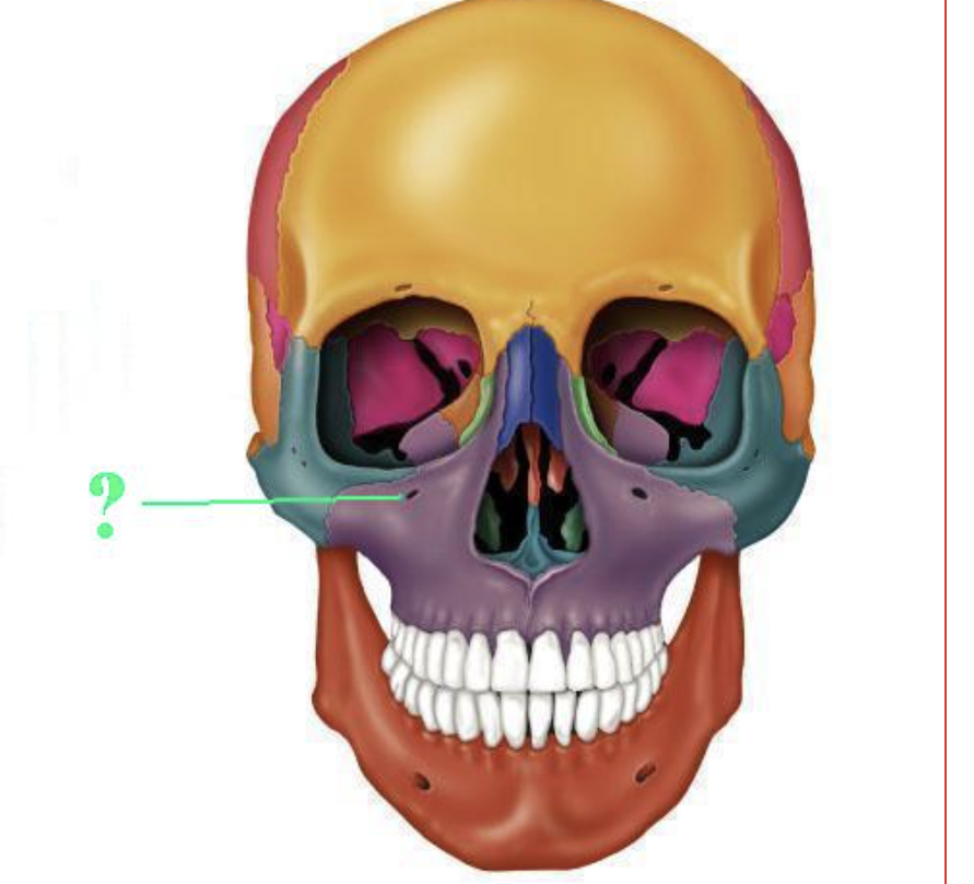
Identify the indicated bone marking (opening).
inftaorbital foramen

Identify the indicated bone marking (opening).
foramen magnum

Identify the indicated bone marking (opening).
vertebral foramen

Identify the indicated bone marking.
body

Identify the indicated type of vertebra.
lumbar
SQUARE BACKSIDE
What part of the clavicle articulates with the sternum?
sternal end
THINK: Sternum = Sternal end
What is the name of the bone of the upper arm?
humerus

Identify the indicated bone.
distal phalanx

Identify the indicated bone.
patella

Identify the indicated bone.
distal phalanx

Identify the indicated bone.
trapezium
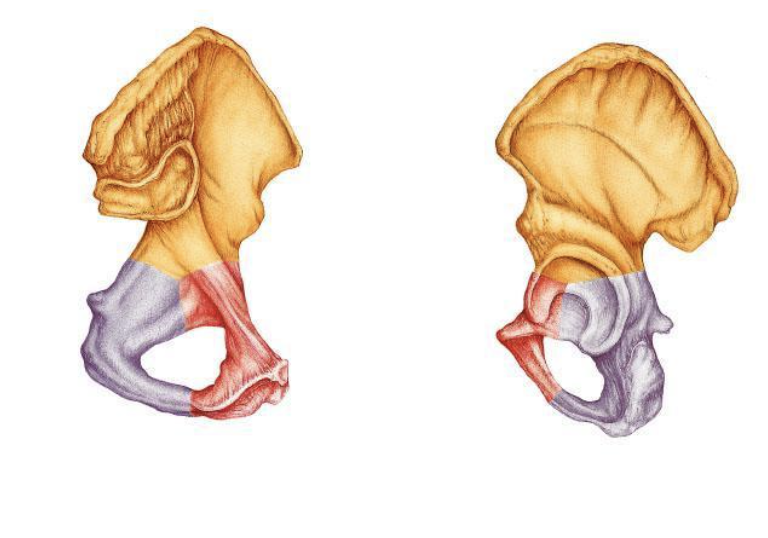
Is the indicated bone from the right of left side of the body?
left
REMEMBER:
if the posterior view, if the acetabulum is to the LEFT of the greater sciatic notch = it is the LEFT OX COXA
VS
if the posterior view, if the acetabulum is to the RIGHT of the greater sciatic notch = it is the RIGHT OX COXA

Is the indicated bone from the right of left side of the body?
left

Identify the indicated bone marking (projection).
coracoid process

Identify the indicated bone marking.
head

Identify the indicated bone marking (projection)?
medial condyle

Identify the indicated bone marking (projection).
medial condyle

Identify the indicated bone marking (surface)?
pubic crest
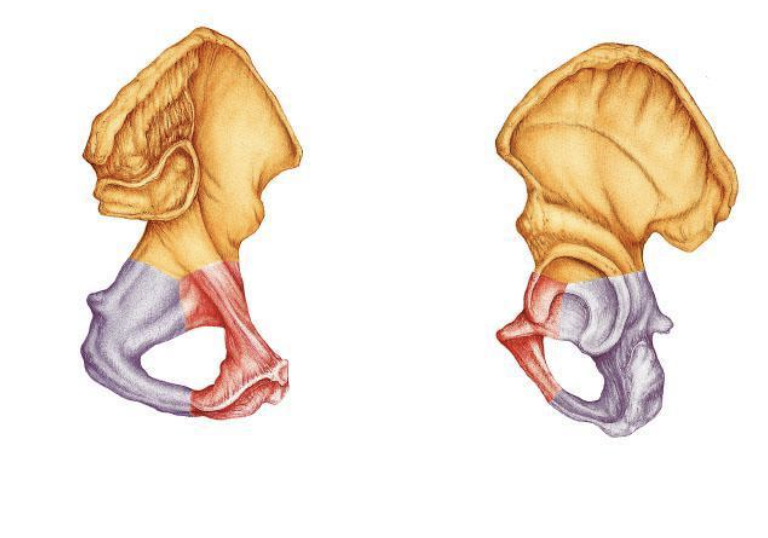
What portion of this bone is indicated by the yellow area?
ilium (big huge portion on top)
(superior) fan-shaped part of the hip bone,
How many phalangeal bones are present on each hand?
14
Movement that results in the movement of a limb toward the midline of the body is called:
adduction

Identify the structure.
posterior cruciate ligament

Identify the structure.
ligamentum teres
OR
ligament of the head of the femur

Identify the indicated layer of a synovial joint.
periosteum
List two of the three structural types of articulations.
fibrous
cartilaginous
synovial
HOW IT IS MADE
List two of the three functional types of articulations.
WHAT IT DOES
synarthroses
amphiarthroses
diarthroses
What structural type of articulation is a symphysis?
cartilaginous
What is the name of the type of synovial joint that has rotational movement around a single axis that accomplishes flexion and/or extension at the joint?
hinge
A site for muscle or ligament attachment that consists of a small rounded projection is a:
tubercle
The clavicle belongs to which of the two main divisions of the skeleton?
appendicular
The tiny channels that radiate from lacunae in compact bone and receive cytoplasmic projections of osteocytes are called:
canaliculi
THINK: tiny Channels = Canaliculi
The type of vertebra that has facets for the articulation of ribs is a:
thoracic
THINK: small, smooth, relatively flat surfaces on a bone where it meets another bone to form a joint.

Identify the indicated bone marking (projection).
mandibular angle
Identify the indicated bone.
parietal
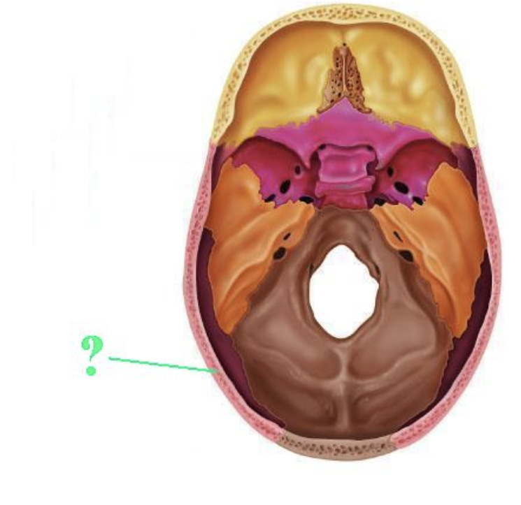

Identify the indicated bone marking (projection).
superior nuchal line
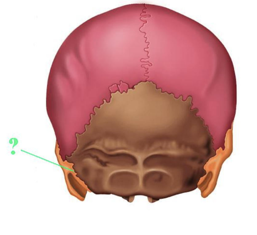
Identify the indicated bone marking (suture).
occipitomastoid suture
What part of the innominate (os coxa) articulates with sacrum?
auricular surface

Identify the indicated bone.
os coxa
hip bone
innominate

Identify the indicated bone.
metaCARPAL
THINK: drive CAR with CARPALS

Identify the indicated bone.
TIGER
CUBS
NEED
M.I.L.C
intermediate cuneiform