M3 Bones
 Call Kai
 Learn
 Practice Test
 Spaced Repetition
 Match
 Flashcards
 Knowt Play
1/239

There's no tags or description
Looks like no tags are added yet.
240 Terms
bone histology
matrix is dense and contains calcium, salts, and collagen

osteon histology
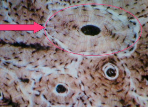
osteocytes histology

lacunae histology

lamellae histology

canaliculi histology

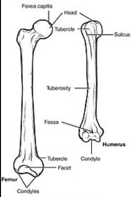
fossa
shallow, basinlike and elongated depression in a bone, often serving as an articular surface
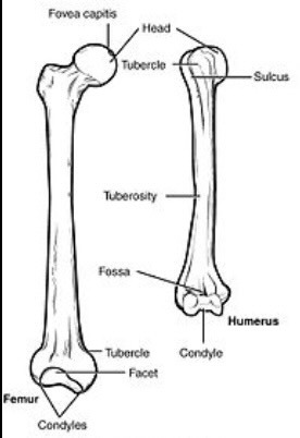
fovea
a small pit in the bone
parietal bone of skull
a bone forming the central and upper back part of each side of the skull

occipital bone of skull
back of head; base of skull

occipital condyle of skull
rounded projections lateral to the foramen magnum that articulate with the first cervical vertebra (atlas)

zygomatic bone of skull
(anterior view)
the arch of bone beneath the eye that forms the prominence of the cheek

zygomatic arch of skull
(lateral view)
cheek bone

frontal bone of skull
(anterior view)
forehead

sphenoid bone of skull
(anterior view)
forms part of the base of the skull and parts of the floor and sides of the orbit

ethmoid bone of skull
(anterior view)
light spongy bone between the eye sockets
forms part of the nasal cavities
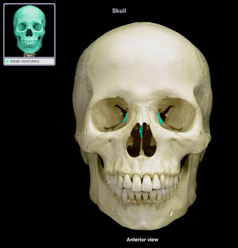
nasal bone of skull
(anterior view)
bridge of nose

vomer bone of skull
(anterior view)

maxilla of skull
upper jaw bone

mandible of skull
lower jaw bone
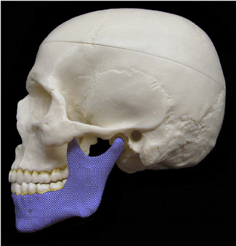
mandible body of skull
main part of the jaw bone, horizontal part, hold lower teeth

mandible mental foramen of skull
allows passage of the mental nerve

mandibular condyle of skull
(lateral view)
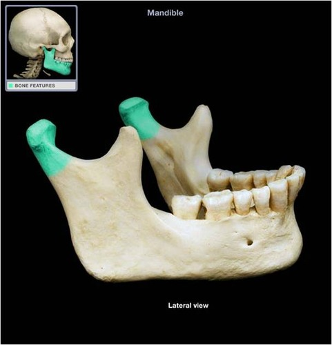
mandibular notch of skull
(lateral view)

mandible coronoid process of skull

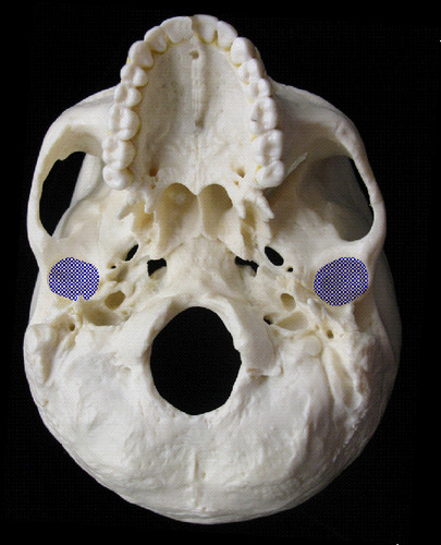
mandibular fossa of skull
the depression in the temporal bone into which the condyle of the mandible fits
external auditory meatus of skull
ear canal

mastoid process of skull
(lateral view)
round projection on the temporal bone behind the ear

styloid process of skull
(lateral view)
pole-like process extending downward from the temporal bone on each side of the skull

temporomandibular joint of skull
the joint formed where the mandible and temporal bone meet

supraorbital foramen of skull
(anterior view)
opening above each orbit (eye holes) allowing blood vessels and nerves to pass

infraorbital foramen of skull
(anterior view)
opening under each orbit (eye hole) carrying the infraorbital nerves and blood vessels in the nasal region

coronal suture of skull
(lateral view)
the suture between the parietal and frontal bones of the skull

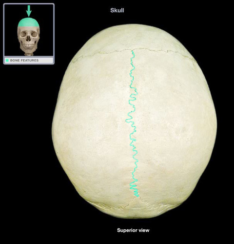
sagittal suture of skull
(superior view)
between parietal bones
squamous suture of skull
(lateral view)
suture between parietal and temporal bones

lambdoid suture of skull
(posterior view)
between parietal bones and occipital bone

palatine bone of skull
(inferior view)
either of two irregularly shaped bones that form the back of the hard palate
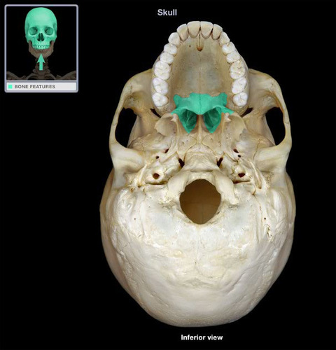
maxilla: palatine process of skull

carotid canal of skull

jugular foramen of skull

foramen lacerum of skull

foramen magnum of skull
large opening at the base of the skull through which the brain connects to the spinal cord

greater wing of the sphenoid bone of skull

lesser wing of the sphenoid bone of skull

stella turcica of sphenoid bone of skull

optic canal of skull

foramen ovale of skull

sphenoid sinus of skull
(lateral view, left side)
sinus above and behind the nose

cribiform plate of skull
(superior view)

olfactory foramina of skull
holes in cribiform plate
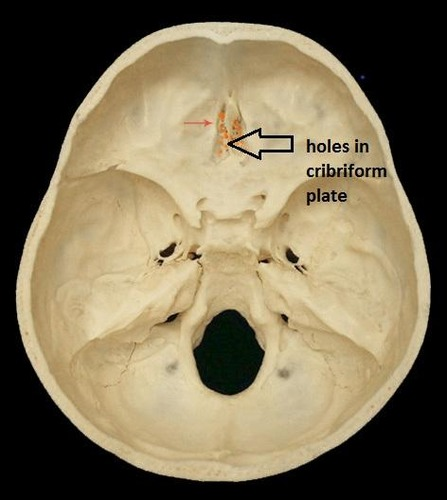
crista galli of skull
(superior view)

internal auditory meatus of skull

superior orbital fissure of skull

inferior orbital fissure of skull

perpendicular plate of the ethmoid bone of skull

frontal sinus of skull
mid-sagittal view

maxillary sinus of skull

ethmoid sinus of skull
superior view
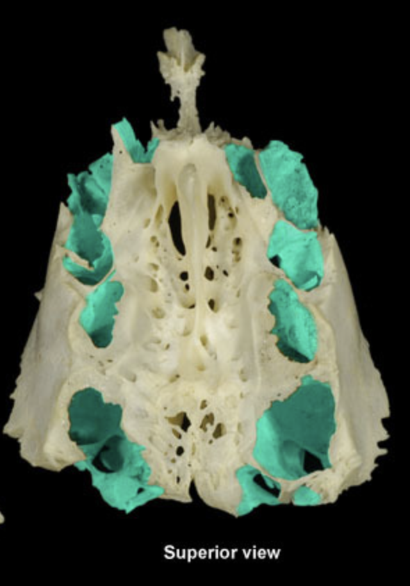
ethmoid sinus of skull
posterior view
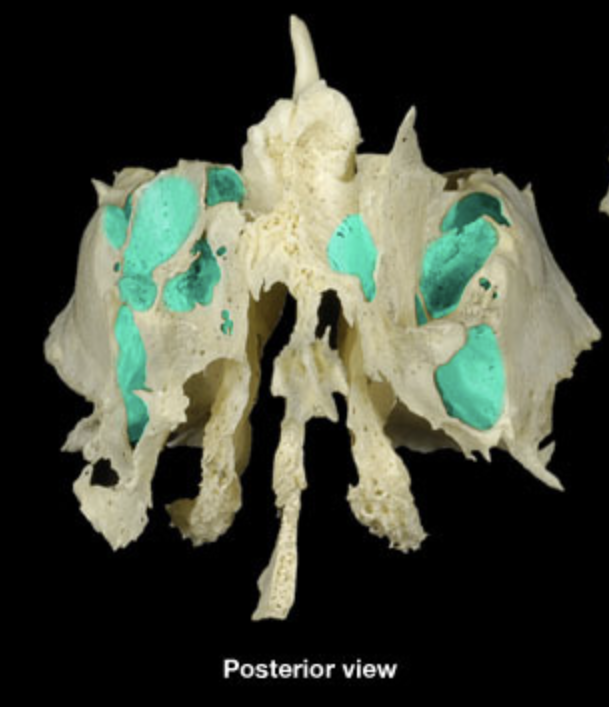
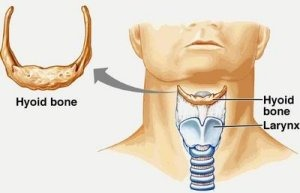
hyoid bone
atlas
superior view
first cervical vertebra (C1), which provides support for the head

axis
posterosuperior view
C2 bone
allows one to pivot neck

cervical region
seven vertebrae (C1-C7)

thoracic region of vertebra
twelve vertebrae (T1-T12)

lumbar region
five vertebrae (L1-L5)

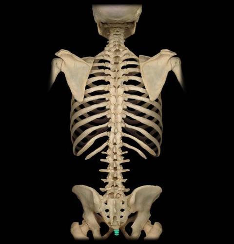
sacrum

coccyx
manubrium of thoracic cage
(anterior view)

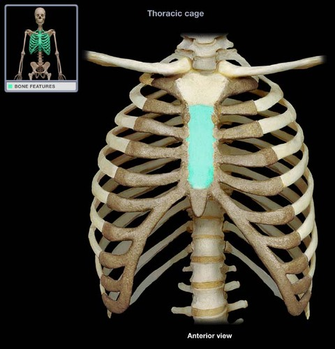
body of thoracic cage
(anterior view)
xiphoid process of thoracic cage
(anterior view)
inferior portion of the sternum

clavicular notch of sternum
(anterior view)

jugular notch of thoracic cage
(anterior view)

sternal angle of thoracic cage
(anterior view)

true ribs of thoracic cage
(anterior view)

false ribs of thoracic cage
(anterior view)

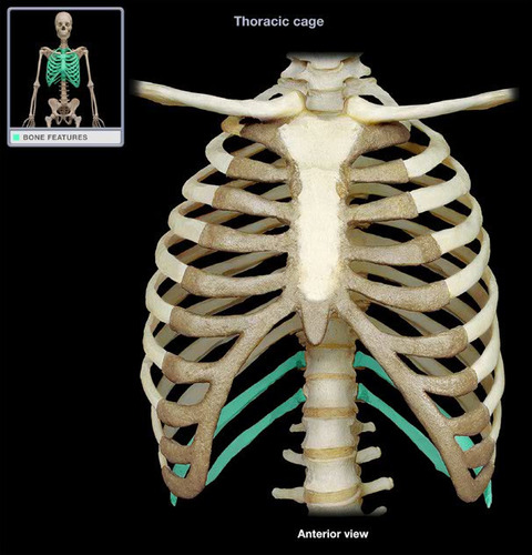
floating ribs of thoracic cage
(anterior view)
head of rib

transverse costal facet (for tubercle of rib) of thoracic vertebra
(superior view)

transverse costal facet (for tubercle of rib) of thoracic vertebra
(lateral view)

superior costal facets of thoracic vertebra
(superior view)

superior costal facets of thoracic vertebra
(lateral view)

inferior costal facets of thoracic vertebra
(lateral view)
cannot be seen from superior view

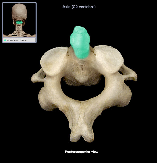
dens or ondontoid process of axis
(posterosuperior view)
transverse foramen of cervical vertebra
(superior view)
only found in the cervical vertebrae and allow passage of the vertebral artery, vein, and nerve

transverse foramen of cervical vertebra
(lateral view)
only found in the cervical vertebrae and allow passage of the vertebral artery, vein, and nerve

transverse process of thoracic vertebra
(superior view)
two lateral projections from the vertebral arch

transverse process of thoracic vertebra
(lateral view)
two lateral projections from the vertebral arch

spinous process of thoracic vertebra
(superior view)
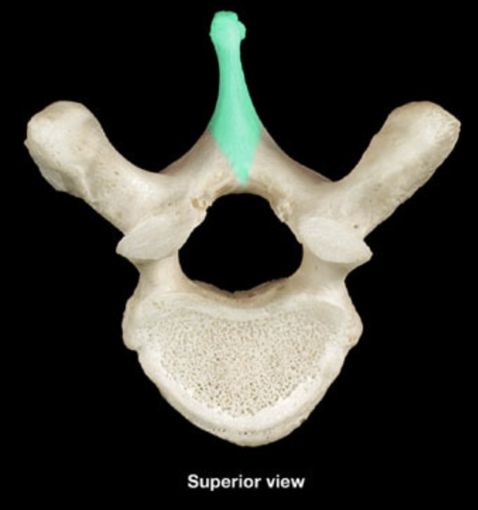
spinous process of thoracic vertebra
(lateral view)

vertebral foramen of thoracic vertebra
(superior view)
cannot be seen in lateral view

vertebral arch of thoracic vertebra
(superior view)
cannot be seen in lateral view

pedicle of thoracic vertebra
(superior view)

pedicle of thoracic vertebra
(lateral view)

lamina of thoracic vertebra
(superior view)

lamina of thoracic vertebra
(lateral view)

body or centrum of lumbar vertebra
(superior view)

body or centrum of lumbar vertebra
(lateral view)

anterior arch of atlas
(superior view)

posterior arch of atlas
(superior view)

parietal bone of fetal skull
