Eukaryotic Exam 2
 Call Kai
 Learn
Learn Practice Test
 Spaced Repetition
 Match
Match Flashcards
 Knowt Play
1/68

Earn XP
Description and Tags
module 5
Name | Mastery | Learn | Test | Matching | Spaced | Call with Kai | Chat |
|---|
No analytics yet
Send a link to your students to track their progress
69 Terms
evolution of aerobic respiration
in the first 2 billion years the atmosphere was made of H2, NH3, and H2O; organisms were anaerobes so they could only do glycolysis and fermentation
about 2.7 billion years ago was the appearance of cyanobacteria using light energy to split water and release O2 into the atmosphere
species evolved that were not only protected from the damaging effects of O2, but possessed metabolic pathways to utilize O2
the O2-dependent prokaryotes and eukaryotes living today evolved from earlier aerobes
in eukaryotes, the utilization of O2 takes place in mitochondrion, which may have evolved from an ancient aerobic bacterium that took up residence inside the cytoplasm of an anaerobic host cell
mitochondria characteristics
big enough to be seen under a light microscope
are surrounded by a semipermeable membrane
double membrane
have very different overall structure; can be sausage-shaped to highly branched, interconnected tubular network
can fuse with one another or split in two
dynamic structure of mitochondria
mitochondria fuse with one another or split in two
the balance between fusion and fission is likely a major determinant of mitochondrial number, length, and degree of interconnection
photo is of mouse fibroblasts with a fluorescent tagged mitochondrial protein; shows how mitochondria can fuse and split

mitochondrial fission
mitochondrial fission is apparently induced by contact with thin tubules from the ER, which can encircle the mitochondrion like a noose
these ER tubules appear to initiate constriction, which is then completed through the action of soluble proteins that are recruited to the outer surface of the mitochondrion from the cytosol

mitochondria structural components
outer mitochondrial membrane
inner mitochondrial membrane, including inner boundary membrane and internal cristal membranes (crystae); joined to one another by narrow connections, but they are functionally distinct
inner membrane and cristal membranes are continuous
intermembrane space
matrix

outer membrane
approx. 50% lipid by weight and a mixture of enzymes involved in diverse activities, like oxidation of epinephrine, degradation of Trp, and elongation of fatty acids
homologous to an outer membrane present as part of the cell wall of certain bacterial cells
both contain porins; integral proteins that have a relatively large internal channel surrounded by a barrel of β strands
the porins of the outer membrane can undergo reversible closure; when they are wide open, the outer membrane is freely permeable to molecules such as ATP, NAD and coenzyme A
inner membrane
100 different polypeptides and a very high protein/lipid ratio (>3":1)
the composition and organization of the IM are the key to the bioenergetic activities of the organelle
rich in cardiolipin; characteristic of bacterial plasma membranes, from which the inner membrane presumably evolved
highly impermeable; all molecules and ions require special transporters to enter the matrix
membranes of gram-negative bacteria
shows to membrane layers; outer and plasma (?)
homologous to the membrane of mitochondria
shows porins: a β-sheet barrel that forms an opening for passage of moderate-sized molecules (metabolites)

mitochondrial matrix
the central compartment of the mitochondria and has a high protein content
proteins are pyruvate dehydrogenase, the enzymes of the tricarboxylic acid cycle, enzymes of fatty acid oxidation, and enzymes required for RNA, DNA, and protein synthesis
where the mitochondrial DNA and ribosomes are located
mitochondrial genome
mitochondria have their own genetic material; a circular DNA in higher plants and animals
this nonchromosomal DNA encodes a small number of mitochondrial proteins that are tightly integrated into the inner membrane, along with proteins encoded by nuclear genes
after a single aerobic bacterium took residence in the cytoplasm of a primitive cell, most of the genes of this symbiotic organism were either lost or transferred to the nucleus of the host cell, leaving only a handful of genes to encode some of the most hydrophobic proteins of the inner membrane
the function of mitochondria
the primary function is to utilize free energy derived from oxidation of organic molecules to produce ATP under aerobic conditions
the reaction in the mitochondria may also serve as a route to interconvert molecules of the different classes of biological molecules
provide intermediates
overview of carbohydrate metabolism
coupling cytosolic glycolysis and pyruvate production to the mitochondrial TCA cycle and ATP formation
if oxygen is present, cell will undergo glycolysis to form pyruvate, which will be transported into the mitochondria to be used to create ATP for energy
in the absence of oxygen, the cell will undergo fermentation to produce NAD+ to be used in glycolysis

glycolysis
the first steps in oxidative metabolism are carried out in glycolysis
produces pyruvate, NADH, and two molecules of ATP
aerobic organisms use O2 to extract more than 30 additional ATPs from pyruvate and NADH
pyruvate is transported across the inner mitochondrial membrane and decarboxylated to form acetyl CoA, which enters the next stage
glycolysis pathway
know the first step, end product, and net reaction (i think)
know overall reaction but I think he means net reaction

net reaction of glycolysis
glucose + 2 NAD+ + 2 ADP +2 Pi → 2 pyruvate + 2 ATP + 2 NADH + 2 H+ + 2 H2O

the oxidative pentose phosphate pathway
generates reducing equivalents in the form of NADPH for reductive biosynthesis reactions within cells
provides the cell with ribose-5-phosphate (R5P) for the synthesis of the nucleotides and nucleic acids
metabolizes dietary pentose sugars derived from the digestion of nucleic acids as well as to rearrange the carbon skeletons of dietary carbohydrates into glycolytic/gluconeogenic intermediates

importance of NADPH
the reactions of fatty acid biosynthesis and steroid biosynthesis utilize large amounts of NADPH
erythrocytes utilize the reactions of the PPP to generate large amounts of NADPH used in the reduction of glutathione
the conversion of ribonucleotides to deoxyribonucleotides (through the action of ribonucleotide reductase) requires NADPH as the electron source
therefore, any rapidly proliferating cell needs large quantities fo NADPH
pyruvate dehydrogenase complex
the pyruvate molecules produced by glycolysis are transported across the inner membrane and into the matrix
in the matrix they are decarboxylated by the pyruvate dehydrogenase complex to form acetyl groups, which are transferred to CoA
complex of sixty peptides constituting three different enzymes:
cubelike cluster: dihydrolipoyl transacetlase molecules
blakc spheres: pyruvate dehydrogenase dimers
small gray sphere: dihydrolipoyl dehydrogenase
net reaction of pyruvate dehydrogenase
pyruvate + HS-CoA + NAD+ → Acetyl-CoA + CO2 +NADH + H+
tricarboxylic acid (TCA) cycle (aka Krebs cycle) pathway

TCA cycle
all of the enzymes of the TCA cycle reside in the soluble phase of the matrix except for succinate dehydrogenase, which is bound to the inner membrane
the first step in the cycle is the condensation of the two-carbon acetyl group with a four-carbon oxaloacetate to form a six-carbon citrate
the two carbons that are removed during the TCA cycle are completely oxidized to CO2 (these two carbons are not the same ones that were brought with the acetyl group)
in the TCA cycle, the chain length of the citrate is decreased one carbon at a time, regenerating the four-carbon oxaloacetate, which can condense with another acetyl CoA
four reduction reactions occur in this cycle, three of which reduce NAD+ to NADH and one reduces FAD to FADH2
net reaction of the TCA cycle
acetyl CoA + 2 H2O + FAD + 3NAD+ + Pi → 2 CO2 + FADH2 + 3NADH + 3H+ + GTP + HS-CoA
catabolic pathways and the TCA cycle
reaction intermediates in the TCA cycle are common compounds generated in other catabolic reactions
for this reason, the TCA cycle is the central metabolic pathway of the cell

the malate-aspartate shuttle
the primary cytoplasmic NADH electron shuttle
mitochondria are not able to import the NADH formed in the cytosol during glycolysis
NADH formed during glycolysis enters the mitochondria via malate-aspartate or glycerol phosphate shuttles
these electrons are indirectly fed into the mitochondrial electron-transport chain and used for ATP formation

the glycerol phosphate shuttle
the secondary cytoplasmic NADH electron shuttle
electron transfer from NADH to DHAP to form glycerol 3-phosphate, then to FAD to form FADH2
enzyme is glycerol 3-phosphate dehydrogenase
energy is lost; energy level of NADH is higher than that of FADH2
oxidative phosphorylation
WATCH LECTURE
a process in which ATP formation is driven by energy that is released from electrons removed during substrate oxidation
energy stored in the high-energy of NADH and FADH2 are released as they are passed through the electron-transport chain

substrate level phosphorylation
a process in which ATP is formed directly by transfer of a phosphate group from a substrate molecule to ATP
oxidation and reduction
redox couple: A/A+ and B+/B
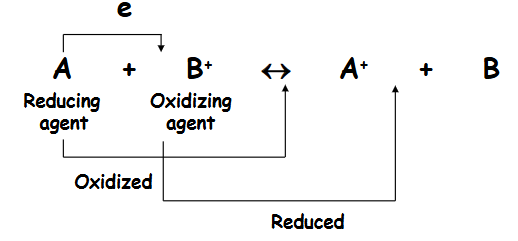
reducing agents

oxidizing agents

oxidation-reduction potential standards
standard conditions: concentrations are 1M for solutes and ions, and 1 atm pressure for gases
standard couple: H+/H2
standard redox potential: E0
E0’ - [H+] is 10-7 (pH of 7)
oxidizing and reducing agents occur as couples, which differ in their number of electrons
the transfer of electrons between a couple causes charge separation that can be measured as an oxidation-reduction (redox) potential
redox potentials in the TCA cycle
electrons are transferred to NAD+ (or FAD+) within in the mitochondrion from substrates of the TCA cycle
isocitrate, malate, a-ketoglutarate, and succinate
most have redox potentials of relatively high negative values; sufficiently high to transfer electrons to NAD+
oxidation of succinate to fumarate (more positive redox potential) proceeds by the reduction of FAD, a coenzyme of greater electron affinity than NAD+
free energy change during electron transport
sorry I didn’t feel like typing all of this

five types of electron carriers in the electron transport chain
flavoproteins, cytochromes, copper atoms, ubiquinone, and iron-sulfur proteins
all are membrane-bound
all of the redox centers within the chain that accept and donate electrons are non-amino acid prosthetic groups that are tightly associated with proteins (except ubiquinone)
flavoproteins
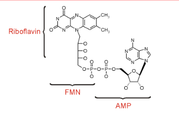
FMN (flavin mononucleotide) is a prosthetic group of some flavoproteins
it is similar in structure to FAD (flavin adenine dinucleotide), but lacking the adenine nucleotide
FMN (like FAD) can accept 2 e- + 2H+ to form FMNH2
why is FMN important
when bound at the active site fo some enzymes, it can accept 1 e- to form the half-reduced semiquinone radical
the semiquinone can accept a second e- to yield FMNH2
since it can accept/donate1 or 2 e-, FMN has an important role in mediating electron transfer between carriers that transfer 2e- (NADH) and those that can accept only 1e- (FE3+)
cytochromes
proteins with heme prosthetic groups
the heme iron can undergo a 1e- transition between ferric and ferrous states
Fe3+ + e- → Fe2+
hemes in the three classes of cytochrome (a,b,c) differ slightly in substituents on the porphyrin ring system
three copper atoms
all three copper atoms are located within a single protein complex fo the inner mitochondrial membrane
the copper atoms can undergo a 1e- transition between cupric and cuprous states
Cu2+ + e- → Cu+
ubiquinone (UG/coenzyme Q)
very hydrophobic
dissolves in the hydrocarbon core of a membrane
includes a long isoprenoid tail, with multiple units having a carbon skeleton comparable to that of isoprene
in human cells, usually n=10
UQ10’s isoprenoid tail is longer than the width of a bilayer
it may be folded to yield a more compact structure, and is postulated to reside in the central domain of a membrane between the two lipid monolayers
the quinone ring can be reduced to quinol in a 2e- reaction
Q + 2e- + 2H+ ←→ QH2
when bound to special sites in respiratory complexes, CoQ can accept 1e- to form a semiquinone radical (Q-); like FMN, CoQ can mediate between 1e- and 2e- donors/acceptors

iron-sulfur proteins
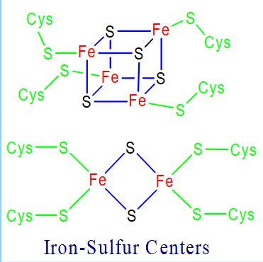
Fe-S centers are prosthetic groups containing 2, 3, 4, or 8 iron atoms complexed to elemental and cysteine S
electron transfer proteins may contain multiple Fe-S centers
4-Fe centers have a tetrahedral structure, with Fe and S atoms alternating as vertices of a cube
Fe-S centers transfer only one electron even if they contain two or more iron atoms, because of the close proximity of the iron atoms
arrangement of electron carriers in the chain
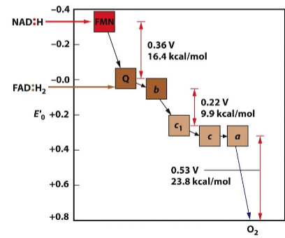
carriers are arranged in the order of increasingly positive redox potential
the electrons are passed from one carrier to the next, losing energy as they move “downhill” along the chain
determination of the sequence of carriers in the chain
the specific sequence of carriers that constitute the electron-transport chain was worked out using a variety of inhibitors that blocked electron transport at specific sites along the route
after an inhibitor was added to cells, the oxidation state of the various electron carriers in the inhibited cells was determined
by identifying reduced and oxidized components in the presence of different inhibitors, the sequences of the carriers could be determined
done by Britton Chance and coworkers at the University of Pennsylvania
tendency for electron transfer
the tendency for electrons to be transferred from one carrier to the next depends on the potential difference between two redox centers; rate of transfer depends on the catalytic activities of the proteins involved
electrons may travel considerable distances (10-20 Å) between adjacent redox centers
may flow through special “tunneling pathways” consisting of a series of covalent and hydrogen bonds that stretch across parts of several amino acid residues
four complexes of the electron transport chain
all distinct (I, II, III, IV)
asymmetric and membrane spanning

cytochrome c and ubiquinone
cytochrome c: a soluble protein in the intermembrane space
ubiquinone: a pool of molecules dissolved in the lipid bilayer
ubiquinone and cytochrome c move within or along the membrane shuttling electrons between large, relatively immobile protein complexes
once inside the complexes, electrons travel along defined pathways between adjacent redox centers whose relative positions are fixed in space
subunits of the complexes

coupling sites
there are three places in which transfer of electrons is accompanied by a major release of free energy; these are coupling sites
the coupling sites occur between carriers that are part of three complexes (I, III, IV)
the energy released at the three sites are conserved by translocation of proton from the matrix across the inner membrane into the inter-membrane space
these three complexes are called proton pumps
what is complex I (NADH dehydrogenase)
the gateway to the ETC, catalyzing the transfer of a pair of electrons from NADH to ubiquinone (UQ) to form ubiquinol (UGH2)
about half of the complex consists of a hydrophilic domain that projects into the matrix, with the remaining hydrophobic portion of the complex embedded in the membrane
together these portions of the complex carry out the two different activities that are required of the ETC (electron transfer and proton translocation)
how does complex I work
electron transfer occurs within the hydrophilic portion of the complex
the passage of a pair of electrons from NADH to ubiquinone is paired with moving four protons from the matrix into the intermembrane space
electron movement induces a conformational change in the complex that causes the lateral movement of helix HL, which leads to tilting of the transmembrane helices
this changes the ionic environment of proton-transferring residues, leading to the movement of protons across the membrane
complex II (succinate dehydrogenase)
consists of four polypeptides: two hydrophobic subunits that anchor the protein in the membrane and two hydrophobic subunits that comprise succinate dehydrogenase
provides a pathway for feeding lower-energy electrons from succinate to FAD to ubiquinone
the path from FADH2 to ubiquinone takes the electrons through three iron-sulfur clusters
complex II contains a heme group, thought to attract escaped electrons to prevent the formation of destructive superoxide radicals
electron transfer through complex II is not accompanied by proton translocation
complex III (cytochrome bc1)
catalyzes the transfer of electrons from ubiquinol to cytochrome c, with four protons translocated across the membrane for every pair of electrons transferred
protons are released into the intermembrane space in two separate steps as a pair of electrons are separated and passed along different pathways through the complex
two protons are derived from the molecule of ubiquinol that entered the complex, and two protons are removed from the matrix and translocated as part of a second molecule of ubiquinol
three of the subunits of complex III contain redox groups: cytochrome b contains two heme b molecules with different redox potentials, cytochrome c1, and an iron-sulfur protein
complex IV (cytochrome oxidase)
electrons are transferred from cytochrome c through a bimetallic copper center (CuA) of subunit II to a heme (a) of subunit I
electrons are passed to a redox center located in subunit I that contains a second heme (a3) and another cooper atom (CuB); the first two electrons reduce the a3-CuB) binuclear center
Fea33+ + CuB2+ +2e- → Fea32+ + CuB+
an O2 binds to the center and accepts the pair of electrons
Fe3+-O- - O- - Cu2+
a third electron was extracted and the binuclear center accepts two protons
one of the O is reduced: Fe4+ = O2- Cu2+ -OH2
a fourth electon and two additional protons are used to form two molecules of water
Fe3+ Cu2+ + 2H2O
four additional protons are pumped across the inner membrane to the intermembrane space
experiment showing that cytochrome oxidase is a proton pump
cytochrome oxidase: a proton pump in synthetic liposomes
the medium becomes acidified following the addition of reduced cytochrome C
translocation of protons across the inner membrane produces electrochemical gradient:
the concentration difference of hydrogen ions (deltapH)
the voltage resulted from the separation of charge (ψ)
the proton-motive force (deltap):
deltap = ψ -2.3(RT/F) deltapH
where 2.3 TR/F is equal to 59 mV at 25 degrees C:
deltap = ψ =59 deltapH
positively charged, lipid-soluble substance (JC-1) is distributed according to the electric potential; the voltage genderated across the inner membrane (inside negative) leads to the accumulation of the dye within the mitochondria
ADD PICTURES!!
F1 and ATPase
during the early 60s, using negative staining technique, Humberto Ferdandez-Moran discovered a layer of spheres attached to the inner side of the inner membrane, projecting from the membrane and attached to it by stalks
a few years later, Efraim Rocker isolated the inner membrane spheres (coupling factor 1 or F1)
he found that the F1 behaved like an ATPase
F1 could be the ATPase because:
enzymes do not affect the equilibrium constant of the reaction they catalyze
enzymes are capable of catalyzing both the forward and reverse reactions
Na+/K+ ATPase
under physiological conditions, Na+/K+ ATPase utilizes the energy from ATP hydrolysis to export Na+ and import K+ against their respective gradients
red blood cells ghosts are prepared with a very high internal [K+] and a very high external [Na+]
under these conditions, K+ moves out of the cell and Na+ moves into the cell
in the presence of ADP and Pi, the movement of the ions can cause ATP to be synthesized
![<ul><li><p>under physiological conditions, Na+/K+ ATPase utilizes the energy from ATP hydrolysis to export Na+ and import K+ against their respective gradients</p></li><li><p>red blood cells ghosts are prepared with a very high internal [K+] and a very high external [Na+]</p></li><li><p>under these conditions, K+ moves out of the cell and Na+ moves into the cell</p></li><li><p>in the presence of ADP and Pi, the movement of the ions can cause ATP to be synthesized</p></li></ul><p></p>](https://assets.knowt.com/user-attachments/4fac3b70-0bbc-44b0-b898-87d7b9098950.png)
structure of ATP Synthase
a spherical F1 head (about 90 Å in diameter)
a basal section, F0, embedded in the inner membrane
a central and spherical stalk connecting F1 and F0
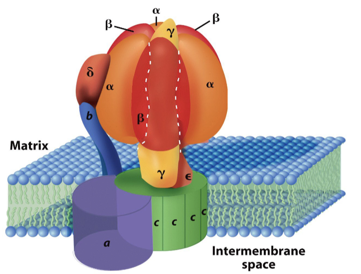
F1 structure
has a stoichiometry of α3β3δγε
the α and β subunits are arranged alternately within the F1 (looks like pieces of an orange)
each of the β subunits contain a catalytic site for ATP synthesis
the γ subunit runs from the outer tip of the F1 head through the central stalk and makes contact with the F0 base piece
F0 structure
has a stoichiometry of ab2c(10-14)
the number of subunits in the c ring varies from 10-14
both the yeast and e. coli ATP synthase have 10 c subunits, whereas the chloroplast one has 14
the F0 base contains a channel through which protons are conducted from the intermembrane space to the matrix
evidence that the F0 base contains a channel through which protons are conducted
intact submitochondrial particles, which contain the ATP synthase embedded in the vesicle membrane, are capable of oxidizing substrates, generating a proton gradient, and synthesizing ATP
if the F1 spheres are removed from the particles, the vesicle membrane can no longer maintain a proton gradient despite continuing substrate oxidation and electron transport

Paul Boyer
proposed a hypothesis for how a proton electrochemical gradient provides the energy required to drive the synthesis of ATP
called the binding change mechanism
the binding change mechanism
the energy released by the movement of a proton is not used to drive ADP phosphorylation directly but principally to change the binding affinity of the active site for the ATP product
enzyme-bound ADP and enzyme-bound Pi readily condense to form a tightly bound ATP molecule without the input of additional energy; instead, energy is required for the release of the tightly bound ATP from the catalytic site
each active site progresses successively through three distinct conformations that have different affinities for substrates and products
the F1 complex has three cat. sites
at any given instant, one site is in the “loose” (L conf.) in which ADP and Pi are loosely bound; a second site is in the “tight” (T conf.) in which nucleotides (ADP+Pi or ATP) are tightly bound; and the third site in the “open” (O conf.) which allows release of ATP
ATP is synthesized by rotational catalysis in which one part of the ATP synthase rotates relative to another parts
α and β subunits rotate relative to the central stalk; the rotation is driven by the movement of protons through the membrane via the channel in the F0 base
binding change mechanism photo

evidence to support the binding change mechanism
in 1994 John Walk and his co-workers published a detailed atomic model of the F1 head
structures corresponding to the L, T, and O conformations were identified in the cat. sites of the 3 β subunits
the γ subunit is perfectly positioned within the ATP synthase to transmit conformational changes from F0 membrane sector to the F1 cat. sites; the γ subunit extends from the F0 sector through the stalk and into the central cavity within the F1 sphere, where it contacts each of the three β subunits differently
in the presence of ATP, the actin filament was observed to rotate counterclockwise
the β subunit was modified to contain 10 histidine residues at its N-terminus, as site located on the matrix face of the F1 head
the γ subunit was modified by replacing one of the serine residues near the end of the stalk with a cysteine residue, which provided a means to attach the fluorescently labeled actin filament
the structure and function of the membrane-bound F0 portion
what is the path taken by protons as they move through the F0 complex and how does this movement lead to the synthesis of ATP? it was postulated that:
the c subunits of the F0 base were assembled into a ring that resides within the lipid bilayer
the c ring is physically bound to the γ subunit of the stalk
the “downhill” movement of protons through the membrane drives the rotation of the ring of c subunits, and the rotation of the c ring of F0 provides the twisting force that drives the rotation of the attached γ subunit, leading to the synthesis and release of ATP
the β subunits are primarily structural components of the ATP synthase
the two elongated β subunits form a peripheral stalk that connects the F1 and F0 portions, and along with the δ subunit of F1, hold the α3β3 subunits in a fixed position while the γ subunit rotates within the center of the complex
each c subunit contains two transmembrane helices connected by a hydrophilic loop that projects toward the F1 head; the hydrophilic loops situated on the tops of the c subunits form a binding site for the bases of the γ and the ε subunits, which together act like a “foot” that is attached to the c ring
as a result of this attachment, rotation of the c ring drives rotation of the attached γ
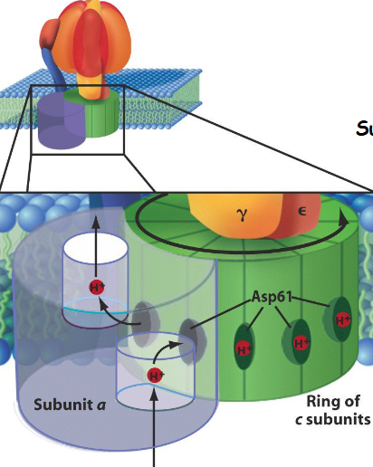
subunit a of the F0 portion
subunit a is stationary; it has two half-channels that are physically separated from one another
one half-channel leads from the intermembrane space into the middle of the a subunit and the other leads from the middle of the a subunit into the matrix
the subunits of the c ring move successively past the stationary a subunit
protons are picked up from the intermembrane space one at a time by each c subunit and carried completely around a circle before they are released into the matrix
each proton moves from the intermembrane space through the half-channel and binds to a negatively charged Asp residue situated at the surface of the c subunit
at the same time, the adjoining c subunit (which was protonated at an earlier step) is aligned with the second half-channel of the a subunit
the Asp releases its associated proton, which diffused into the matrix
after dissociation of the proton, the c subunit returns to its original conformation
binding of the proton to the carboxy group generates a major conformational change in the c subunit that causes that subunit to rotate approx. 30 degrees in a counterclockwise direction
the movement brings the adjoining c subunit, which has just released the proton, into alignment with the other half-channel and repeat the cycle
how many molecules of ATP comes from ATP synthase
if a c ring is composed of 12 subunits, it will require 4 protons to move the ring 120 degrees, which would drive a corresponding rotation of the γ subunit 120 degrees and lead to the release of one newly synthesized ATP by the F1 complex
therefore, the translocation of 12 protons would lead to the fill 360 rotation of the c ring and γ subunit, and the synthesis and release of 3 molecules of ATP
other processes driven by proton-motive force
uptake of ADP/Pi in exchange for ATP and H+; ATP/ADP exchange is accomplished by the adenine nucleotide translocase (ANT)
pull Ca2+ ions into the mitochondrion
drive mitochondrial fusion
specifically targeting polypeptides to enter the mitochondrion from the matrix
pumping pyruvate into the matrix (symport)
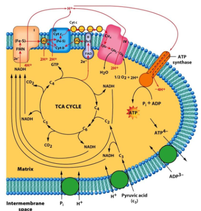
photo shows a summary of the major activities during aerobic respiration in a mitochondrion
peroxisomes
membrane-bound vesicles that contain oxidative enzymes
the oxidize very long chain fatty acids, and synthesize plasmalogens (a class of phospholipids)
they form by splitting from pre-existing organelles, import preformed proteins, and engage in oxidative metabolism
hydrogen peroxide, and reactive and toxic compound, is formed in peroxisomes and is broken down by the enzyme catalase
peroxisomes contain enzymes to carry out the two-step reduction of molecular oxygen to water
glyoxysome
a specialized type of peroxisome in plant seedlings
plant seedlings rely on stored fatty acids to provide the energy and material to form a new plant
one of the primary metabolic activities in these germinating seedlings is the conversation of stored fatty acids to carbohydrate
citrate is converted into glucose by a series of enzymes of the glyoxylate cycle localized in the glyoxysome