Skeletal System Ch. 7
 Call Kai
 Learn
Learn Practice Test
 Spaced Repetition
 Match
Match Flashcards
 Knowt Play
1/47

There's no tags or description
Looks like no tags are added yet.
Name | Mastery | Learn | Test | Matching | Spaced | Call with Kai | Chat |
|---|
No analytics yet
Send a link to your students to track their progress
48 Terms
What are the three major regions of the axial skeleton? how many bones are there in the axial skeleton?
Three major regions are: Skull, Vertebral column, and Thoracic cage
There are 80 bones in the axial skeleton
What is the name of the bones in the cranium and facial bones?
Bones of cranium: neurocranium
Facial bones: Viscerocranium
What are the four suture of the skull? What is a suture of the skull and what is it function?
Coronal Suture: separate the frontal bone from the two parietal bones
Sagittal suture: Runs down the top center of the skull, separates the parietal bones
Lambdoid suture: Curves across the back, separating the parietal bones and the occipital bone
Squamous suture: located on the sides, separating the temporal bone to the parietal bone
Sutures are an immobile joint
Dense fibrous connective tissue
Add strength
List the bones found in the anterior skull
Frontal bone, Parietal bone, Nasal bone, Sphenoid bone (greater wing), Temporal bone, Ethmoid bone, Lacrimal bone (tear), Zygomatic bone (cheek), Infraorbital foramen (hole under the orbital socket), Maxilla (above the teeth), Mandible (jaw), Ethmoid bone (middle nasal concha, Perpindicular plate), Inferior nasal conch, and Vomer

What bones are seen on the posterior view of the skull?
Parietal bone, Occiptal bone, Superior nuchal line, Occiptal condyle, Inferior nuchal line

What bones and landmarks are found in the lateral view of the skull?
Parietal bone, Temporal bone, Coronal suture, Lambdoid suture, Squamous suture, Occipital bone, Zygomatic process, External acoustic meatus, Mastoid process, Styloid process, Condylar process, Mandibular notch, Mandibular ramus, Mandibular angle, Coronoid process, Mental foramen, Mandible, Alveolar processes, Maxilla, Zygomatic bone, Nasal bone, Lacrimal fossa, Lacrimal bone, Ethmoid bone, Sphenoid bone (greater wing), Frontal Bone

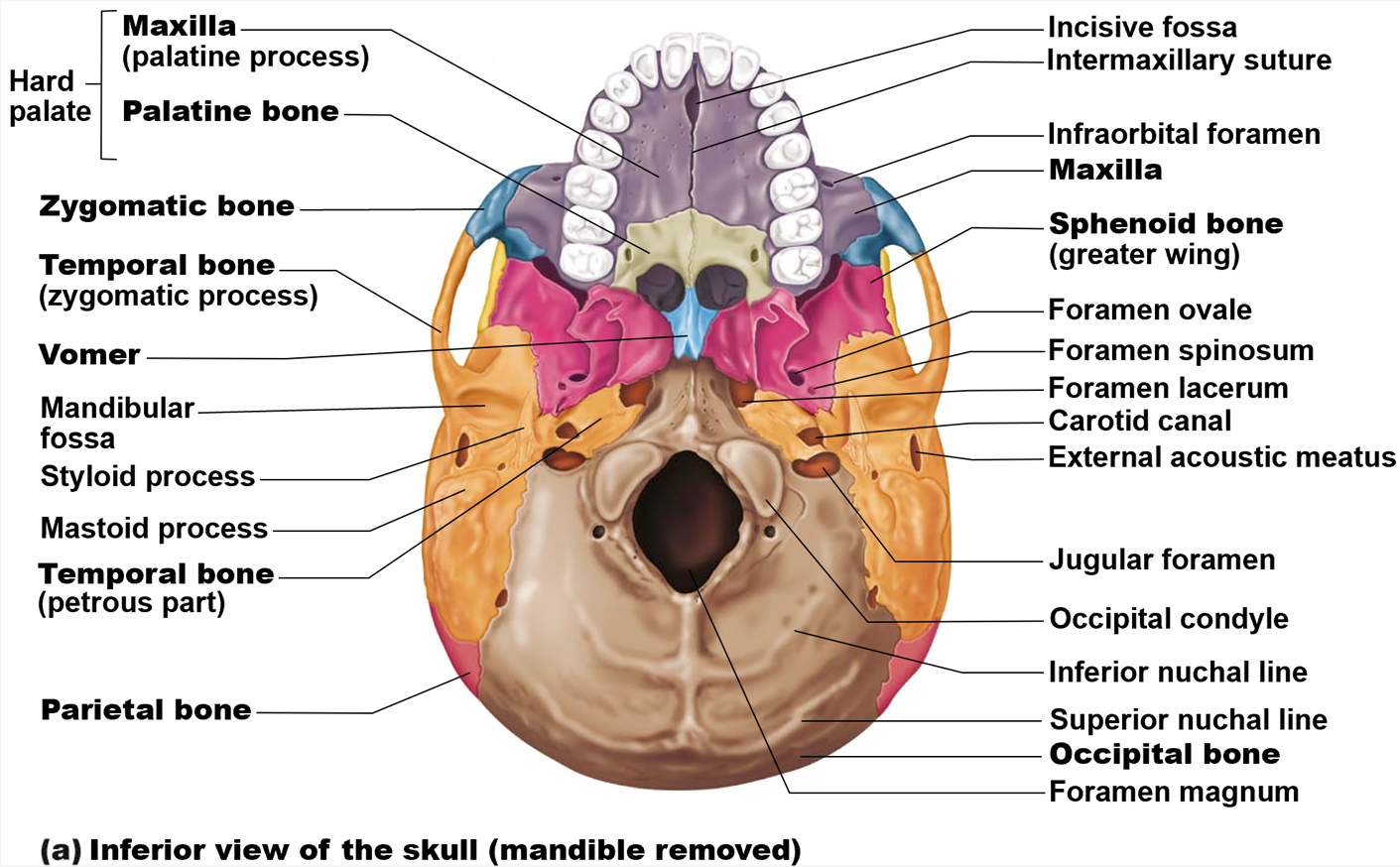
Name the bones and landmarks seen in the skull from an inferior view of the skull.
Maxilla (palatine process), Palatine bone, Zygomatic bone, Temporal bone (zygomatic process), Vomer, Mandibular fossa, Styloid process, Mastoid process, Temporal bone, Parietal bone, Incisive fossa, Intermaxillary suture, Infraorbital foramen, Sphenoid bone (greater wing), Foramen ovale, Foramen spinosum, Foramen lacerum, Carotid canal, External acoustic meatus, Jugular foramen, Occipital condyle, Inferior nuchal line, Superior nuchal line, Occipital bone, Foramen magnum
Name the bones and landmarks seen in a midsagittal section of the skull.
Temporal Bone, Occiptal bone, Parietal bone, Frontal bone, Sphenoid bone, Nasal bone, Ethmoid bone, Vomer, Maxilla, Mandible, Palatine bone.
Landmarks: Squamous suture, Lamboid suture, Internal acoustic meatus, Sella turcica of sphenoid bone, Pterygoid process of sphenoid bone, Palatine process of maxilla, Alveolar processes, Sphenoidal sinus, Crista galli, Frontal sinus, Coronal suture

Label the landmarks of the foramina of the skull and all of the cranial nerves.
Cribiform Plate [Olfactory (I) Nerve fibres], Optic Canal [Optic Nerve (II) including (central artery of retina] opthlamic artery, Superior Orbital Fissure [Oculomotor Nerve (III), Trochlear Nerve (IV), Opthalmic division of Trigeminal (V), Abducens Nerve (VI), Superior Opthalmic vein], Foramen Rotundum [Maxillary Division of Trigeminal Nerve (V)], Foramen Ovale [Mandibular Division of Trigeminal Nerve (V)], Foramen Spinosum [Middle Menigeal Artery and Vein], Carotid Canal (Internal Carotid Artery), Foramen Lacerum, Internal Acoustic Meatus [Facial Nerve (VII) (Including intermediate nerve), Vestibulocochlear Nerve (VIII), Labyrinthine Artery), Jugular Foramen [Glossopharyngeal nerve (IX), Vagus Nerve (X), Accessory Nerve (XI), Sigmoid Sinus → Internal Jugular Vein], Hypoglossal Canal [Hypoglossal Nerve (XII)], Foramen Magnum [Vertebral Arteries, Medulla of Brain, Spinal roots of Accessory (XI) Nerve]
![<p><strong>Cribiform Plate </strong>[Olfactory (I) Nerve fibres], <strong>Optic Canal</strong> [Optic Nerve (II) including (central artery of retina] opthlamic artery, <strong>Superior Orbital Fissure</strong> [Oculomotor Nerve (III), Trochlear Nerve (IV), Opthalmic division of Trigeminal (V), Abducens Nerve (VI), Superior Opthalmic vein], <strong>Foramen Rotundum </strong>[Maxillary Division of Trigeminal Nerve (V)], <strong>Foramen Ovale </strong>[Mandibular Division of Trigeminal Nerve (V)], <strong>Foramen Spinosum </strong>[Middle Menigeal Artery and Vein], <strong>Carotid Canal </strong>(Internal Carotid Artery), <strong>Foramen Lacerum, Internal Acoustic Meatus </strong>[Facial Nerve (VII) (Including intermediate nerve), Vestibulocochlear Nerve (VIII), Labyrinthine Artery), <strong>Jugular Foramen </strong>[Glossopharyngeal nerve (IX), Vagus Nerve (X), Accessory Nerve (XI), Sigmoid Sinus → Internal Jugular Vein], <strong>Hypoglossal Canal </strong>[Hypoglossal Nerve (XII)], <strong>Foramen Magnum </strong>[Vertebral Arteries, Medulla of Brain, Spinal roots of Accessory (XI) Nerve]<strong> </strong></p>](https://assets.knowt.com/user-attachments/b14c2ca4-b7cc-4e68-ac40-a6ba53b05636.png)
What is found in the neurocranium?
Cranial Cavity
Interior occupied by brain
Calvaria
Skull Cap
Cranial Fossa
Anterior
Middle
Posterior
8 bones

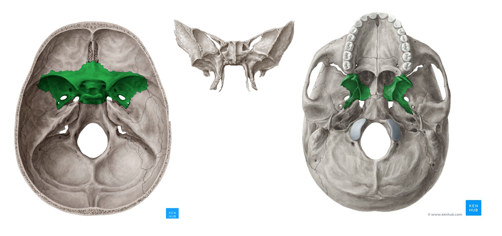
Sphenoid bone

Ethmoid Bone
What forms the skull, how many bones are there, how many paired and unpaired bones?
Skull is formed with the upper and lower jaws, the nose, nasal cavity and nasal septum, and the orbit.
14 bones, with six paired bones and two unpaired bones
The paired bones: maxilla, palatine, zygomatic, nasal, lacrimal, and inferior nasal conchae bones
The unpaired bones: vomer and mandible bones
Although classified with the brain-case bones, the ethmoid bone also contributes to the nasal septum and the walls of the nasal cavity and orbit.
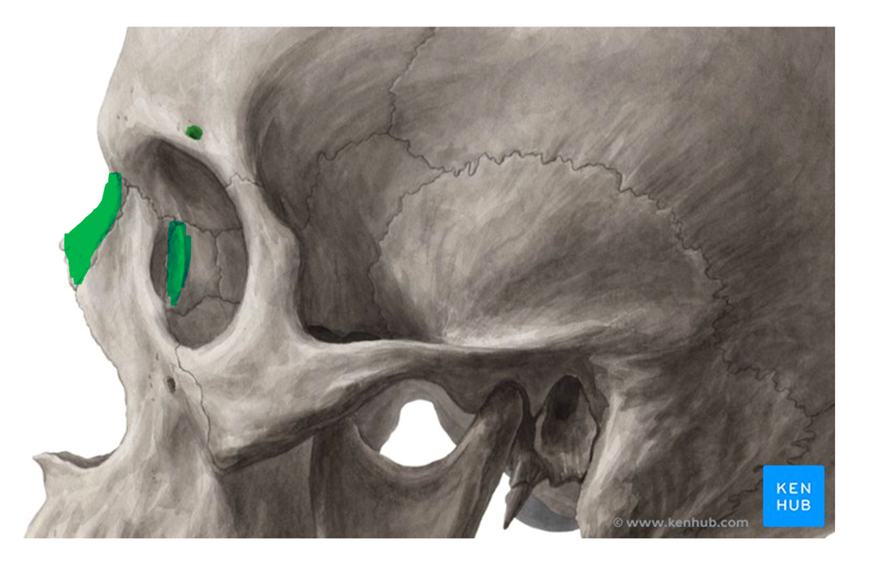
Nasal and Lacrimal Bone


Zygomatic Bones

Vomer Bone


Inferior Nasal Conchae Bones


Maxillae and Palatine Bones


Mandible Bone
What bones form the bony orbit?
Frontal bone, Sphenoid bone, Lacrimal bone, Ethmoid bone, Maxilla bone, Zygomatic bone

What bones are part of the nasal septum?
Perpendicular plate of ethmoid bone, Vomer bone
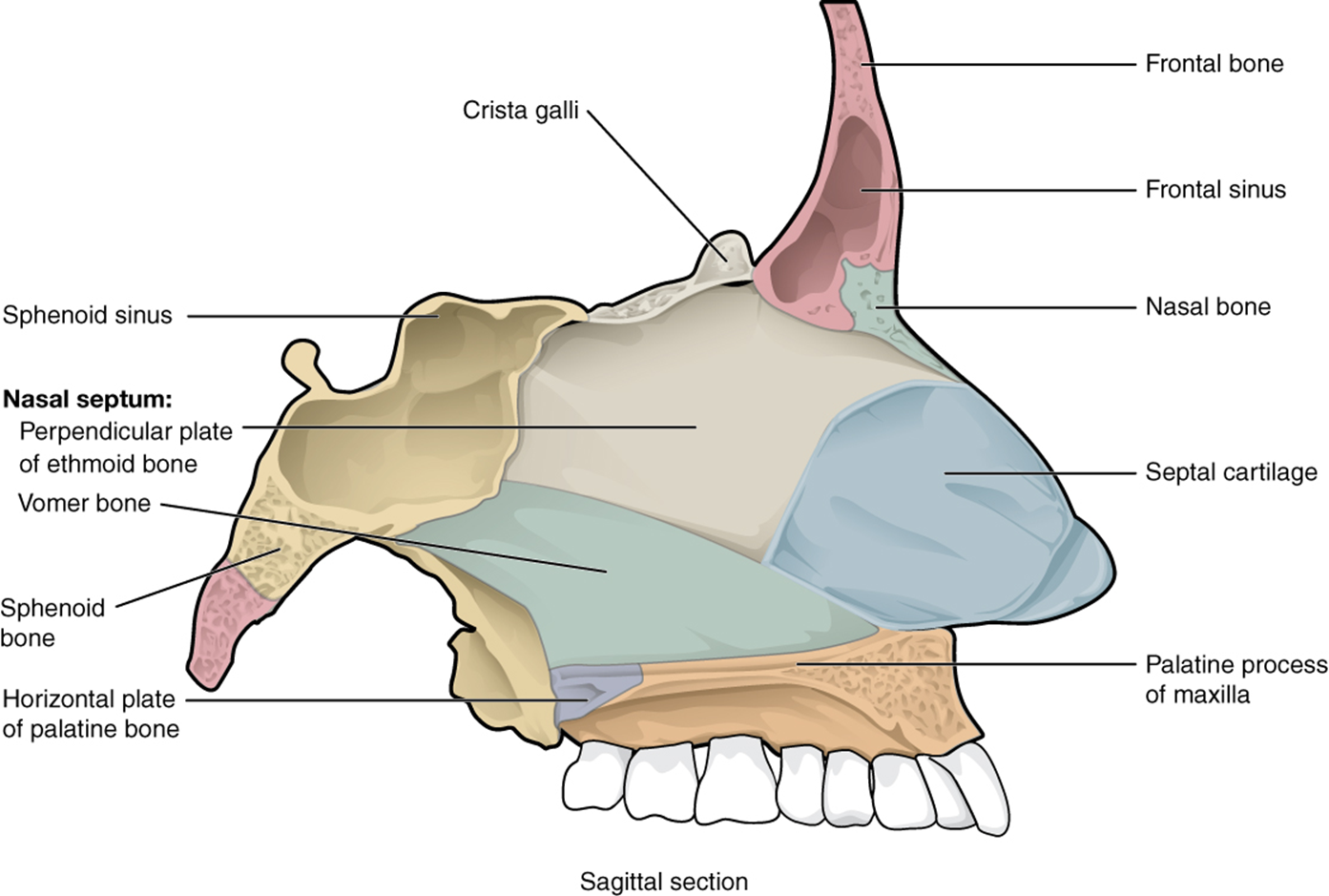
What are the 4 paranasal sinuses?
Frontal
Located between eyebros
Maxillary
Largest, Paired
Commonly involved with infections
Sphenoid
Midline sinus
Ethmoid air cells
Behind superior nasal conchae
Air filled spaces
Reduce bone mass
Resonance to voice
Where is the Hyoid bone?

How many vertebrae does the vertebral column have? How many in the cervical, thoracic, lumbar, sacrum and coccyx?
There are 33 vertebrae
24 vertebrae + sacrum (5 fused vertebrae) + Coccyx (4 fused vertebrae)
Cervical curvature: C1-C7
Thoracic curvature: T1-T12
Lumbar curvature: L1-L5
What is the C1 (cervical) called and what does it do
C1 is called Atlas. C1 supports the skull and moves head up and down
Why does the vertebral column have curvatures? What are the primary and secondary curvatures?
Increase the resilience and flexibility of the spine, and ability to absorb shock
Two posteriorly convex curvatures (primary)
Thoracic and sacral (pelvic)
Two posteriorly concave curvatures (secondary)
Cervical and lumbar
What are the 3 abnormal curvatures of the spine?
Scoliosis (abnormal lateral curve)
Kyphosis (hunchback)
Lordosis (swayback) (e.g., pregnant women)
What are the landmark structure of a vertebra?
Spinous process, Transverse process, Vertebral foramen, Body (centrum)
Lamina, Pedicle, Superior articular facet and process

How many Cervical Vertebra are there? What is the C1 and C2 of the Cervical Vertebra called? What does the C2 have to distinguish itself from others.
There are 7 total cervical vertebra. C1 is called atlas, C2 is called axis. The C2 has the dens of axis that other cervical vertebrae doesn’t have.
What are the function of atlas (C1) and axis (C2)?
The atlas (C1) connect directly to the base of the skull and allows the head to move up and down (nodding - yes motion). The atlas also has a transverse ligament that sit inside the ring that holds the dens of axis. The axis (C2) has the dens of axis that fits inside the ring of C1. The function of axis (C2) is the pivot joint allowing the head to turn left and right (side to side; “no” motion).
How many vertebrae does the thoracic have? What landmarks are found in the thoracic vertebrae?
There are 12 total thoracic vertebrae.
The thoracic vertebrae landmarks are: Spinous process, Transverse process, Superior articular process, Transverse costal facet (for tubercle of rib), Intervertebral disc, Body, Interior costal facet (for head of rib), and Inferior articular process.
How many vertebrae does the lumbar have? What are the landmarks of the lumbar?
The lumbar has 5 vertebrae.
The landmarks of the lumbar are: Superior articular process, Transverse process, Spinous Process, Body, Intervertebral disc, and Inferior articular process.
How many fused vertebrae does the Sacrum and Coccyx have?
Sacrum has 5 fused vertebrae. Coccyx has 4 fused vertebrae.
What are the ligaments found in the spine?
Anterior and posterior longitudinal ligaments: From neck to sacrum; Stabilize and protects the spine from over bending and run downs the bodies of the spine.
Ligamentum Flavum: Connects adjacent vertebrae
Short ligaments: connect each vertebra to those above and below
Supraspinous ligament, ligamentum flavum,
What is intervertebral disc made up of?
Cushion like pad composed of two parts
Nucleus pulposus: innergelatinous nucleus that gives the disk its elasticity and compressibility
Anulus fibrosus: Outer collar composed of collagen and fibrocartilage
Intervertebral foramen: space/hole found between two vertebrae
What is the sternum made up of?
Sternum: Manubrium (Superior), Body, Xiphoid process (Inferior)
How many ribs are there? What is the bone structure of a rib? What are the 3 rib types?
Ribs are curved and flattened bone
Ribs are found at the T1 to T12
The three types of ribs are: true ribs, false ribs and floating ribs
True ribs are found at T1-T7
False ribs are found at T8-T10
Floating ribs are found at T11 & T12
What make up the appendicular skeleton?
Bones of the limbs and their girdles
Pectoral girdle attaches the upper limb to the body trunk
Pelvic girdle secures the lower limbs
What makes up the Pectoral girdle?
Clavicle, Scapula, Acromioclavicular joint
Clavicle: Acromial end (lateral - more rounded), Sternal end (medial - more flat)

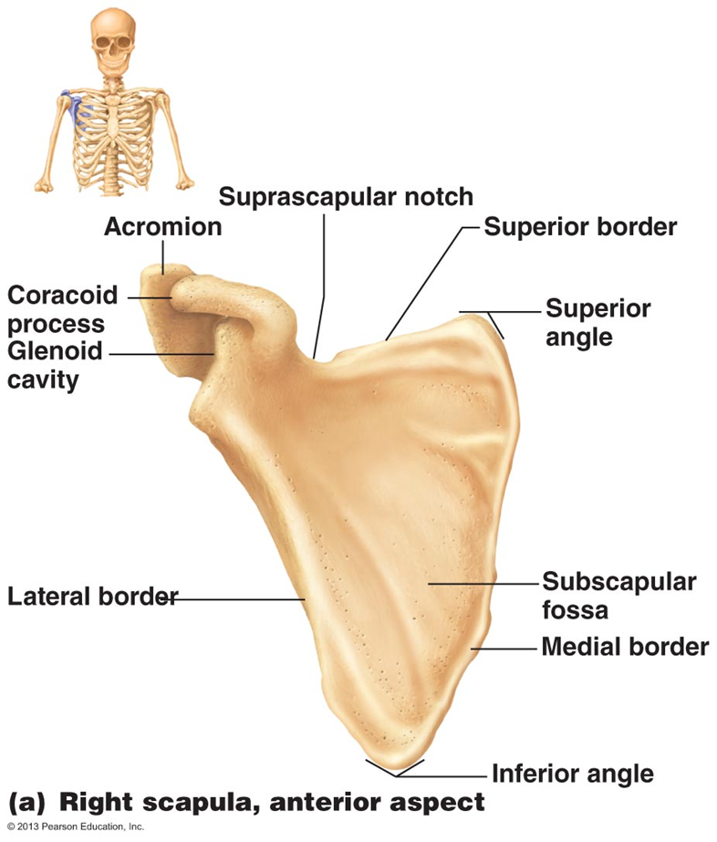
What are the landmarks of the Scapula?
Acromion, Suprascapular notch, Spine of the scapula (connect to the acromion), Superior border, Superior angle, Subscapular fossa, Medial border, inferior angle, lateral border, Glenoid Cavity, and Coracoid Process
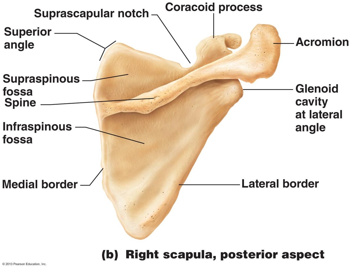
Posterior of the scapula with landmarks
Acromion, Coracoid process, Suprascapular notch, superior angle, Supraspinous foss, Spine of the scapula, Infraspinous fossa, medial border, lateral border, Glenoid cavity at lateral angle
What are all the landmark of the Humerus?
Head of humerus, Anatomical neck, Surgical neck, Radial groove, Deltoid tuberosity, Tochlea, Capitulum, Olecranon fossa, Lateral epicondyle, Medial epicondyle, Coronoid fossa

How does the Humerus connect to the radius and ulna?
Head of radius connect to Capitulum
Coronoid process of ulna connect to Trochlea
Olecranon (elbow) fits into the olecranon fossa (humerus)
Trochlear notch locks into the trochlea (humerus)

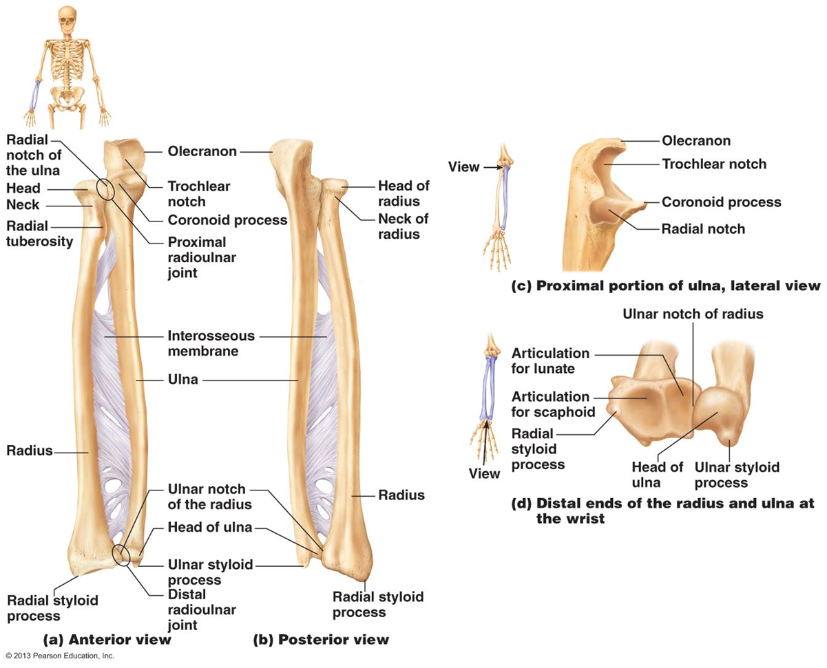
What are the landmarks of Radius?
Head of radius, Neck of radius, Radial tuberosity, Radial styloid process