Lecture 7: Protein Purification and Identification
 Call Kai
 Learn
Learn Practice Test
 Spaced Repetition
 Match
Match Flashcards
 Knowt Play
1/19

There's no tags or description
Looks like no tags are added yet.
Name | Mastery | Learn | Test | Matching | Spaced | Call with Kai | Chat |
|---|
No analytics yet
Send a link to your students to track their progress
20 Terms
How to collect protein samples
four important consideration
Choosing the correct cell type or tissue is important for obtaining an adequate quantity and quality of sample.
Important considerations:
Protein folding
Post-translational modifications (PTM)
Protein-protein interactions (separating to prevent conformational change)
Co-factors
Molecular biology can help
Genetically engineer useful tags or sequences into your POI (to make the protein easier to find or grown).
Express your POI in a specific cell type or organism (e.g. E. coli).
Fusion proteins
what is this process about?
Why is it useful (possible additions to the proteins (5))
Fuse protein domains together using molecular biology to purify and obtain the POI.
Fully functional protein or a small amino acid sequence called a protein tag (gene).
E.g. His tag (6-10 AA)
E.g. FLAG tag (positively and charged AA, binding of anti-FLAG to pull out of solution)
E.g. green fluorescent protein: for visualization (GFP)
Adds useful features to proteins being studied
Fluorescence for visualization
E.g green fluorescent protein
Tags for affinity purification
Obtaining proteins from other non POI
Tags for Western blot detection
Antibodies for different tags
Enzymes for activity assays
May catalyze certain reaction to indicate the protein is present
Fused complexes for crystallization
Looking at the overall structure
Example picture
Dark grey: the POI
N-terminu: flag tag
C-terminus: 6 histine rage (light gret)
Thiroedoxin: help with solubility
After purification, an exonuclease will cleave off the specific sites to remove the fusion proteins

Cell lysis
what is it
What cells is involved and what do they generate
Three common methods to do this in a lab
Two things to consider before doing it
Lysing cells generates a crude extract.
Heterogenous mixture of proteins and cellular contents
To be able to obtain the proteins from within the cells
Common Methods:
Mechanical/physical methods:
Grinding (peddle), Sonication (high frequency), Vortexing with glass beads (break apart membranes)
Osmotic Pressure (<20 mM NaCl)
Water to burst the cells
Chemical basis (e.g. detergents)
E.g. Detergents
Important considerations:
Lysing cells may release proteases – enzymes (designed to break down unwanted proteins, and maybe your POI) )that may degrade your protein of interest (POI).
Solution? Protease inhibitors (ability to bind to the proteases and prevent functionality to break down your POI)
Conditions (pH/temp/detergents) may alter your protein’s structure and lead to denaturation.
Solution? Buffers + Ice (for pH issues)
Regulate the temp
Importance of protein purification
there are many proteins in a mammalian cell (about 1 to 3 billion)
Must isolate the particular protein from other cellular component in order to study it - very important when doing in vitro (inside a test tube rather than studying the organism as a whole)
A heterogenous population will contain other things that may affect the protein of interest
Purifying a protein is a must step for understanding the protein structure/function
Centrifugation purpose
what is the main purpose of a centrifugation
Used to separate the supernatant (your POI including anything that is light) of soluble materials from a pellet of other large organelles or insoluble precipitate
Depend on what you study, you would either take the supernatant or pellet
Allows for the isolation of a particular organelle
Centrifugation can be combined with filters to separate by size

Chromatography
what is the purpose of chromatography
Chromatography: Differential partitioning of a molecule between a mobile (buffer) and stationary (resin; column) phase.
E.g. resin or beads for the stationary phase
Proteins can be purified based on differences in their chemical properties:
Size or shape: Size-Exclusion/Gel Filtration chromatography
Charge: Ion Exchange Chromatography
Binding interactions: Affinity chromatography
Hydrophobicity: RP-HPLC (Reverse Phase High Pressure Liquid Chromatography)
Size exclusion chromatography
Proteins are separated based on size and shape
Columns contain a resin of porous beads
Smaller proteins enter the beads and move through the resin slowly (e.g. red beads) (more tubes used and came out later than the larger molecules)
Larger proteins bypass the beads and move through the resin quickly (e.g. yellow molecules) (more than 100 KDa = passes through)
Wait for timing, when your POI comes out or if they are completed excluded (depending on the size of the POI)
Considerations:
Small but elongated proteins may appear larger – they may not enter the beads and elute faster
Calibration with proteins of known MW is required
MW = Molecular Weight
Vo = void volume
Anything larger than the column’s fractional range (i.e. what doesn’t fit in the pores) goes straight through - bigger molecules
Ve = elution volume of a molecule - the smaller molecules
Vt = total volume of the column
Issue: all molecules are colourless
Use UV absorbance to see when the POI comes out

Ion-exchange chromatography
Separates molecules based on their net charge
Recall the isoelectric point (pI) is the pH when a polypeptide is neutral
Cation exchange resins attract and bind positively charged polypeptides
Anion exchange resins attract and bind negatively charged polypeptides
Proteins can be eluted by increasing the salt concentration or changing the pH
Movement
Negatively charged or neutral go through the resin (column)
Positively charged does not go through

Affinity Chromatography
Proteins are attracted to the column based on their affinity for specific molecules or chemical groups.
The resin contains molecules or ligands that are complementary to specific proteins in the sample.
Interact via non-covalent interactions
The bound protein is released from the resin by passing a solution containing free molecules to compete for binding.
Useful for concentrating proteins in a small volume

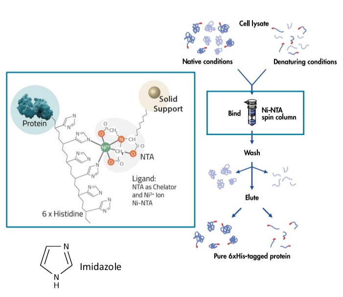
His-tag and Nickel-NTA resin
Nickle (Ni2+) can coordinate binding of histidine side chains to NTA (nitrilotriacetic acid), but only when the His side chains are close to one another
A his-tag (6-10 His residues) can be added to proteins to help with purification
Strong positive charge helps in binding
The tagged-protein binds to the column, other proteins will not
Free imidazole can be used to elute the POI
Competing interactions to results in eludating the POI
Immunoprecipitation
Immunoprecipitation is a form of affinity purification
Antibodies that specifically bind to a POI or tag are used to trap a POI in a resin
The POI contains the antigen
Protein complexes can be precipitated out of solution (co-immunoprecipitation; Co-IP)
Denature all proteins bound to the resin, as well as any interacting proteins (potentially your POI)
Consideration: can only be used if specific antibodies are available or a tag is added to the POI or “bait”

High Pressure Liquid Chromatography
Uses very fine beads and high-pressure pumps to move a sample through the column
Achieves higher resolution of peaks
Resin choice determines separation basis, usually silica covered in hydrocarbons
Known as Reverse Phase HPLC when separating basedon hydrophobicity
In RP-HPLC, hydrophobic compounds move more slowly through the column and have a longer retention time (any compounds that cannot interact will move faster through the column - staying in the mobile phase)
Issues: only use for smaller proteins
Larger proteins may cannot go through at all

Dialysis for protein purification
Dialysis can be used to remove small molecules (e.g. salts, H+, ions, imidazole)
Samples are placed in a semi-permeable dialysis bag (i.e. a filter) and incubated with a buffer.
Diffusion allows molecules to move from the sample into the buffer.
Only for smaller molecules can diffuse out (buffer has less salt - to increase it through diffusion), repeat the step until everything we are not interested in are out of the sample - used until after
Often used after chromatography
Can dilute out (i.e. remove) small molecules used in chromatography that might interfere with other experiments or later assays.
Can also be used to change the pH of the buffer (buffer exchange).
Ranking of specifity

Measuring protein
Biomolecules absorb light at characteristic wavelengths
Can be measured using a spectrophotometer
Most proteins are colourless and do not absorb visible light (380-750 nm)
However, amino acids absorb light in the UV spectrum (100-380 nm)
The concentration of protein in solution can be measured based on the absorbance at 280 nm, arising from aromatic amino acids (e.g. peptide bonds, aromatic rings)

Protein absorbance
Stains like Coomassie Blue bind to proteins and absorb light in the visual spectrum
Rings can be able to absorb the light effectively and only basic Amino acids
Can also be used to visualize proteins or quantify the concentration of protein in a Bradford assay
Beer-Lambert Law
issue with this
can only determine concentration, but not if your POI is in thtere

SDS-page - separating proteins
what is SDS + another alternative?
Sodium Dodecyl Sulfate (SDS) –Polyacrylamide Gel Electrophoresis
A amphipathic molecule (some negative and positive polarity regions)
SDS is a detergent that can denature proteins
Disrupt non-covalent interactions
1 molecule of SDS binds approx. every 2 amino acids
Gives a polypeptides a uniform (-) charge relative to its mass
Beta-mercaptoethanol (BME) can also be used to reduce disulfide bonds
The polyacrylamide gel creates a mesh or sieve of cross-linked molecules that separate subunits based on size
Under denaturing (SDS) and reducing (BME) conditions polypeptide chains will have the same mass-to-charge ratio and migrate in the gel towards the anode
Larger proteins will move through the pores slower, allowing for separation of the proteins
Protein size can be deduced by comparing to MW markers
Identity needs to be confirmed by immunoblotting or mass. spec.

SDS-page - visualizing proteins
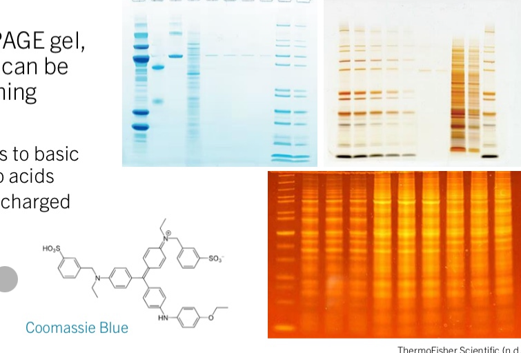
Now separated in the PAGE gel, the colourless proteins can be visualized through staining
Protein stains include:
Coomassie Blue – binds to basic and hydrophobic amino acids
Silver Stains – binds to charged amino acids
Fluorescent Dyes
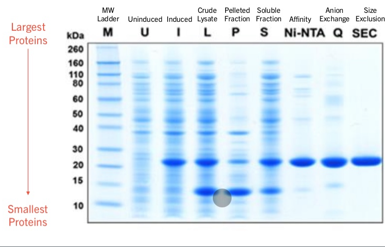
How to determine the purity of your sample?
need to do multiple purification processes to get the protein you want
