HPHY 321 Lab Exam 2( New Material)
 Call Kai
 Learn
 Practice Test
 Spaced Repetition
 Match
 Flashcards
 Knowt Play
1/141

There's no tags or description
Looks like no tags are added yet.
142 Terms
Olfactory Nerve

Optic Nerve

White Matter

Grey Matter

Anterior horn of grey matter
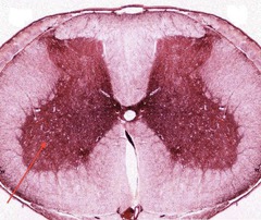
Posterior horn of grey matter

Lateral horn of grey matter
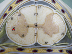
Ventral Root

Dorsal Root

Spinal Nerve
Location: Intervertebral Foramen
Union of Dorsal and Ventral Roots

Dorsal Root Ganglion
Contains cell bodies for afferent (sensory) neurons

Dorsal Ramus

Ventral Ramus

Gray Ramus Communicans

Gray Ramus Communicans
...
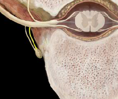
White Ramus Communicans

Sympathetic Ganglion

Sympathetic Trunk

Superior Cervical Ganglion

Middle Cervical Ganglion

Inferior Cervical Ganglion
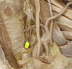
Central Canal

Anterior Median Fissure

Posterior Median Sulcus

Conus Medullaris
Inferior end of spinal cord -
Upper Lumbar Vertebral Canal

Cauda Equina
Bundle of dorsal and ventral roots for spinal nerves below L2

Filum Terminale
From tip of Conus Medullaris to attach to coccyx

Vertebral Artery

Basilar Artery
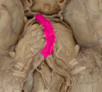
Posterior Cerebral Artery

Posterior Communicating Cerebral Artery
(Connects internal carotid to basilar artery)

Middle Cerebral Artery

Anterior Cerebral Artery
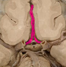
Anterior Communicating Cerebral Artery

Internal Carotid Artery
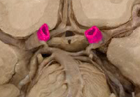
Internal Carotid Artery(Image)
Pointing to the larger of the two

External Carotid Artery

Common Carotid Artery
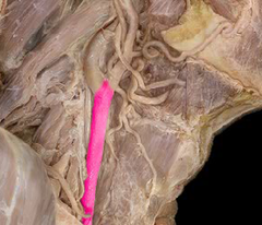
Superior Sagittal Sinus
(Found where the falx cerebri sits

Inferior Sagittal Sinus
(right aboe the corpur callosum)

Superior Sagittal Sinus(model)

Inferior Sagittal Sinus

Straight Sinus
(above the cerebellum in the tentorium cerebelli)

olfactory, optic, vestibulocochlear,
Which Nerves are only sensory
oculomotor, trochlear, abducent, accessory, hypoglossal
Which nerves are only motor
trigeminal, facial, glossopharyngeal, vagus
which nerves are both sensory and motor
Confluence of Sinuses
(@ internal occipital protuberance, terminates @ transverse sinuses, drainage for superior sagittal, straight, & occipital sinus.)

Transverse Sinus
(lies in the transverse groove)

Sigmoid Sinus
(sigmoid groove)

Sigmoid sinus

Occipital Sinus

Occipital sinus

Internal Jugular Vein

Optic Nerve(Model)

Oculomotor Nerve( Enters the superior orbital fissure)
What is this nerve and where does it enter?

Oculomotor Nerve(Model)
Model is tricky but it is a nerve that appears to come out of the pons

Trochlear Nerve( goes through the superior orbital fissure)
Extends from the inferior colliculus and extends through which canal/foramen in the skull

Trigeminal Nerve(superior orbital fissure, foramen rotundum, and the foramen ovale)
Goes through which canal(s)?

Ophthalmic branch, Maxillary branch, and the mandibular branch
What are the three branches of the trigeminal nerve
Abducent nerve(through the superior orbital fissure)
Between the medulla oblongata and the pons. Where does this nerve travel through

Facial Nerve(internal auditory meatus)
Where does this nerve travel through and what is it?

Vestibulocochlear Nerve( through the internal auditory meatus)
Comes from the middle cerebellar peduncle. Where does it travel through?

Glossopharyngeal Nerve(jugular foramen)
What is this nerve and where does it travel

Vagus nerve
The thicker of the three that enter the jugular foramen

Accessory Nerve(Jugular foramen)
This nerve travels down towards the spinal cord. Where does it go through in the skull

Hypoglossal Nerve

oculomotor, trochlear, and a branch of the trigeminal, as well as the abducent
Which nerves travel through the superior orbital fissure
facial and vestibulocochlear
Which nerves travel through the internal auditory meatus
Vagus, glossopharyngeal, and the accessory
(If you are going to VAGUS, remember your lipgloss( GLOSSOPHARYNGEAL), a jug(JUGULAR FORAMEN) of wine, and your accessories(Accessory))
Which nerves travel through the jugular foramen
Sclera and the cornea and the conjunctiva
What does the fibrous tunic of the eye include
Sclera of the eye
(Where the muscles of the eye attach)

Sclera of the fibrous tunic

Cornea of the fibrous tunic

Cornea of the eye

Conjunctiva of the eye of the fibrous tunic

Conjunctiva of the fibrous tunic(picture)

choroid, iris, pupil, ciliary body, zonular fibers
What is found in the vascular tunic
Choroid of the vascular tunic( cow eye)

Choroid of the vascular tunic

Iris of the vascular tunic of the eye

Pupil of the vascular tunic of the eye

Ciliary body of the vascular tunic of the eye
(controls dilation)

Ciliary body of the vascular tunic of the eye

Zonular fibers
( Connect the ciliary bodies to the lens)
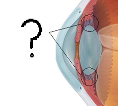
Retina, ora serrate, optic disc, fovea centralis, and the optic nerve
What makes up the sensory tunic of the eye
Retina of the sensory tunic

Retina of the sensory tunic(Cow eye)

Ora serrate of the sensory tunic
(junction between retina and ciliary body)

Optic disc of the sensory tunic
(in front of the optic nerve)

Fovea centralis of the sensory tunic

Fovea centralis of the sensory tunic

Optic nerve

Anterior chamber of the eye
2?
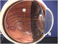
Posterior Chamber of the eye
B?

Lens of the eye

Vitreous chamber of the eye

Vitreous body of the vitreous chamber
(The gel)

Lacrimal gland of the lacrimal apparatus
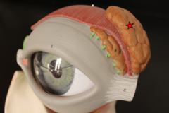
Excretory duct of the lacrimal gland

Lacrimal Sac
