BIOS 255 Exam 2 Review with expert curated questions and answers-Chamberlain
 Call Kai
 Learn
Learn Practice Test
 Spaced Repetition
 Match
Match Flashcards
 Knowt Play
1/70

There's no tags or description
Looks like no tags are added yet.
Name | Mastery | Learn | Test | Matching | Spaced | Call with Kai | Chat |
|---|
No analytics yet
Send a link to your students to track their progress
71 Terms
Tunica externa (tunica adventitia)
epithelial cells, connective tissue, elastic fibers
Tunica media
elastic fibers and smooth muscle (usually the largest layer- controls the diameter)
Tunica intima (interna)
endothelial cells, connective tissue
Aneurysm
is a weak point in the wall of a vessel, bulging sac that pulsates with each heartbeat.
Capillaries
vessels that allow for the exchange of substances between blood and interstitial space
Sinusoidal
large openings allow the passage of larger molecules such as proteins and formed elements; found in the liver
Veins
vessels that carry blood TOWARDS from the heart
Carotid bodies
responsible for monitoring blood pH
Blood pressure (BP)
is the force exerted by blood against a vessel’s wall as it flows through. • BP = systolic pressure/diastolic pressure
Systolic blood pressure
refers to the pressure in the arteries when the heart contracts and pumps blood into the circulation.
Diastolic blood pressure
• represents the pressure in the arteries when the heart is at rest between beats.
Pulse Pressure
systolic pressure - diastolic pressure
Mean Arterial Pressure: MAP
diastolic pressure + 1/3pulse pressure
Arteriosclerosis
stiffening of the arteriole walls due to degradation of elastic tissues.
Atherosclerosis
stiffening of blood vessel walls due to lipid deposits.
Vascular Resistance (R)
• Three factors determine systemic vascular resistance: blood vessel diameter, blood vessel length, blood viscosity
Blood vessel diameter
CONSTRICTED VS DIALATED
▪ A decrease in vessel diameter increases resistance and requires an increase in pressure to maintain the original flow rate.
Blood vessel length
An increase in blood vessel length increases resistance and require an increase in blood pressure to maintain the original flow rate
Blood viscosity
An increase in viscosity results in thicker blood that is harder to move and requires an increase in blood pressure to maintain the original flow rate
BHP = Blood hydrostatic pressure
facilitates the filtration of fluid and small molecules out of the blood capillaries and into tissues
Vasoconstriction
Definition: Vasoconstriction is the narrowing of blood vessel diameter due to contraction of the smooth muscle in the vessel wall.
Effect on Blood Flow: Decreases blood flow to the area downstream of the constricted vessel.
Effect on Blood Pressure: Increases resistance to blood flow, which leads to an increase in blood pressure. The heart must work harder to push blood through the narrower space.
How it Happens: Often triggered by the sympathetic nervous system, certain hormones (like angiotensin II and catecholamines), or reflexes responding to low blood pressure or stress.
Vasodilation
Definition: Vasodilation is the widening of blood vessel diameter due to relaxation of the smooth muscle in the vessel wall.
Effect on Blood Flow: Increases blood flow to the area downstream of the dilated vessel.
Effect on Blood Pressure: Decreases resistance to blood flow, which leads to a decrease in blood pressure. Blood moves more easily through the wider vessel.
How it Happens: Can be triggered by local chemicals (like histamine or prostaglandins), decreased sympathetic stimulation, or certain hormones (like natriuretic peptides).
EDEMA
accumulation of excess fluids in a tissue
Increased capillary filtration
increase in hydrostatic pressure or an increase in capillary permeability may increase the amount of water that filters out into surrounding tissues
Reduced capillary reabsorption
lack of albumin in blood plasma reduces the reabsorption of tissue fluid (Remember osmosis: wherever solutes are, water follows!).
Obstructed lymphatic drainage
If the lymphatic vessels have a blockage, then lymph is unable to return to the bloodstream and accumulates in tissue fluids.
Flow of blood into the right atrium
axillary vein, subclavian vein, brachiocephalic vein, superior vena cava, right atrium
Hepatic portal vein
carries oxygen-poor but nutrient-rich blood from the intestines to the liver
Internal jugular vein
drains blood from most of the brain
need to know
Blood in the axillary artery will flow to the Brachial artery
need to know
Axillary vein and Hepatic vein carry deoxygenated blood
Circle of Willis
The internal carotid artery is a key artery that contributes along with vertebral artery to a loop of vessels located at the base of the brain
Celiac Trunk
a major unpaired artery that branches off the abdominal aorta just below the diaphragm
Immune System
cell population that inhibits all organs and defends the body from agents of disease
Lymphatic System
network of organs, tissues, and vessels densely populated with immune system cells
Fluid recovery
85% of tissue fluid is reabsorbed by blood capillaries and put into venous circulation; remaining 15% is reabsorbed by the lymphatic system
Immunity
lymphatic fluid is inspected for foreign invaders as it passes through lymph nodes
Lipid absorption
lymphatic vessels called lacteals in the small intestine absorb dietary lipids and transport them to the bloodstream
Lymph: recovered extracellular fluid
Clear, colorless fluid similar to blood plasma, but much less protein; same composition as interstitial fluid
Lymphatic Organs: Primary
-site where B & T cells are generated and mature
- red bone marrow and thymus

Lymphatic Organs: Secondary
-mature cells migrate to play a role in immunity
-lymph nodes, tonsils, and spleen
Lymphatic Vessels
• Vein-like vessels that transport lymph
• Penetrate nearly every body tissue except cartilage, cornea, bone, and bone marrow
• Closely associated with blood capillary beds
Right lymphatic duct
receives lymph from right arm, right head, and right thorax → empties into right subclavian vein
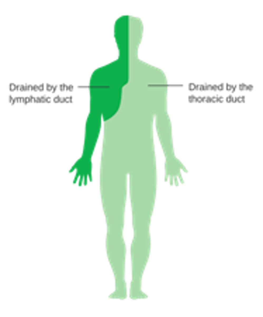
Thoracic duct
larger and longer, receive lymph from below diaphragm, left arm, left head, left neck, and left thorax → empties into left subclavian vein

Lymphatic Route
Lymphatic Capillaries >Collecting Vessels: thin walls, many valves >Lymph Trunks:
lumbar, intestinal, bronchomediastinal, subclavian, jugular >Collecting Ducts - right
lymphatic duct, thoracic duct (left lymphatic duct)> Subclavian vein(left)
Mucosa-associated lymphatic tissue (MALT)
respiratory, digestive, urinary, and reproductive tracts
Lymphatic nodules (follicles)
temporary dense masses of lymphatic cells (lymphocytes and macrophages) that form in response to pathogens
Tonsils
found in pharynx and oral cavity - helps to trap inhaled or ingested foreign substances
Pharyngeal tonsils
wall of the nasopharynx
Palatine tonsils
pair at posterior oral cavity, tonsilitis
Lingual tonsils
pair at root of tongue
Spleen
largest lymphatic tissue in the body; upper right quadrant; surrounded by a capsule
Red Pulp
blood reservoir; helps to rid the body of old or damaged RBCs
White Pulp
consists of B-cells, T-cells, and macrophages and functions similar to a large lymph node
Thymus
site of T cell maturation
Cervical (neck)
monitor lymph from head and neck
Axillary (armpit)
monitor lymph from upper limb and female breast
Thoracic (chest)
monitor lymph from mediastinum, lungs, and airway
Abdominal (posterior abdominopelvic wall)
monitor lymph from the urinary and reproductive systems
Intestinal/Mesenteric (adjacent to intestines)
monitor lymph from the digestive tract
Inguinal (groin)
monitor lymph from the entire lower limb
Popliteal (back of knee)
monitor lymph from the foot and leg proper
Arteries
vessels that carry blood AWAY from the heart
Carotid sinuses
helps control blood pressure and HR
Aortic Bodies
a small cluster of chemoreceptors located near the aortic arch, responsible for sensing changes in blood oxygen and carbon dioxide levels, as well as pH
portal system
•consists of blood vessels that have a capillary bed at either end.
Hypophyseal portal
connects the hypothalamus to the anterior pituitary
Hepatic portal
connects the lower GI tract to the liver
Factors affecting blood pressure
1. cardiac output
2. Blood volume
3. Vascular resistance or resistance to flow
thrombus
a blood clot that forms within a blood vessel and remains attached to the vessel wall.
brachiocephalic trunk
•delivers blood towards the upper right limb via the right subclavian artery branch and blood towards the head via the right common carotid artery.
