Veterinary Anatomy and Physiology: The Nervous System and Special Senses
 Call Kai
 Learn
Learn Practice Test
 Spaced Repetition
 Match
Match Flashcards
 Knowt Play
1/319

Earn XP
Description and Tags
Comprehensive vocabulary flashcards covering the cellular structure, neurophysiology, central nervous system, peripheral and autonomic divisions, spinal reflexes, meninges, and special senses based on veterinary anatomy and physiology lecture materials.
Name | Mastery | Learn | Test | Matching | Spaced | Call with Kai | Chat |
|---|
No analytics yet
Send a link to your students to track their progress
320 Terms
Nervous System
A communication network that enables an animal to adjust itself or its parts to changes in the external and internal environments. Sensory → integrated → Motor
Sensory Components of the Nervous System
The functional division responsible for detecting changes in the internal and external environments (hot stove sense in finger)
Integrative Components of the Nervous System
The functional division that processes sensory data coupled with information stored in memory (spinal cord/brain integrates and tells what to do)
Motor Components of the Nervous System
The functional division that provides an appropriate response to processed sensory and stored information (withdrawal hand)
Neurons
Excitable nerve cells that COMMUNICATE nerve impulses and join with other cells via synapses.
Glial Cells (Neuroglia)
Non-neuronal SUPPORT cells that provide essential service to neurons and their microenvironment.
Glial Cell to Neuron Ratio
A 10-fold greater number of glial cells compared to neurons in the human brain.
Neuron consists of
Dendrites, cell body, axon (travels in that order too)
Neuron Cell Body (Soma)
The metabolic core of the nerve cell containing the nucleus from which processes extend.
Dendrites
Highly branched cellular processes that conduct nerve impulses TOWARD the cell body and provide extensive receptive sites.
Axon
A single specialized neuronal process that conducts action potentials away from the cell body, 1 per neuron, may have myelin sheath
Nerve Fiber
Another name for axon, visible part of nerve
Neurilemma
Plasma membrane of neuron
Axolemma
The specialized plasma membrane that covers the axon of a neuron.
Nodes of Ranvier
Regularly spaced interruptions in the myelin sheath along an axon where the axolemma is exposed to extracellular fluid, does this because it needs the fluid for power (electricity), longer nerves have lots
Telodendritic Zone
The terminal branching at the end of an axon, includes synapse
Neuron Polarity
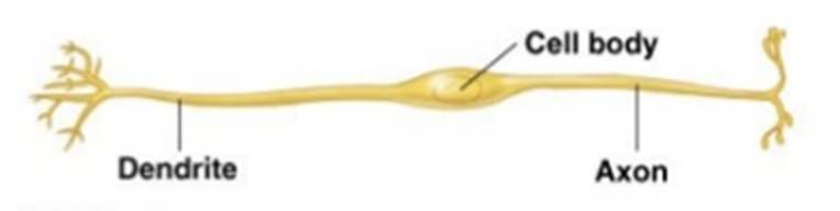
The classification of a neuron based on the number of poles or processes extending from its cell body (dendrites), in mammals can be dipolar or multipolar
Bipolar Neuron
A neuron with two distinct processes extending from opposite sides of the cell body: one dendrite and one axon, found in retina of the eye and olfactory region)
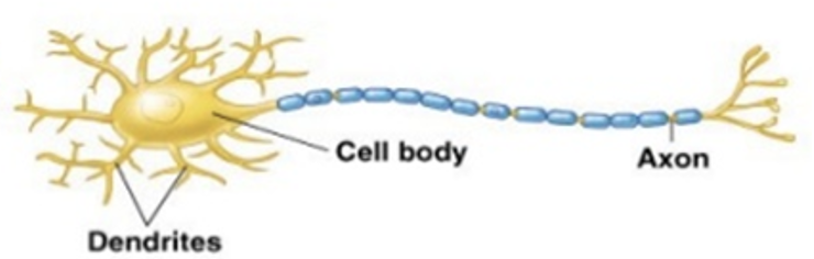
Multipolar Neuron
A neuron possessing one axon and multiple branching dendrites extending from the cell body, most neurons in CNS
Nucleus (Neuroanatomy)
A localized cluster of nerve cell bodies situated within the brain or spinal cord (CNS).
Ganglion
A localized cluster of nerve cell bodies situated outside the brain or spinal cord (PNS).
Tract (Fasciculus)
A bundle of parallel nerve fibers having a common origin and termination within the brain or spinal cord (CNS).
Nerve
A macroscopic bundle of neuron fibers located outside the brain and spinal cord (PNS).
Synapse
The specialized junction (between nerve and something else) where the terminal bud of a neuron communicates chemically with another neuron or effector cell without physical contact.
Synaptic Gap (Synaptic Cleft)
The fluid-filled microscopic space existing between communicating cells across which neurotransmitters diffuse, nerves never have true physical contact
Synapse characteristics
One-way conduction (the presynaptic neuron to the postsynaptic cell), facilitation (repeated impulses provide for easier subsequent transmission), easier to fatigue, chemicals inolved
Presynaptic Neuron
The transmitting neuron that conducts action potentials toward the synapse and releases neurotransmitters.
Postsynaptic Cell
The receiving neuron or target effector cell situated across the synaptic cleft from the terminal bulb, area between (cleft) is filled with interstitial fluid
Synapse vesicles
Membrane-bound structures in the presynaptic neuron that store neurotransmitters until they are released into the synaptic cleft.
Terminal Bulb
The distal expanded swelling of an axon terminal that fits into an indentation of the postsynaptic cell membrane.
Oligodendrocytes
Glial cells that produce the insulating myelin sheath surrounding nerve fibers in the CNS
Schwann Cells
Glial cells that form the myelin sheath surrounding peripheral nerve fibers in the PNS
Astrocytes
The most numerous, star-shaped glial cells that provide structural support, nourish neurons from blood vessels, and release glutamate.
Ependymal Cells
Ciliated glial cells that line brain ventricles and the central canal of the spinal cord, joining with capillaries to form the choroid plexus; help make cerebral spinal fluid
Microglia
Phagocytic glial cells of vascular origin that proliferate in the CNS during inflammation or cellular injury; They recycle debri and defend from invaders
Myelin sheath
Surround fiber and serve as electrical insulator, formed by oligodendrocytes in CNS and Schwann calls in PNS, faster when myelinated
Unmyelinated Nerve Fibers
Axons that lack a lipid sheath and maintain direct contact with extracellular fluid along their entire length.
Saltatory Conduction
The rapid mechanism of impulse propagation in myelinated fibers where action potentials leap from one node of Ranvier to the next.
Schwann Cell Myelination Architecture
The structural arrangement where one single Schwann cell wraps its cytoplasm repeatedly around a single segment of one axon.
Oligodendrocyte Myelination Architecture
One wraps around many axons
Node of Ranvier in CNS
An axonal region where the axolemma is broadly exposed directly to the extracellular space without overlapping glial processes, need electrolyte to fire so takes those from surrounding fluid, fast and brings saltatory conduction
Node of Ranvier in PNS
An axonal junction where the outer cytoplasmic processes of adjacent Schwann cells overlap, restricting extracellular exposure.
Central Nervous System (CNS)
The primary control center of the nervous system consisting of the brain and spinal cord.
Peripheral Nervous System (PNS)
The communication network outside the brain and spinal cord consisting of cranial nerves and spinal nerves (limbs and organs), has somatic and autonomic nervous systems
Somatic Nervous System
The functional division of the nervous system under voluntary control that innervates skeletal muscle.
Autonomic Nervous System (ANS)
The visceral motor division of the nervous system providing involuntary control over glands, smooth muscle, and cardiac muscle.
Sympathetic Nervous System
The ANS division activated during emergency or stress to initiate 'fight-or-flight' physiological responses; eyes dialate, heart rate increases
Parasympathetic Nervous System
The ANS division responsible for vegetative, restful 'rest-and-digest' homeostatic maintenance
Enteric Nervous System
The intrinsic ANS division dedicated entirely to monitoring and regulating gastrointestinal tract functions.
Sphingomyelin
The specific white lipid component found in high concentrations within the myelin sheath.
Brain
The encephalic organ of the CNS providing memory, central processing for problem-solving, and sensory input-output capability; divided into brain stem, cerebellum, and cerebrum (specific spots) also into forebrain, midbrain, and hindbrain)

Forebrain (F)
The rostral primary division of the brain comprising the cerebral cortex, cerebral striata (basal nuceli), rhinencephalon (olfactory brain), thalamus, hypothalamus, and epithalamus

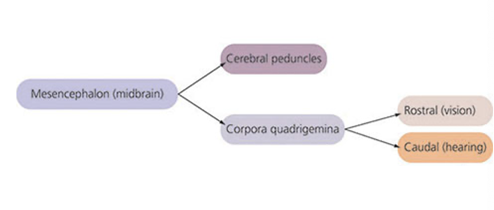
Midbrain (Mesencephalon) (M)
The short central segment of the brain stem located between the diencephalon and the pons; has cerebral peduncles and corpora quadrigemina
Hindbrain (Rhombencephalon) (H)
The caudal primary division of the brain consisting of the cerebellum, pons, and medulla oblongata.

Cerebrum
The largest brain division consisting of the paired right and left cerebral hemispheres.
Cerebellum
The hindbrain structure responsible for coordinating voluntary movement, balancing physical status, and regulating posture.
Brainstem
Most important part, primitive, heart rate and breathing, required for life, has interbrain, midbrain, pons, and medulla oblongata, and spinal cord
Cerebral Hemispheres (F)
Paired expanses of the cerebrum composed of an outer cortex of gray matter, a central mass of white matter, and basal nuclei.
Parts of the forebrain
Cerebral cortex, basal nuclei, rhinecephalon, white matter, thalamus, hypothalamus, and epithalamus
What does each hemisphere have (F)
gray matter, cerebral cortex (central mass), medullary substance (nerve fiber), and basal nuclei (complex movements like walking)
Cerebral Cortex (CeCo) (F)
The superficial layer of gray matter over the cerebral hemispheres, concerned with consciousness and high-level association; size associated with intelligence level, gives association and association center, has motor area (control opposite hemispheres thus opposite side of body), and sensory area, and has specific fibers
White matter
Myelinated, grey matter is unmyelinated
White matter fibers of the cerebral cortex (CeCo) (F)
Association, commisural, and projection
Association Fibers (CeCo) (F)
Cerebral white matter fibers that connect different cortex areas within the same cerebral hemisphere.
Commissural Fibers (CeCo) (F)
White matter nerve tracts that cross the midline to connect corresponding areas of the right and left cerebral hemispheres, makes sure both sides working the same
Projection Fibers (CeCo) (F)
Long white matter fiber tracts that connect the cerebral cortex with lower parts of the brain and the spinal cord, comes from brain and goes down
Cerebral cortex parts (CeCo) (F)
Frontal lobe, parietal, occipital, and temporal lobes.
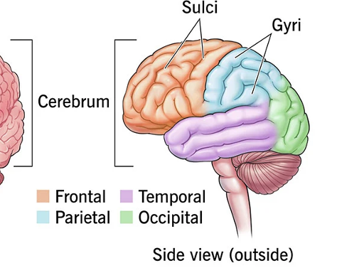
Frontal Lobe (CeCo) (F)
The rostral cerebral lobe dedicated to conscious, thinking, and planning.
Parietal Lobe (CeCo) (F)
The dorsal cerebral lobe responsible for receiving and processing somatic sensory information from the body, process senses
Temporal Lobe (CeCo) (F)
The lateral cerebral lobe responsible for hearing reception and speech interpretation, hearing/speech
Occipital Lobe (CeCo) (F)
The caudal cerebral lobe responsible for processing visual input received from the eyes.
Association area (CeCo) (F)
Sense → association (nostalgia)
Somesthetic Area (CeCo) (F)
The cortical body sense region that receives tactile, thermal, and pain input from the skin, taste sensations, and proprioception from joints and muscles; area that receives
Broca’s area (CeCo) (F)
A region in the frontal lobe associated with speech production
Wernicke’s area (CeCo) (F)
A region in the frontal lobe associated with speech understand
Rhinencephalon (Rh) (F)
Olfactory sense, under the brain to the front
Basal Nuclei (Cerebral Striata) (BN) (F)
Subcortical aggregates of gray matter in the cerebrum that coordinate complex, semi-voluntary movements like walking and running (writing, typing, walking)
Thalamus (F)
A large diencephalic relay center that processes and directs sensory impulses from the body to the cerebral cortex
Info → spinal cord → ___ → specific area
Hypothalamus (F)
The ventral diencephalic structure (below thalamus) that houses the pituitary gland and regulates autonomic functions, neurosecretion, and homeostasis, first thing to release a hormone
Pituitary Gland (Hypophysis) (F)
The primary master endocrine gland positioned immediately ventral to and regulated by the hypothalamus.
Epithalamus (F)
The dorsal diencephalic segment housing an olfactory correlation center and the endocrine pineal gland.
Pineal Gland (F)
A neurosecretory organ in the epithalamus that secretes melatonin
Midbrain parts (M)
Cerebral peduncles and Corpora quadrigemina
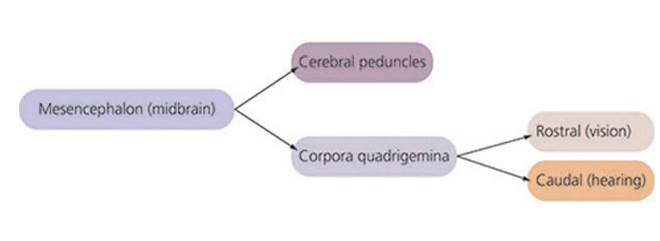
Cerebral Peduncles (M)
Massive fiber tracts on the ventral midbrain that continue pathways to and from the spinal cord to relay and control movement, continuation of fiber/nuclei from spinal cord
Corpora Quadrigemina (M)
Four dorsal midbrain elevations consisting of the superior and inferior colliculi.
Superior (Rostral) Colliculi (M)
The rostral pair of midbrain eminences of the corpora quadrigemina serving as visual reflex centers.
Inferior (Caudal) Colliculi (M)
The caudal pair of midbrain eminences of the corpora quadrigemina serving as auditory reflex centers.
Hindbrain parts (H)
Cerebellum, Pons, and Medulla oblongata

Cerebellar Sensory Inputs (H)
Afferent signals delivered to the cerebellum from proprioceptors, the inner ear equilibrium apparatus, the visual cortex, and the motor cortex (receives info, gives fire to motor control)
Proprioceptive receptors (H)
In cerebellum, tells where body is located in space, throughout all limbs
Cerebellum side effect (H)
Ipsilateral (left side effects left side)
Feline Cerebellar Hypoplasia (H)
A congenital condition caused by underdevelopment of the cerebellum in cats, producing hypermetria, ataxia, and tremors.
Hypermetria (H)
A cerebellar defect characterized by exaggerated voluntary movements that overshoot their intended goal ('goose stepping').
Ataxia (H)
Failure of smooth muscular coordination resulting in clumsiness, imbalance, and irregular gait.
Intention Tremor (H)
Involuntary trembling or quivering that appears during purposeful, voluntary movement and ceases at rest.
Brain Stem (BrSt) (H)
The stalk-like base of the brain comprising the midbrain, pons, and medulla oblongata; visceral function (heartbeat, blood vessels, etc)
Pons (BrSt) (H)
The rostral part of the hindbrain stem bridging the medulla oblongata to midbrain and cerebellum, containing vital fiber tracts. (2nd hit)
Medulla Oblongata (BrSt) (H)
The most caudal brainstem region containing vital autonomic reflex centers and cranial nerve nuclei. (1st hit)
Medullary Vasomotor Center (BrSt) (H)
A vital reflex center in the medulla oblongata that regulates blood vessel smooth muscle tone and blood pressure.
Medullary Cardiac Center (BrSt) (H)
A reflex center within the medulla oblongata that regulates the rate and force of heart contractions.