O/S Radio Midterm
 Call Kai
 Learn
 Practice Test
 Spaced Repetition
 Match
 Flashcards
 Knowt Play
1/133

There's no tags or description
Looks like no tags are added yet.
134 Terms
Rheumatoid arthritis dx (at least 4)
Morning stiffness
Arthritis in three or more joints
Symmetrical arthritis
Rheumatoid nodules
Serum rheumatoid factor
Radiographic changes
Synovitis articular involvement
Fairly symmetrical
Bilateral joint involvement
Sparing of DIP joints of hands
MCP, PIP and wrist joint involvement
Synovitis tendon and ligament involvement
Palmar flexor tendinitis
Rotator cuff tendinitis
Atlantoaxial ligament involvement

rheumatoid arthritis

rheumatoid arthritis

rheumatoid arthritis

rheumatoid arthritis

rheumatoid pleurisy
rheumatoid pleurisy radiographic features
Small to moderate effusion
Unilateral
Often chronic
rheumatoid interstitial fibrosis and pneumonitis radiographic features
Linear opacities
Bases
Honeycombing
rheumatoid interstitial fibrosis and pneumonitis CT findings
Reticular opacities
Subpleural zones
Thickened interlobular septa
Honeycombing

rheumatoid interstitial fibrosis

necrobiotic (rheumatoid) nodule
Multiple
Well-circumscribed
Periphery of lung
Cavitation is common
Thick, smooth walls
Change in size

systemic lupus erythematosus - water bottle sign - pericardial effusion

SLE - pericardial effusion

SLE - atelactasis
Subsegmental
Generally in bases
May be migratory and fleeting
Associated with pleural effusion or diaphragm dysfxn

(right = normal)
SLE - intestinal vasculitis
Esophagus to colon
CT findings
Dilated bowel
Wall thickening
Target sign
Engorged mesenteric vessels (comb sign)
Ascites

SLE - CNS vasculitis

SLE - Jaccouds arthropathy

SLE - soft tissue calcifications
Scleroderma
Progressive systemic sclerosis
Widespread fibrosis and atrophy
Skin
Lung (2nd most common site)
GI tract (esophagus most common)
Limited form and diffuse form
Females > Males
Age 40 to 70 years
Scleroderma - skin, GI tract, lung
Skin
Fingers and hands
Raynaud’s phenomenon
Subcutaneous calcifications
GI tract
Esophageal motility dysfunction
Small bowel dysfunction
Lung
Isolated pulmonary hypertension
Interstitial lung disease
CREST syndrome
Calcinosis
Raynaud’s phenomenon
Esophageal dysmotility
Sclerodactyly
Telangiectasia

scleroderma - raynauds missing tip finger

scleroderma calcification in the fingers and hands

Scleroderma: lung fibrosis (2nd most common site)

scleroderma: fibrosis
Linear opacities
Bases
Calcinosis of soft tissues

Scleroderma - esophagus
Proximal third unaffected
Incompetent LES
Patulous G-E junction
No normal peristalsis
Resultant reflux
Eventual structure

scleroderma - esophagus - “bird’s beak”, achalasia

scleroderma - small bowel
Narrow separation between folds “hide bound”
Small-bowel dilatation and air-fluid levels
Wide-mouthed diverticula
Polymyositis/dermatomyositis
Idiopathic inflammatory muscle disease
Peak incidence in childhood and late adulthood
Woman > Men (2:1)
? Viral infection with altered immune response
Minority of cases a/w malignancy
polymyositis
weakness
Muscle enzyme elevation
Characteristic EMG
Inflammatory infiltrates on Bx
dermatomyositis
sxs of polymyositis, with characteristic rash
skin in polymyositis/dermatomyositis
Erythematous patches: face, neck, chest, extensors surfaces
Heliotrope lids: violet discoloration/swelling of eyelids
Gottron’s sign: heaped-up erythematous papules over MCP /PIP joints
muscle in polymyositis/dermatomyositis
Gradual/steady progression of muscle weakness
Skeletal muscles, including diaphragm, pharynx, upper esophagus
lung in polymyositis/dermatomyositis
Dyspnea on exertion
Non-productive cough
Aspiration pneumonitis

air space consolidation???
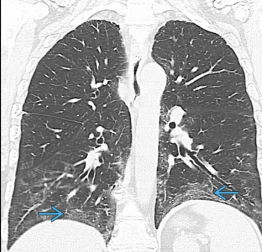
polymyositis CT findings
Linear abnormalities
Ground-glass attenuation
Air-space consolidation
mainly in the middle and lower lung zones
peribronchial and subpleural distribution
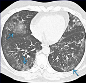
polymyositis
Chronic interstitial pneumonitis
Aspiration pneumonia
Due to hypotonic esophagus/pharynx
Hypostatic pneumonia
Due to chest wall involvement/hypovent
Diffuse soft tissue calcifications
ankylosing spondylitis
Autoimmune disease of unknown pathogenesis
Primarily affects the axial skeleton
Male > Female (3:1)
Involvement of thoracic spine is most common
Pleuro-pulmonary manifestations in 1-2%

ankylosing spondylitis - bamboo spine

ankylosing spondylitis - anterior syndesmophytes

ankylosing spondylitis - fused SI joints

ankylosing spondylitis - Radiographic findings
Reticulonodular opacities in the lung apices
Become confluent as the disease progresses
Upper-lobe fibrotic and bullous disease
can mimic tuberculosis

ankylosing spondylitis - CT findings
Peripheral interstitial lung disease
Bronchiectasis
Paraseptal emphysema
Apical fibrosis
Sjogren syndrome
Idiopathic , autoimmune disorder
Characterized by dry mouth and dry eyes
Woman > Men (9:1)
Infiltration of exocrine glands by immunoglobulin-producing lymphocytes
Salivary and lacrimal glands most frequently involved
Extraglandular involvement occurs in 5%–10% of cases
Primary Sjogren’s Syndrome
Skin: Dry skin, Raynaud’s, purpura
Lung: Recurrent infection & interstitial fibrosis
GI: Angular cheilitis, oral candidiasis, beefy glositis
Kidney: RTA, interstitial nephritis
Endocrine: chronic thyroiditis
Secondary Sjogren’s Syndrome
Associated with other rheumatic disorders (RA, SLE, scleroderma)
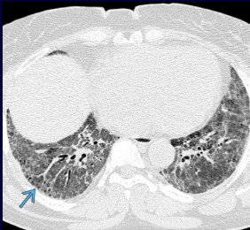
Sjogren Syndrome
bronchiectasis - all the small holes in the periphery of the lung
lower lung predominant
bronchiolar inflammation
increased parenchymal lines
pulmonary fibrosis (10-15% of cases)

sjogren syndrome - lymphocytic interstitial pneumonitis
reticulo-nodular pattern, especially lower lobes
pulmonary cyst (arrow pointing at in later aspect of lower lobe)

right = normal
sjogren’s syndrome - big inflamed parotid glands
AIDS classification
infects CD4+ T-cell lymphocytes
AIDS defined as ≤200 CD4 cells ***

cerebral atrophy - AIDS
Most common post-mortem finding in patients who die of AIDS
Poor correlation between severity of imaging abnormality and degree of cognitive impairment
Widened sulci and cisterns (CSF spaces)
Enlarged ventricles
Mass lesions - Mass effect present
Toxoplasmosis
Primary CNS Lymphoma
Cryptococcal disease
Other infections including Herpes simplex/zoster, M. tuberculosis, M. avium intracellulare, T. pallidum, Fungi (Aspergillosis, Candidiasis, Nocardiosis, Coccidioidomycosis), Amebiasis
Infarction
Mass lesions - mass effect absent
PML (progressive multifocal leukoencephalopathy)
HIV encephalitis
CMV encephalitis
Cerebral Atrophy
toxoplasmosis
Most common opportunistic CNS (central nervous system) infection in AIDS
Caused by Toxoplasma gondii (reservoir: infected cats)
Radiographic features:
Solitary or multiple ring-enhancing lesions on post-contrast imaging (MRI and CT)
Target appearance
Marked surrounding edema

toxoplasmosis - most common opportunistic CNS infection in AIDS
target appearance

primary CNS lymphoma - AIDS
2nd most common focal brain lesion in adults with AIDS
typically low CD4 counts <50
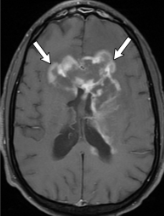
primary CNS lymphoma - AIDS
toxoplasmosis vs CNS lymphoma ***
Toxoplasmosis
More likely to present with multiple ring-enhancing intracranial masses
Random distribution
Empiric treatment with anti-protozoan drugs and reassessment of lesions on follow up imaging is often used to distinguish between the two
Nuclear medicine thallium scan appears as a cold lesion
CNS lymphoma
Lymphoma is more likely to present with a single intracranial mass than toxoplasmosis
Though lymphoma can present as multiple ring enhancing lesions in immunocompromised PTs
Periventricular location and subependymal spread
Nuclear medicine thallium scan appears as a hot lesion

progressive multifocal leukoencephalopathy
bilateral and asymmetrical

HIV encephalopathy
bilateral and symmetric
most common pulmonary infection
streptococcal pneumonia ***
infectious pulmonary disease in HIV/AIDS
pyogenic bacterial pneumonia (strep)
pneumocystis pneumonia (PCP)
myobacterium tuberculosis
non-infectious pulmonary diseases in HIV/AIDS
asthma
emphysema
kaposi sarcoma
pneumocystic jirovecii pneumonia (PCP)
upper lobe air-filled cysts or pneumatoceles —> cause pneumothorax ***
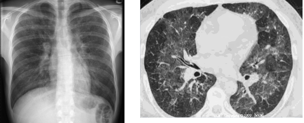
pneumocystis jirovecii pneumonia ***
fine reticular markings on the periphery of the lungs

Mycobacterial infection - post primary TB
upper lung zone consolidations with cavitation
CD4 <350
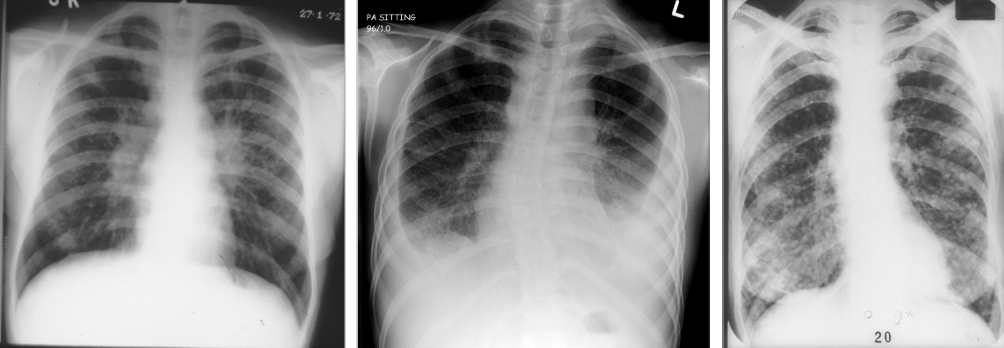
Mycobacterial infection - primary TB
patchy areas of consolidation in any lung zone, effusions
CD4 <200
hilar and/mediastinal adenopathy

mycobacterial infection - miliary TB
hematogenous spread of diffuse tiny nodules, all lung zones

fungal pneumonia
dysphagia
candidiasis, viral esophagitis (herpes, CMV, HIV), KS
GI manifestations - diarrhea
cryptosporidiosis, giardia, mycobacteria
GI manifestations - colitis
CMV, mycobacteria, C. diff
GI manifestations - cholangitis
CMV and/or cryptosporidiosis
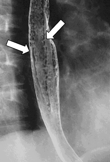
esophagitis - candidiasis
“shaggy” mucosa from plaques

esophagitis - HSV ulcers
“target” shaped small, superficial ulcers

HIV ulcers
larger and deeper ulcers compared to Herpes

acalculus cholecystitis (acute inflammation of the gallbladder that occurs without the presence of gallstones)

biliary disease

biliary disease

biliary disease
focal stenosis

pancreatic disease
HIV & AIDS - The Renal System
may be an early, unsuspected manifestation of HIV infection
most patients are asymptomatic
sonographic findings include enlarge echogenic kidneys with poor corticomedullary differentiation
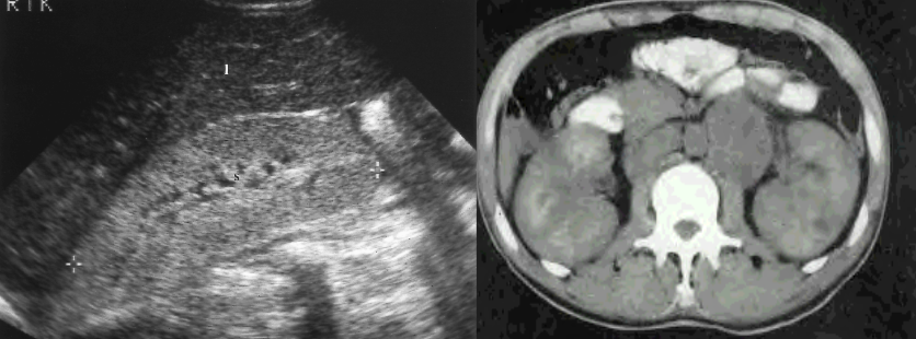
HIV nephropathy
study of choice to identify renal stones ***
non-contrast CT scan
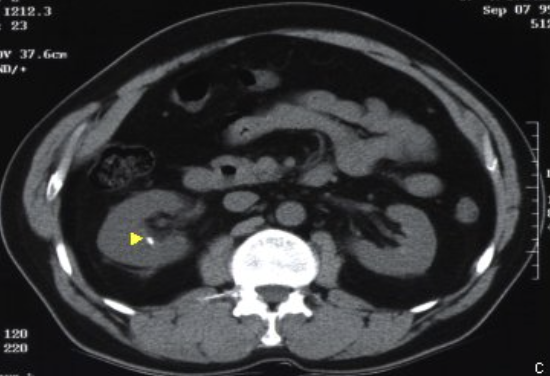
***
renal colic ***

cellulitis
Swelling, calor, pain
Staph and strep most common
Subcutaneous fat stranding

necrotizing fasciitis

soft tissue abscess

pyomyositis
Staph most common
Infection of the muscle
Can lead to abscess formation and myonecrosis

osteomyelitis
Hematogenous spread, direct extension, direct inoculation
Staph most common
Tibia, wrist, femoral heads, ribs, spine TL junction
X-ray changes not evident for 10-14 days***
Nuclear medicine (MDP, Ga-67, In-111)
MRI with contrast (decreased T1 signal)
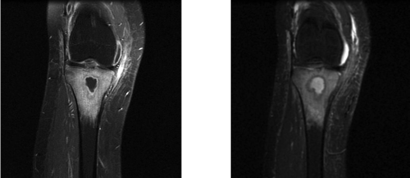
osteomyelitis with brodie abscess
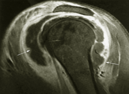
septic arthritis
Important to keep in the differential diagnosis with a mono-articular process
Early diagnosis with aspiration and culture is key to correct diagnosis and determining bug sensitivity
Can be aggressive and lead to early destruction of cartilage and bone
kaposi sarcoma
AIDS defining malignancy
Associated with human herpesvirus 8 (HHV-8)
Low-grade vascular tumor that involves the lymphovascular system

kaposi sarcoma - skin manifestation
cutaneous and subcutaneous nodules
kaposi sarcoma - GI manifestations
involves the liver, spleen, and bowel
liver and spleen - multiple nodules
bowel - early disease consists of flat lesions that are not demonstrated on barium studies
Lesions coalesce, enlarge and become more nodular
Extension into the muscular wall🡪 large bulky polypoid masses and irregular fold thickening
higher risk of developing malignancies

kaposi sarcoma
left = small bowel wall thickening
right = large bowel involvement

kaposi sarcoma - GI manifestation
lobulated mass involving the rectosigmoid

kaposi sarcoma - pulmonary manifestation
nodules, linear opacities, 40% have pleural effusion, adenopathy
present with cough, fever, shortness of breath, hemoptysis and chest pain
occurs in 45% kaposi sarcoma cases