unit 3 sensory system
 Call Kai
 Learn
 Practice Test
 Spaced Repetition
 Match
 Flashcards
 Knowt Play
1/72

Earn XP
Description and Tags
biol 2404 acc
73 Terms
This is the conscious interpretation of sensory information.
sensory perception
These are sensory structures that respond to specific stimuli from the external or internal environment.
sensory receptors
Usually, stimulus energy is converted into ___. These trigger __ which send information to the brain for interpretation.
receptor potentials; action potentials
Hair cells ___ when the hairs bend towards the kinocilium and they ___ when the hairs (stereocilia) bend away from the kinocilium.
depolarize; hyperpolarize
Which of these receptors is not matched with its proper function?
gustatory receptors; detect odorant molecules
These sensory structures are located only in the head.
special senses
All of these are sensory receptors for general senses except
olfactory receptors
These receptors respond to mechanical forces. They are used to detect touch, stretch, and position.
mechanoreceptors
These receptors respond to excess stimuli that might be harmful. They are used to detect pain.
nociceptors
These receptors respond to changes in temperature. They are used to detect hot and cold temperatures.
thermoreceptors
These receptors respond to photons of light. They are used to detect vision.
photoreceptors
These receptors respond to chemicals dissolved in solution. They are used to detect smell and taste.
chemoreceptors
Nerves formed by the axons of olfactory receptor cells pass through openings in the cribriform plate of the ethmoid bone. They synapse with cells in the olfactory __.
bulbs
Axons of the olfactory receptor cells form this cranial nerve.
CN I, Olfactory nerve
Olfactory messages are sent to the thalamus and primary olfactory cortex by way of the olfactory __.
tracts
The paired olfactory receptors are found in the __ region of the nasal cavity.
superior
Olfactory receptors detect odorant molecules dissolved in water (mucus).
True
Humans can detect all these distinct taste sensations except
spicy
Gustatory receptors are found in __ located on the tongue, pharynx, and larynx.
taste buds
This cranial nerve transmits gustatory information from the anterior two-thirds of the tongue.
CN VII, Facial nerve
This cranial nerve transmits gustatory information from the pharynx and larynx.
CN X, Vagus nerve
This cranial nerve transmits gustatory information from the posterior one-third of the tongue.
CN IX, Glossopharyngeal nerve
This is a set of six muscles that attach to the outer eyeball and move the eyeball.
extrinsic eye muscles
These glands produce tears.
lacrimal glands
This transparent mucous membrane produces lubricating mucus. It covers the outer surface of the eye (except the cornea) and inner eyelid surfaces.
conjunctiva
This is the gelatinous fluid that is found in the posterior cavity.
vitreous humor
This is the fluid-filled space in front of the lens.
anterior cavity
This is the watery fluid that is found in the anterior cavity.
aqueous humor
This is the fluid-filled space behind the lens.
posterior cavity
The sclera and cornea make up this outer layer of the eye.
fibrous tunic
The iris, ciliary body, and choroid make up this middle layer of the eye.
vascular tunic
This is the neural tunic with pigmented cells, rods, cones, bipolar cells, and ganglion cells.
retina
This is the blind spot where the optic nerve exits the eye. There are no photoreceptors here.
optic disc
This is the center of focus in the macula lutea of the retina. It contains large numbers of cones.
fovea centralis
This structure changes shape to allow precise focusing of light on the retina.
lens
This is the correct pathway of light waves through the eye.
cornea, anterior cavity, pupil, lens, vitreous humor, retina

Identify the structure labeled “B” on the drawing of the retinal layers below.
bipolar cell

Identify the structure labeled “C” on the drawing of the retinal layers below.
rod

Identify the structure labeled “A” on the drawing of the retinal layers below.
ganglion cell

Identify the structure labeled “D” on the drawing of the retinal layers below.
cone
These are the photoreceptors involved in black-and-white vision
rods
These photoreceptors have little convergence in the retinal pathway.
cones
These are the photoreceptors responsible for vision in dim light.
rods
These photoreceptors have high convergence in the retinal pathway.
rods
These are the photoreceptors responsible for vision in bright light.
cones
These are the photoreceptors involved in color vision.
cones

Identify the cranial nerve labeled (4) in the visual pathway flow chart below.
CN II, Optic nerve

Identify the structure labeled (5) in the visual pathway flow chart below.
optic chiasma

Identify the structure labeled (2) in the visual pathway flow chart below.
bipolar cells

Identify the structure labeled (3) in the visual pathway flow chart below.
ganglion cells

Identify the structure labeled (6) in the visual pathway flow chart below.
optic tract
When the eye focuses on closer objects, the lens becomes ____.
round
When the eye focuses on distant objects, the lens becomes ____.
flat
These ligaments from the ciliary muscles attach to the lens, allowing it to change shape and focus light on the fovea centralis.
suspensory ligaments
What is the correct order of auditory ossicles, starting with the tympanic membrane?
malleus, incus, stapes
The auditory tube connects the
middle ear and nasopharynx
Which structures are found in the outer ear?
pinna, external auditory canal, tympanic membrane
Which structures are found in the middle ear?
malleus, incus, stapes
The oval window and round window separate the
middle ear and inner ear
The tympanic membrane separates the
outer ear and middle ear
Which structures are found in the inner ear?
membranous and bony labyrinths
Perilymph is the fluid that fills up the space
within the membranous labyrinth
Which structures make up the bony labyrinth?
cochlea, semicircular canals, vestibule
Endolymph is the fluid that fills up the space
within the membranous labyrinth
Which structures make up the membranous labyrinth?
cochlear duct, semicircular ducts, saccule, utricle
Which fluid touches the hair cell receptors?
endolymph
The hair cells in these receptor organs are responsible for hearing.
organ of Corti in cochlear duct
The hair cells in these receptor organs are responsible for static equilibrium.
maculae in saccule and utricle
The hair cells in these receptor organs are responsible for dynamic equilibrium.
ampulla in semicircular ducts

Identify the cranial nerve labeled (3) on the auditory pathway flow chart below.
CN VIII, Vestibulocochlear nerve

Identify the structure labeled (2) on the auditory pathway flow chart below.
sensory neurons
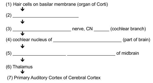
Identify the structure of the midbrain labeled (5) on the auditory pathway flow chart below.
inferior colliculi

Identify the part of the brain labeled (4) on the auditory pathway flow chart below.
medulla oblongata