Unit 3 Ovarian Pathology Study Flashcards
 Call Kai
 Learn
Learn Practice Test
 Spaced Repetition
 Match
Match Flashcards
 Knowt Play
1/141

There's no tags or description
Looks like no tags are added yet.
Name | Mastery | Learn | Test | Matching | Spaced | Call with Kai | Chat |
|---|
No analytics yet
Send a link to your students to track their progress
142 Terms
What are the sonographic criteria for a simple cyst?
Anechoic, thin-walled, and PAE
True or False. All women continue to have normal-sized ovaries after menopause.
False
How would you expect ovaries to appear in a postmenopausal patient?
Atrophic w/o follicles
Abnormal ovaries that are suggestive of malignancy are defined as appearing __________ and ___________.
Enlarged and echogenic
When an ovarian cyst measures ______, it is recommended having it removed surgically.
>7cm
Are Corpus Luteum cysts unilateral or bilateral? Prone to what? Size? May accompany?
Unilateral, Prone to hemorrhage, and may grow 1 to 10cm in size, may accompany intrauterine pregnancy
What are the clinical symptoms of corpus luteum cysts?
Pain (major symptom), Irregular menstrual cycle, Mimic ectopic pregnancy, and prone to rupture
What is the “ring of fire.”
Corpus luteum has peripheral rim of color around wall on color Doppler
Sonographically, what does corpus luteum cyst look like?
“Cystic” type of lesion, thick walled, may have internal echoes secondary to hemorrhage, ring of fire

What is this an image of?
Corpus Luteum Cyst

What is this an image of?
Corpus Luteum Cyst / Hemorrhagic Cyst
What sign/symptoms does patient usually present with when a cyst hemorrhages?
Acute onset pelvic pain
What is the sonographic appearance of acute hemorrhagic cyst?
Usually hyperechoic, may mimic solid mass
Usually has smooth posterior wall and shows PAE indicating its cystic component
Internal pattern becomes more complex as undergoes hemolysis
How does internal pattern change as clot undergoes hemolysis?
Internal pattern becomes more complex with reticular type pattern. Interconnecting lines believed to be result of fibrin strands, concave outer margin, no color doppler
Which type of cyst is associated with high level of human chorionic gonadotropin?
Theca-Lutein Cysts
Theca-Lutein cysts are usually ______, _________, and _______________.
Large, bilateral, and miltiloculated
Patient with theca lutein cyst may present with clinical symptoms of what?
Nausea and vomiting
What are the sonographic findings associated with theca lutein cysts? What may occur with these cysts?
Multilocular cysts in both ovaries, may undergo hemorrhage, rupture, and torsion
Theca lutein cyst are typically seen with what type of patients?
Patients with GTD

What is this an image of?
Theca Lutein Cyst
Ovarian hyper stimulation syndrome frequent complication of what?
Ovulation induction (fertility treatment)
What are the signs/symptoms of mild form of OHS?
Pelvic discomfort, but no significant weight gain
Sonographically, ovaries enlarge, but measure less than 5cm in diameter
What are the signs/symptoms of moderate form OHS?
Nausea/vomiting, patient gains weight 5-10lbs
Sonographically, ovarian enlargement between 5-12cm
What are the signs/symptoms of severe form OHS?
Severe pelvic pain
Abdominal distention
Weight gain more than 10 lbs
What is the sonographic appearance of severe OHS?
Notably enlarged ovaries greater than 12cm in diameter, made up of numerous large, thin-walled cysts which may replace ovarian tissue, ascites and pleural effusion

What are these images of?
Ovarian Hyper Stimulation
What percentage of adnexal masses are paraovarian cysts?
10-20%
Where are paraovarian cysts located?
In broad ligament, superior to uterine fundus
Paraovarian cysts are most common in what decades of life?
3rd and 4th decades
What are clinical symptoms for paraovarian cysts?
Asymptomatic, pain with large cysts
What is the sonographic appearance of paraovarian cysts?
Simple cyst adjacent to ovary and may have nodular areas and occasional septations
How is paraovarian cyst diagnosed?
Only possible by demonstrating normal ipsilateral ovary close to but separate from the cyst
Patients with history of ________________, ________, _____, and ___________________ tend to have peritoneal inclusion cysts.
Abdominal surgery, trauma, PID, and endometriosis
How are peritoneal inclusion cysts formed?
Adhesions trap peritoneal fluid around ovaries, resulting in large adnexal mass
What is the clinical presentation of peritoneal inclusion cysts?
Pelvic pain and/or pelvic mass
What is sonographic appearance of peritoneal inclusion cysts?
Multi-loculated cystic adnexal masses
The presence of an intact ovary amid septations and fluid
What is this an image of? Figure 29.27
What is endometriosis?
Common condition in which functioning endometrial tissue is present outside the uterus
Know three locations in pelvis where ectopic tissue may be found.
Anywhere in pelvis;
Ovary, fallopian tube, broad ligament, external surface of uterus, scattered over peritoneum, cul-de-sac, bowel, and bladder
What are the 2 forms of endometriosis?
Diffuse: consists of endometrial plantings within peritoneum, leads to disorganization of pelvic anatomy with appearance similar to PID or chronic ectopic pregnancy
Localized: consists of discrete mass called endometrioma, (chocolate cyst) and frequently found in multiple sites
Which is the more common form of endometriosis?
Localized form
True or False. Diffuse endometriosis is commonly diagnosed with sonography.
False, rarely diagnosed by sonography
The localized form of endometriosis is known as ______________ or ________________.
Endometrioma or chocolate cyst
What clinical symptoms would a patient with endometriosis possibly present with?
Pelvic pain, dysmenorrhea, dyspareunia, dyschezia, urinary symptoms and infertility
Describe the appearance of an endometrioma.
Well-defined unilocular or multilocular predominantly cystic mass, containing diffuse homogeneous, low-level internal echoes, small, linear, hyperechoic foci may be present in wall of cyst. Likely cholesterol deposits, patterns can range from anechoic to solid, depends on amount of blood and its organization
Is endometrioma usually symptomatic or asymptomatic?
Symptomatic, chronic discomfort with menses
What type of disorder is polycystic ovarian syndrome? What does it result in? Common cause of what 2 things?
Endocrine disorder
An elevated level of male hormones (androgens) and infrequent or absent ovulation (anovulation)
Infertility and higher-than-usual rate of early pregnancy loss
What is the classical Stein-Leventhal syndrome?
Oligomenorrhea or amenorrhea, hirsutism, obesity
What age group does polycystic ovarian syndrome affect?
Late teens and early 20’s
What is the sonographic appearance of polycystic ovarian syndrome?
Bilaterally enlarged polycystic ovaries with increased stromal echogenicity and multiple tiny cysts around periphery of ovary
Ovary is usually round instead of oval
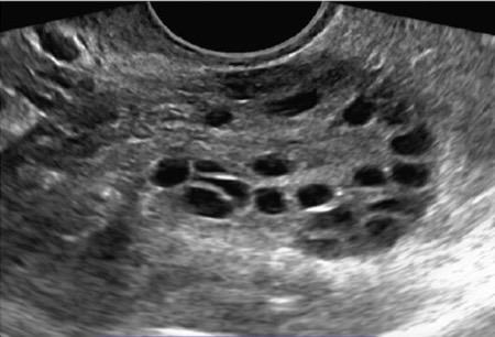
What are these images of?
Polycystic ovary
What are diagnostic criteria for polycystic ovarian syndrome?
12 or more follicles measure 2 to 9mm or ovarian volume over 10mL and blood work
What is an acute abdominal condition requiring prompt diagnosis and surgical intervention?
Ovarian torsion
Which ovary is more likely to torso?
Right ovary
Ovarian torsion causes _______ and __________________ with subsequent ___________.
Edema, loss of arterial flow, and infarction
What other structure can typically be affected by ovarian torsion?
Fallopian tube
A tossed ovary is usually _____ in diameter.
>4cm
Torsion usually occurs in who?
Children and younger females with mobile adnexa, preexisting ovarian cyst or mass, or pregnenacy
What are clinical symptoms of ovarian torsion?
Acute severe unilateral pain, intermittent pain may precede acute pain by weeks, pain may mimic appendicitis if on right side, pt may have fever, nausea, and vomiting, and palpable mass
Is the chance of getting ovarian torsion in the opposite ovary increased or decreased?
Increased
True to False. An ovarian torsion and a mass in a postmenopausal patient is more likely to be malignant.
True
What are the sonographic findings associated with ovarian torsion?
Hypoechoic enlarged ovary, absent blood flow on Doppler examination, and free fluid in cul-de-sac

What is this an image of?
Ovarian torsion
Why does ovarian cancer have the highest mortality rate of all gynecological cancers?
Late diagnosis due to few clinical symptoms
Which risk factor for ovarian cancer is most important?
Family history
True or False. Serum CA 125 is helpful in diagnosing all types and stages of ovarian cancer.
False
What percentage of ovarian neoplasms do epithelial neoplasms make up (benign or malignant)
65-75% of all ovarian tumors
90% of ovarian malignant tumors
Know how to appearance of benign lesions differs from a malignant lesion.
Benign-well defined anechoic lesions
Malignant-lesions with irregular walls, thick irregular septations, mural nodules, and solid vascular internal elements
Be able to list the 5 categories of epithelial-stromal cell tumors.
Serous, Mucinous, Endometrioid, Clear cell, and Transitional cell
Which epithelial-stromal tumor is the most common?
Serous tumors
Which epithelial-stromal tumor is the 2nd most common?
Mucinous tumors
What is the sonographic appearance of serous cystadenoma?
Large, thin-walled, unilocular cystic masses, may contain thin septations
What is the sonographic appearance of serous cystadenocarcinoma?
Large, multilocular cystic masses with papillary projections and septations, walls and septations thickened, echogenic solid material may be visualized in cyst, and ascites often present
What is the sonographic appearance of mucinous cystadenoma?
Huge cystic masses, measuring up to 30cm filing the entire pelvis and abdomen, multiple thin septa with low-level echoes caused by mucoid material, and infrequently, papillary projections may be present
What is the sonographic appearance of mucinous cystadenocarcinoma?
Large, multiloculated cystic masses with papillary projections and echogenic material, can have appearance similar to serous cystadenocarcinoma
What is pseudomyxoma peritonei? What is the sonographic appearance?
When a mucinous tumor or mucocele ruptures, it can spread to the peritoneum; similar to ascites or may have multiple septations in the fluid, filling the pelvis and abdomen. Low level echoes may be visualized
Pseudomyxoma peritonei can occur with which tumors?
Mucinous cystadenoma and cystadenocarcinoma
Which epithelial tumor has a better prognosis than other epithelial malignancies and why?
Endometroid tumor, diagnosis at an earlier stage
What is the sonographic appearance of Endometrioid tumors?
Typically cystic mass with papillary projections, some are predominantly solid mass that may have areas of hemorrhage or necrosis
Clear cell tumors are considered to be of _______________________ origin.
Endometroid carcinoma
True or False. Clear cell tumors are almost always malignant.
True
What is another name for transitional cell tumor?
Brenner tumor
Transitional cell tumors are…
Uncommon and almost always benign, usually asymptomatic, mostly discovered incidentally by sonography or surgery
What is the sonographic appearance of transitional cell tumors? What are they composed of?
Hypoechoic solid mass, outer wall may have calcification, cystic component is uncommon, if present, usually a coexistent cystadenoma and composed of dense fibrous tissue
True to False. A cystic component is common with Brenner tumor?
False
List the malignant epithelial tumors.
Serous cystadenocarcinoma, mucinous cystadenocarcinoma, Endometroid, and clear cell
List the benign epithelial tumors.
Serous cystadenoma, mucinous cystadenoma, and transitional cell tumor

What is this an image of?
Serous Cystadenocarcinoma

What are these an image of?
Mucinous Cystadenocarcinoma

What is this an image of?
Endometroid Tumor
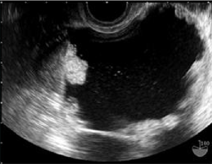
What is this an image of?
Clear Cell Tumor

What is this an image of?
Serous Cystadenoma

What is this image of?
Mucinous Cystadenoma

What is this an image of?
Transitional Cell Tumor / Brenner Tumor
Serous cystadenomas make up what % of all benign ovarian neoplasms?
25%
Serous cystadenocarcinomas make up what % of all malignant ovarian neoplasms?
50%
What decades of life are mucinous cystadenomas most common?
3rd to 6th decades of age
What decades of life are micinous cystadenocarcinomas most common?
4th to 7th decades of age
What decades of life are Endometroid tumors most common?
5th and 6th decades of age
What decades of life are clear cell tumors most common?
5th to 7th decades of life