Equine MSK 1 - Lameness and Imaging
 Call Kai
 Learn
Learn Practice Test
 Spaced Repetition
 Match
Match Flashcards
 Knowt Play
1/311

There's no tags or description
Looks like no tags are added yet.
Name | Mastery | Learn | Test | Matching | Spaced | Call with Kai | Chat |
|---|
No analytics yet
Send a link to your students to track their progress
312 Terms
Equine Lameness Examination, Diagnostic Approach and Imaging
- Be able to recognize Equine lameness (i.e. pick the lame leg/s)
- Know the AAEP Lameness grading system and its application
- Understand, describe and perform a comprehensive lameness examination including taking relevant history, distant and close inspections, musculoskeletal palpation.
- Understand, describe and perform apropriate manipulative tests (e.g. hoof testers, percussion, limb flexion tests etc)
- Understand, describe and execute commonly performed local anaesthesia techniques (i.e. nerve and joint blocks)
- Understand, describe and execute commonly performed imaging modalities and techniques used in equine lameness diagnosis
- Describe, execute and interpret commonly performed radiographic and ultrasonographic images in particular
- Apply pre-requisite knowledge of musculoskeletal anatomy and function to equine lameness and construct a comprehensive differential diagnosis list for lameness originating from specific anatomic regions
Lame forelimb is identified by?
Head lift when weight bearing on lame limb
Lame hindlimb is identified by?
Hip hike/greater range of motion on lame limb
Also
- sometimes,
butt of tail lifts up on lame limb
- hinf limb on lunge; inside leg should normally be brought in under body but if not, and instead tow stabbing and shortened crainal phase of stride, indicates hind limb lameness
- Owner may describe it as horse not wanting to stop or perform its transitions as it normally would

Summary of Grades of Lameness (AAEP)
Grade 0 - Not detectably lame
Grade 1 - Inconsistently lame at the trot on the circle
Grade 2 - Inconsistent on straight line at the trot but consistently lame on the circle
Grade 3 - Consistently lame on straight line at the trot
Grade 4 - Lame at walk
Grade 5 - Non-weight bearing lame
Steps to investigating lameness
History - use, recent weather events, age
Examine from a distance
- run eyes over systemically
Examine gait in hand/lunge, stand horse square
- conformation/angular limb deformities
- hoof assymetry: smaller is more likely to be lame
- pelvic symmetry
- obvious swellings
Palpation of joints, tendons and ligaments
- presence vs absence of digital pulse
- heat, cold
- pain response
- oedema / effusion of joint, tendon, digital sheath, bursa
Hoof testers and percussion
Flexion tests
Nerve and joint blocks

Spavin flexion test exacerbates pain in which joints?
hock, stifle, hip joint
Wind galls/wind puffs are?
digital tendon sheath inflammation and subsequent joint effusion above the fetlock joint.

When lunging a horse, the lameness is more apparent on?
the inside leg
Where to palpate digital pulse?
Palpate the neurovascular bundle at the palmar/plantar abaxial borders of the proximal sesamoid bones. Above, below, medial or lateral. all work

Which structures are you feeling when you feel
- Hard swellings
- Firm swellings
- Fluid filled swellings
- bone (osteophytes)
- soft tissue
- effusions, blood, oedema, pus
how to differentiate fluid swellings as oedema?
how to differentiate fluid swellings as cellulitis ?
Oedema - in subcut tissue, and pitting
Cellulitis - similar to oedema, but associated with inflammation: heat, pain, potentially broken skin and exudative
what is crepitus ?
bubbly, creaky, crunchy, grinding, clicking

What is a symptom of subcut emphysema (air in subcut space)?
crepitus - bubbling crackle sound when palpated.

how to systemicatically use hoof testers
Heel - quatre - toe - toe- quatre - across heel - frog:heel - frog:heel
Assess for flinch/pain response and repeat;. Compare to other hoof.
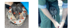
Purpose of flexion tests
Increasing intra-articular and subchondral bone pressure to exacerbate a clinical or subclinical lameness
what is static vs dynamic flexion test?
Static flexion: flexing without trotting off
Dynamic flexion test - Flex and hold for 60 secs and trot off
Name the flexion tests done on the forelimb vs hindlimb
Forelimb
-Distal limb flexion: coffin, pastern, fetlock
- Carpal
- Shoulder, elbow
Hindlimb
- Distal limb flexion: coffin, pastern, fetlock
- Spavin - hock, stifle, hip
*more in prac, MSAT guarentee*
For perineural nerve blocks, list:
- agent
- volume
- needle size
- aseptic?
Prilocaine/mepivicaine
Volume:
- 1.5 ml/injection for most
- 2-3ml/ injection for low 4 and low 6
Needle: 25g, 5/8 inch (orange)
Aseptic? no - just alcohol wipes
For intra-articular nerve blocks, list:
- agent
- volume
- needle size
- aseptic?
Prilocaine/mepivicaine?
Volume depends on joint , typically 5mL
Needle: 19-21g, 1.5 inch (green)
Aseptic? yes

List 3 perineural vs 3 joint blocks for the forelimb
Perineural
- Palmar digital nerve block PDNB (distal limb)
- Abaxial sesamoid nerve block ABSNB (distal limb)
- Low 4 point nerve block (fetlock)
Intra-articular joint blocks of
-coffin (DIP),
- fetlock
- carpus
4 types of hindlimb nerve blocks
Perineural
- Palmar digital nerve block PDNB (distal limb)
- Abaxial sesamoid nerve block ABSNB (distal limb)
- Low 6 point nerve block (fetlock)
Intra-articular joint blocks of
-coffin (DIP),
- fetlock
- tarsus
How does the approach for ABSNB differ in forelimb to hindlimb?
1) medial nerves are injected from the contralateral side of the horse
2) may have to block off additional dorsal metatarsal nerves as well

What are the 4 nerve/joing block options for the distal limb
Palmar digital nerve block (PDNB)
- common FL, rare HL
Abaxial Sesamoid nerve block (ABSNB)
- common FL, rare HL
Distal interphalangeal (DIP) joint block
Navicular Bursa block (Nbursa)
- rare
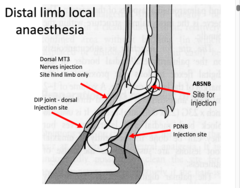
PDNB blocks?
Blocks:
- DIP Joint
- Navicular apparatus
- P3
- Heel/sole
- Toe/sole eventually
- Skin
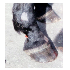
ABSNB blocks?
Blocks:
- Mid pastern (P1) and distal (entire foot) including skin sensation
- DIP joint too but not commonly used for this
- NOTE In the hindlimb, dorsal metatarsal nerves, which do not get blocked by this injection, may extend along dorsal P1, meaning dorsal P1 is not blocked out

DIP blocks?
DIP joint + sole + Nbone
*Similar to PDNB but no loss of skin sensation*

List 3 LA for fetlock region
Intra-articular fetlock joint block
- Good for IA structures
Low 4 point block
- forelimb
Low 6 point block
- hind limb
Low 4 and low 6 blocks?
Fetlock joint, sesamoids, suspensory ligament branches and below

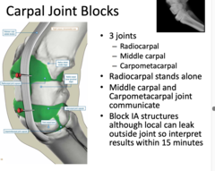
Carpal joint blocks which joints of the carpus
Radiocarpal joint and Middle carpal joint
(intercarpal joint communicates with middle carpal)
What are the 4 Standard Equine Radiographic views
DP (dorsopalmar/plantar) or CD (craniocaudal)
LM (lateral-medial)
DLPMO (dorsolateral to palmaromedial oblique)
DMPLO (dorsomedial to palmarolateral oblique)
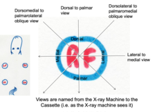
above vs below radiocarpal joint
craniocaudal, dorsopalmar
reference for radiographic terminology
• OA - osteoarthritis (same as degenerative joint disease)
• OC fragment / F# - osteochondral fragment ('bone chip') - composed of bone and cartilage
• Periarticular osteophyte - bone spur in or around joint margins
• Enthesiophyte - new bone formation within tendon, ligament or joint capsule insertions
• Periosteal new bone - new bone production due to periosteal lifting or trauma
• Opacity/Radiopacity - the degree of 'whiteness' of the object being radiographed -> sclerosis
• Lucency/Radiolucency - degree of 'blackness' of object being radiographed -> lysis / malacia
• Osteomyelitis - septic inflammation of bone marrow
• Sequestrum - sclerotic, necrotic fragment of bone surrounded by a radiolucent zone (the involucrum)
• Boney modelling - any macriscopic change in bone density

term for increased blackness vs increased whiteness
blackness = radiolucency = lysis/malacia of bone
whiteness = increased opacity = sclerosis

sclerotic, necrotic bone fragment surrouneded by radiolucent zone
sequestrum (is surrounded by the involucrum)

Name of inflammation of bone vs bone marrow
osteitis
osteomyelitits
Define enthesiophyte
new bone formation within tendon, ligament or joint capsule insertions
boney modelling vs remodelling
modelling - any macroscopic change to bone - more macroscopic - not coupled - ie endochondral ossification
remodelling - consistent process of resorption and formation of bone - more microstructures
4 descriptive terminologies for ultrasound
isoechoic - uniform, normal image
hypoechoic - less echogenicity than expected (fluid)
anechoic - complete abscence (black = fluid, gas)
hyperechoic - increased echogenicity (white, sclerosis)

low vs high frequency ultrasound probe re resolution and depth penetration
low frequency probes have deeper penetration of tissue but poorer resolution
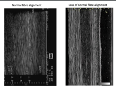
fibre alignment
Prognostic indicator
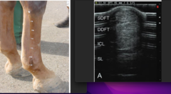
What tendons will you see on ultrasound image of metatarsal/canon bone region
SDFT
DDFT
ICL = inferior check ligament (proximal only)
SL = suspensory ligament
Linear and curved array transducers
linear for limbs for parallel fibres, curved for chest/abdomen
what is a stand off probe?
rubber covering that helps to see more superficial structures
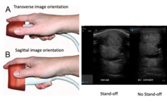
how to view fibre alignment of tendon?
rotate probe 90°, from transverse to sagittal image
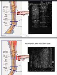
How to read severity of tendon/ligament lesions
• Tendon and lesion crosssectional area
• Lesion severity / type
• Lesion location & length
• Fibre alignment / pattern
• Echogenicity
*MORE ON THIS LATER QLETS*
CSA reference balue of SDFT, DDFT anf SL
SFT

How to grade lesions by ultrasound;
Assess the lesion, not the entire tendon!! Within the lesion, assess echogenicity
• Type 0
- Isoechoic - normal tendon / no lesion
• Type 1 - Slightly hypoechoic but mostly echoic
• Type 2 - 50% echoic & 50% anechoic
• Type 3 - Mostly Anechoic
• Type 4 - Completely Anechoic (core lesion)

photo of type 4 lesion
completely anechoic
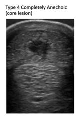
How to grade fibre alignment scores?
Score 0 - ≥ 75% parallel fibres within target image/path
• Score 1 - 50-75% parallel fibres within target image/path •
Score 2 - 25-50% parallel fibres within target image/path •
Score 3 - 25% parallel fibres within target image/path
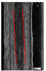
Most useful ultrasound scores?
CSA is best indicator of severity of lesion, whilst FAS is best indicator for prognosis.
*the higher the scores, the more sever the injury*
If conventional imaging (Radiograph, ultrasound) is unable to yield results, what other imaging modalities do you use?
scintigraphy, MRI, CT
- all are more sensitive to bone and soft tissue injury but awkward to run and expensive
- MRI and CT only from hoof to carpus/tarsus
Quality of shoeing/trim
- Size, fit of shoe
- Nail placement - parallel to ground surface; last nail not beyond widest point of hoof
- Adequate width of shoe at quarters to allow for heel expansion
4 hallmark signs of lameness originating at the HOOF
1. Increased or asymmetric digital pulses
2. Increased heat in hoof capsule
3. Pain on palpation of coronet
4. Pain on hoof testers (SF pain) or hoof percussion (deeper pain- navicular structures or P3)
Distal limb sites for palpation
Hoof capsule for asymmetric heat
• Palpate Digital pulses
• Coronet - pain, swelling, discharge
• Collateral cartilages - Pain, calcification
• Palmar pastern - SDFT, DDFT, Distal Sesamoidean ligts
• Coffin/DIP joint effusion
Coffin join effusion
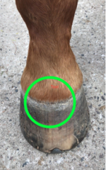
Distal limb nerve blocks in order of commonality
PDNB - first choice
ABSNB - second choice but less specific to foot
DIP joint - but requires aseptic techinique
Navicular bursa blockk - difficult

Hind limb dorsal area may still have innervation on ABSNB because?
Dorsal metatarsal nerves can sometimes extend to the coronet thus innervating that area even if ABSNB has been blocked off.

Causes of distal limb lameness
Bony
• Calcification of collateral cartilages / Side bone
• Fractures of P3 or Navicular bone
• Septic pedal osteitis
• Traumatic pedal osteitis
• Caudal heel pain/Navicular disease
• DIP/Coffin joint Osteoarthritis
• Desmitis of DIP joint collateral ligaments
• SDFT branch desmitis
• Laminitis
Soft tissue
- hoof abscess
- seedy toe/white line disease
- sole /heel bruising
- corn
- hoof wall cracks
- thrush
- canker
- keratoma
- sheared hell (lateral/medial imbalance)
- desmitis of coffin joint collateral ligaments
- tendonitis of SDFT
- penetrating/traumatic injuries
Soft tissue causes of distal limb lameness
- hoof abscess
- seedy toe/white line disease
- sole /heel bruising
- corn
- hoof wall cracks
- thrush
- canker
- keratoma
- sheared hell (lateral/medial imbalance)
- desmitis of coffin joint collateral ligaments
- tendonitis of SDFT
- penetrating/traumatic injuries
6 ddx for 4-5/5 lameness
Subsolar abscess
Penetrating sole injury
Septic synovitis
Cellulitis
Fracture
Neurogenic injury
Subsolar abscess - what is this?
An abscess penetrating deep into the sole of the hoof, underneath the hoof capsule (usually occur at junction of white line and hoof wall)
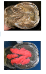
Appear as black dots. When trimmed back, exude pus.

Subsolar abscess
- 3 important hx events
- csx
*in red*
Hx important
- Recent wet weather, softens sole making it more easily penetrated
- Shod recently - nail bind (too close to white line)
- Subclinical seedy toe
Clinical signs
- Acute sudden onset severe, 4-5/5 lameness
- Hallmark distal limb signs (bounding digital pulse, alpable Heat hoof wall/coronet , +ve hoof testers)

subsolar abscess treatment
Make an adequate drainage hole
Sugar-iodine poultice hoof poultice maintained for 7-10 days or until drainage strops and lameness has improved
Antibiotcs usualy not necessary unless concurrent cellulitis.
what extra measure should you consider with subsolar abscess treatment?
tetanus vaccination
What 2 sequalae may occur seconday to unresolved hoof abscess
1) Eruption at coronet dude to inadequate drainage
2) Invasion into bone and septic pedal osteitis
Subsolar abscess that has erupted at coronet due to inadequate drainage

Septic osteitis of P3 - what are the 2 causes?
Subsolar abscess or Penetrating wounds
Septic osteitis of P3
- 4 CSx
Persistent lameness (severe )
+
discharging tract
+
Under-run / separated sole
+
Concurrent distal limb cellulitis, swelling around coronet
which nerve block is reccomended to abolish lameness in cases of septic pedal osteitis
ABSNB
How to diagnose septic osteitis?
radiographs
what does radiology of septic osteitis show?
osseous sequestrum surrounded by involucrum
- osteolysis by osteoclasts (radioluscent)
- demineralization
- irregular margins (periosteum)
- involucrum (gas opacity)

Treatment of Septic Osteitis?
Surgery and antibiotics
Post surgery - bandage/fill decifit and apply hospital plate shoe (photo) to elevate sole off ground and protect sole surface until site is cornifed

What is seedy toe/white line disease?
Separation of wall and white line resulting in foreign material impaction - usually at toe and might extend proximally over time.

What are 3 key aetiologies of seedy toe
1) underlying chronic subclinical laminitis
2) inadequate hoof care
3) wet environments
2 steps for seedy toe tx *in red*
1) Resection of diseased wall and laminae with care.
2) Heart bar or straight Bar shoe with wall clips to stabilize wall (if you dont stabalsie the wall, it may split further and cause verttical wall cracks)

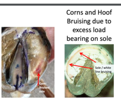
What are corns? aetiology?
Deep bruising of sole, often adjacent to bar/heel region and secondary to ill-fitting shoe or prolonged showing interval where shoe pressure on sole at heels

treatment of corns
resection of bruised tissue, soft bedding with poultice until soleis firm, then apply wide webbed seated out shoe with no sole pressure
Thin soles;
Thoroughbred horses after wet season on hard ground -> soles become so soft you can see the pedal bone

What is the recomended shoes for poor hoof conformation / generalised sole soreness/bruising and WHY *red*
Wide web deep seated out aluminium shoe
- reduce pressure on the soles by distirbuting force evenly throughout the hoof walll, over a larger area than traditional shows. Deep seated = no chance soles will touch ground
- aluminium is light weight

Vertical hoof wall cracks type 1 vs 2 definition and treatment
Type 1
- Doesnt involve coronet
- TX: resect to normal laminae, bar shoe + wall clips
Type 2
- involves coronet
- TX: complete hoof wall strip, bar shoe + wall clips
List come causes of vertical hoof wall cracks
Management - poor farriery, poor hoof care
Trauma - to coronet or laminar
Previous disease - seedy toe, hoof abscess, laminitis
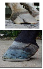
What do you call thick black purtrid discharge ?
Where mostly located?
Tx?
Thrush - in frog sulci and heel bulbs
Tx
- debride, iodine, dry environment

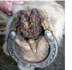
Canker; Define, Aetiology, Tx
Proliferative degeneration of frog, solar corium and digital cushion.
Suspected as sarcoid variant.
Tx - radical debridement
Hoof radiography
- preparing the hoof
Clean all dirt and debris from central/lateral sulci of frog
Clean off hoof wall
Trim small amount of hoof as needed
Remove shoes
Pack sulci with material to eliminate gas shadows
Solar surface elevated onto a block
Standard hoof views
1. Lateromedial view (LM)
2. Dorsopalmar / plantar (horizontal beam)
3. High Coronary - angle the beam 45 to 65° proximally
4. Upright pedal - angle the foot (tippy toes)
5. Special navicular view
6. 2x obliques
High coronary view
Like a DP but angle beam 45-65 degrees proximo-distally.

Upright pedal view
Tippy toes

Special navicular view / skyline view
Taken from Palmar-Dorso angled proximo-distally

High coronary view to visualise P3
vs navicular bone
1. 50° Centred at the coronet for evaluation of P3
2. 60° Centred 1-2 cm proximal to coronet for evaluation of navicular bone - collimated and grid
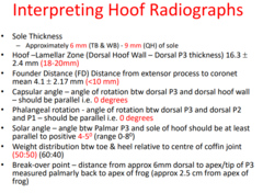
Interpreting hoof radiographs
Dorsal hoof wall thickness
Sole thickness
Weight distribution
Breakover point
P3 sole angle
Founder Distance (FD) Distance from extensor process
Capsular angle
Phalangeal rotation
Solar angle
Weight distribution btw toe & heel rel
What are 8 assessments of the distal limb on LM radiographs
1. Hoof lamellar zone
2. Sole thickness
3. Heel-toe weight bearing
4. Breakover point
5. P3:sole angle
6 Capsular rotation
7. Phalangeal rotation
8. P3 sinking
Dorsal hoof wall thickness / Hoof-lamellar zone
- what is this and what is the value range
Thickness of the dorsal hoof wall.
In a normal horse, distal and proximal measurements should be the same, and less than 20mm.

What is assymetry or >20mm hoof-lamellar zone on radiograph indicative of?
Laminitis
Sole thickness value range
6-9mm

Weight distribution values and how to measure
Find centre of coffin joint relative to P2.
Vertical line down to measure how much heel vs toe you have.
Want 60:40 or 50:50

Break over point - how to measure
Measrue 6mm dorsally from tip of P3.
From there, measure distance to thumbtack that has been inserted into apex of the frog.
Measure this distance. This distance can then be transcribed onto the sole surface by measuring from the apex of the frog dorsally - this is the pint at which breakover should occur. So trim toe back to this distance measured dorsally from apex of frog.

How to measure P3:sole angle
Measure the angle of P3 relative to sole of hoof in LM view.
Want 0 to +5 degrees (not a negative palmar angle as this impacts breakover and puts pressure on flexor tendons)

Angle rotation of P3 relative to hoof wall - capsular rotation
- should be?
Line of dorsal P3 to angle of hoof wall.
Want them to be parallel. ie angle should be 0.
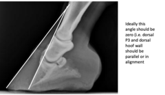
What does increased capsular location indicate?
laminitis
Angle rotation of P3 relative to P1 and P2 - phalangeal rotation
Line of dorsal P3 to angle of other pastern bones. Useful for chronic laminitis that may not be evident in capsular rotation.
Want them to be parallel. ie angle should be 0.
