Paediatrics Matrix
 Call Kai
 Learn
Learn Practice Test
 Spaced Repetition
 Match
Match Flashcards
 Knowt Play
1/154

There's no tags or description
Looks like no tags are added yet.
Name | Mastery | Learn | Test | Matching | Spaced | Call with Kai | Chat |
|---|
No analytics yet
Send a link to your students to track their progress
155 Terms
Congenital Heart Defects
Disruption of normal sequence of cardiac morphology leads to the formation of pathological connections (shunts) b/w right and left heart chambers → blood flows from high pressure to low pressure
Shunts:
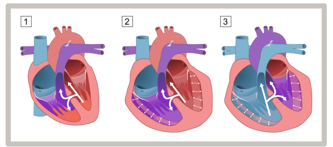
Left-to-Right Shunt: oxygenated blood from lungs shunts back into pulmonary circulation via ASD, VSD or PDA → pulmonary HTN → RV pressure overload → RH Hypertrophy/HF (but no cyanosis)
Right-to-Left Shunt: blood flows from RH to LH via shunt → deoxygenated blood enters circulation → cyanosis
Eisenmenger Syndrome: pulmonary arterial HTN
Prolonged pulmonary HTN from L-t-R shunt → constriction → permanent pulmonary vessel remodelling → irreversible pulmonary HTN
RV hypertrophy compensates pulmonay HTN → ↑ in RV pressure exceeds LV pressure → blood flow reversal → cyanosis
In pts w PDA, cyanosis is in lower extremeties
Mx: lung/heart-lung transplant
Sx: exercise intolerance (fatigue, pallor, diaphoresis, tachycardia, dyspnoea, grunt/nasal flaring), failure to thrive, recurrent bronchopulmonary infections
RHF: hepatic venous congestion (w hepatomegaly)
LHF: tachypnoea, pulmonary oedema, ↓ cardiac output (↓ BP, pallor, sweats, cool extrem., syncope)

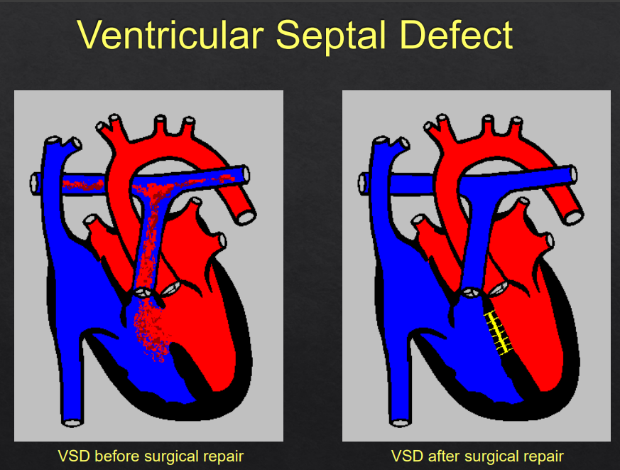
Ventricular Septal Defect Ix (R1)
Simple Acyanotic CHD, Most common CHD
Left-to-Right Shunt → RV Hypertrophy → Pulmonary HTN → ↓ Cardiac Output → LV Hypertrophy
Occurs isolated or w other defects (AVSD, ToF, TGA)
RF: Down Syndrome, Intrauterine Inf. (TORCH etc), Maternal Diabetes
Sx: Cough, noisy breathing/breathing difficulties, recurrent bronchopulmonary inf., CHF, exercise intolerance, swelling/oedema, failure to thrive/LOW, short stature, crying/irritable/unwell, dysmorphic features, newborn resp. distress
Small defects = asymp., Medium/Large defects = HF by age 2-3m
Ix:
First Line: Clinical Exam = pansystolic or harsh holosystolic murmur over L sternal border, mid-diastolic murmur; CXR = cardiomegaly, overload; ECG = LV/RV hypertrophy (↑ QRS, LAD, RAD, prolong PR, RBBB)
Gold Standard: Echocardiogram (size, location, L-t-R blood flow etc) + Doppler US (L-t-R blood flow)
Ventricular Septal Defect Mx (R1)
Mx: nutrition, diuretics, ACEi, Surgical Closure
First Line: Echocardiogram monitoring (small defects often heal spontaneously)
Long Term: Surgery (VSD closure if sx)
Indications: children <1yr w pulmonary HTN, children not improved w medical therapy
Cx: pulmonary HTN, HF, cyanotic disease (TOF, truncus arteriosus), arrhythmias, Eisenmenger syndrome, infective endocarditis, aortic regurgitation
Assoc. Conditions: pulmonary atresia, tetralogy of Fallot, Truncus Arteriosus
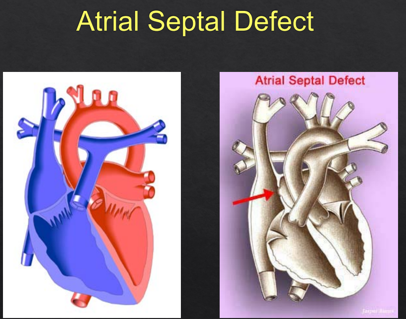
Atrial Septal Defect Ix (R1)
Simple Acyanotic CHD, Second most common CHD
ASD I (Ostium primum atrial septal defect) = 15-20%, usually accompanied by other heart defects
ASD II (Ostium secundum atrial septal defect) = 70%, usually isolated
ASD → O2 blood shunting from LA to RA → ↑ O2 sat. in RA → ↑ O2 sat. in RV/pulmonary artery
RF: F>M, chromosomal assoc. (Downs Syndrome etc), fetal alcohol syndrome, Holt-Oran (hand-heart) synd.
Sx: Exertional dyspnoea/fatigue, recurrent bronchopul. inf. supraventricular arrhythmias, Cough, noisy breathing/breathing difficulties, swelling/oedema, failure to thrive/LOW, short stature, crying/irritable/unwell, dysmorphic features, newborn resp. distress
Often minimal sx until adulthood (acyanotic), small defects = asymp.
Ix:
First Line: Clinical Exam = systolic ejection murmur, widely split fixed S2; CXR = enlarged right heart and pulmonary artery; ECG = RV hypertrophy (RAD, RBBB)
Gold Standard: Echocardiograph (size, shunt volume, global cardiac function, interatrial communication etc)
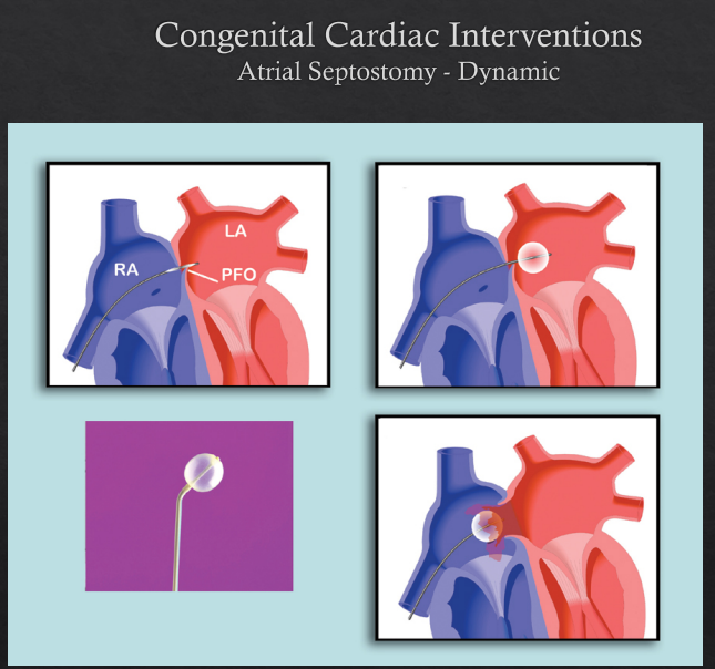
Atrial Septal Defect Mx (R1)
Mx: nutrition, diuretics, ACEi, Surgical Closure
First Line: Echocardiogram monitoring (often spontaneous closure)
Long Term: surgery (atrial septosomy, ASD closure)
Cx: HF, pulmonary HTN (chronic left-to-tight shunt and AV valve regurg.), Embolism/ischaemic stroke
Assoc. Conditions: pulmonary atresia, Total Anomalous Pulmonary Venous Return
Cardiac Failure Ix (R1)
Abnormality of cardiac structure/function leading to failure of the heart to deliver oxygen at a functional rate
Due to over-circulation or pump failure
RF: CHD/large left-to-right shunts/valve regurg. (ASD, VSD, PDA), Infections
Commonly due to structural heart disease and reversible conditions
Sx: Tachypnoea, Tachycar, Cool Extremeties/Cyanosis, Reduced/Unequal Pulses, Cough, swelling/oedema, noisy breathing/breathing difficulties, failure to thrive/LOW, crying/irritable/unwell, Gallop Rhythm, Hepatomegaly
May be asymp.
Ix:
First Line: Clinical Exam, ↑ BNP, CXR (cardiomegaly, ↑ pulmonary markings), ECG (sinus tachy, LBBB, MIs)
Gold Standard: Echocardiograph (ventricular function, valve lesions, shunts etc)

Cardiac Failure Mx (R1)
Mx:
First Line: Stabilize cardiopulmonary function (Diuretics, Dopamine, Ventilation/O2, Correct Metabolic Acidosis); Devices = ICD; Transplant (end-stage HF)
Neonate: prostaglandins (maintain ductal patency)
Congestion: diuretics (furosemide)
↑ Contractility: ionotropes (dopamine, dobutamine)
↓ Afterload: vasodilators (milrinone)
Cardiac Failure: diuretics, digoxin, ACEis (captopril = better in neonates, enalapril = >2yrs), spironolactone, BB (carvedilol, metoprolol), inotropes
Watch children on ACEi for ↓ renal func. and hypotension
Long Term: surgery
Cx: pulmonary HTN, cardiogenic shock, chronic ventricular dysfunction\, staged palliation/transplant
Rheumatic Fever (R2)
Multisystem inflammation resulting from an autoimmune response to Streptococcus pyogenes (GAS) usually after recurrent infection (pharyngitis or impetigo)
RF: Children age 5-14yrs, Aboriginal Origin, poverty, overcrowding, genetics, immunosuppression/↓ immunity
Sx: Fever/PUO, Cough, noisy breathing/breathing difficulties, swelling/oedema, failure to thrive/LOW, limp/joint pain, rash/skin changes
Carditis: SOB, exercise intolerance, tachycardia, apical systolic/basal diastolic murmurs, HF
Most commonly affects mitral valve = mitral murmur
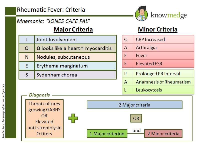
Ix: Clinical Exam (Duckett Jones ARF Criteria), Throat Swab, ECG (PR Prolongation), Echocardiograph (valvulitis, ventricular dysfunction), ESR/CRP, Antistreptolysin O Titre
Major Criteria: carditis, polyarthritis, chorea, erythema marginatum, subcut. nodules
Mx: IM benzathine benzylpenicillin
Acute: Aspirin, Oral Penicillin V/IM Benzyl Penicillin G
Secondary Prevention: IM Bezathine Penicillin (every 3-4wks until 21yrs if no carditis or 40yrs if carditis present)
Arthritis/Severe Arthralgia: naproxen, ibuprofen, aspirin
Cx: Chronic Rheumatic Heart Disease (valvular scarring, HF, AF, embolisms, need valvular transplants)
Rheumatic Heart Disease: damage to the cardiac valves accumulating over recurrent episodes
Patent Ductus Arteriosus (R2)
Simple Acyanotic CHD
Failure of the ductus arteriosus to completely close postnatally
RF: Down syndrome, fetal acohol syndrome, congenital rubella
Sx: Cough, noisy breathing/breathing difficulties, swelling/oedema, failure to thrive/LOW, short stature, crying/irritable/unwell, newborn resp. distress
Small = asymp., Large = wide pulse pressure
Murmur = continuous ‘machinery’ murmur
PDA = Prolonged Deafening Auscultations
Ix: Clinical Exam, CXR (↑ pulmonary blood flow, aortic knob), ECG (LAD), Echocardiogram (visualise ductus and quantify shunt, L cardiac enlargement)
Mx: Observation, Surgery/Interventional (Closure, Coil Embolisation, Amplatzer Ductal Occluder via catheter)
Cx: HF, pulmonary HTN, endarteritis, infective endocarditis, Eisenmenger syndrome, cyanosis
Tetralogy of Fallot/TOF (R2)
Complex Cyanotic CHD
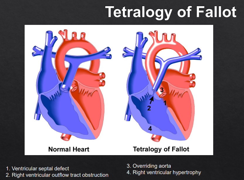
Simultaneous occurrence of four defects:
RV Outflow Tract Obstruction (RVOTO) due to pulmonary infundibular stenosis
RV Hypertrophy (RVH)
Ventricular Septal Defect (VSD)
Overriding Aorta (aorta displaced above VSD)
R-t-L shunting → blood flow RH to LH → deoxygenated blood → cyanosis
ToF: A ventricular septal defect → blocked RV outflow → Aorta override → RV hypertrophy
RF: DiGeorge syndrome, Down synd., Maternal Diabetes, ETOH consumption, phenylketonuria
Sx: Clinical Cyanosis = ‘Blue Baby’ (pale grey/blue skin), Hypoxemia, Failure to thrive/LOW, short stature, fits/faints/funny turns
Tetrad: VSD, RV Outflow Obstruction, Overriding Aorta, RV Hypertrophy, Cyanosis, ‘Boot-Shaped’ Heart on CXR
Clinical Cyanosis, Harsh Systolic Murmur
Untreated Children tend to Squat: ↑ SVR → ↓ R-t-L shunt
Ix: Clinical Exam (Harsh Systolic Murmur), CXR (‘Boot-Shaped’ Heart), ECG (RAD), Echocardiogram (RVOT obstruction, VSD severity), ↓ SPO2
Mx: requires early surgical intervention (shunt or repair: to ↑ pulmonary blood flow or repair TOF/close VSD) - Bad Prog without surgery! (age 1-3yrs)
Pharm: Prostaglandin E1 infusion (Alprostadil) = prevents closure of ductus arteriosus = intentional shunt → mix de/oxygenated blood
Administer O2, Morphine
Cx: Residual RVOT obstruction, pulmonary regurg., arrhythmias, long‑term RV dysfunction

Transposition of Great Arteries (R2)
Complex Cyanotic CHD
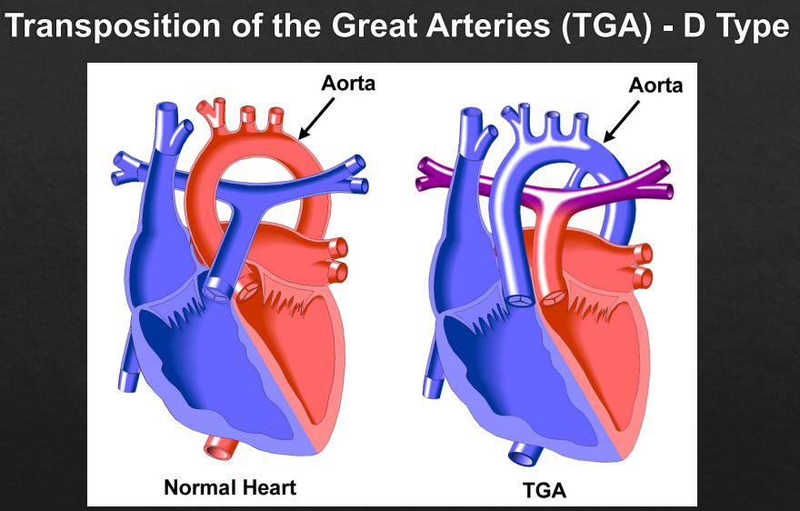
Anatomical reversal of the aorta and pulmonary artery
R-t-L shunting → blood flow RH to LH → deoxygenated blood → cyanosis
Aorta connects to RV rather than LV, receives cyanotic blood, whilst pulmonary arteries connect to LV
RF: Maternal Diabetes, DiGeorge syndrome
Sx: Clinical Cyanosis = ‘Blue Baby’ (pale grey/blue skin), Hypoxemia, Cough, noisy breathing/breathing difficulties, failure to thrive/LOW, newborn resp. distress
Severe Neonatal Cyanosis unresponsive to O2 (due to parallel circulations → survival depends on mixing via ASD, VSD, or PDA)
Ix: Clinical Exam (systolic murmur at left sternal border), CXR (‘Egg on a String’ heart), ECG, Echocardiogram (aorta arising from RV and pulmonary artery from LV)
Mx: requires early surgical intervention (transposition repair - switches Aorta and Pulmonary Arteries, Atrial septostomy to improve mixing); Maintain PDA w prostaglandin where needed
Pharm: Prostaglandin E1 infusion (Alprostadil) = prevents closure of ductus arteriosus = intentional shunt → mix de/oxygenated blood
Cx: coronary insufficiency, ventricular dysfunction, arrhythmias
Poor Prog without Tx (will die within first year)
Infective Endocarditis (R2)
Infection involving the endocardial surface of the heart, incl. valvular structures, chordae tendineae, sites of septal defects, mural endocardium
RF: valvular/congenital heart disease (RHD, CHD), TAVI/valve implantation, ToF
Staphylococcus, Streptococcus, Enterococcus = pathogens associated w endocarditis from TAVI
Sx: Fever/PUO, embolism/splinter haemorrhage, swelling/oedema, pallor, bruising/abnormal bleeding, fits/faints/funny turns, crying/irritable/unwell, eye issues, newborn resp. distress
Murmur (type is valve-dependent)
Ix: Clinical Exam, blood cultures x3, CRP/ERC, ECG, Echocardiogram: TOE>TTE (vegetations, valve destruction)
Duke Diagnostic Criteria
Mx: IV abx, HF mx, Surgery (valve repair/replacement)
Abx: nonsevere = cephalosporin-based abx; severe = non–beta-lactam abx (fluclox, cefazolin)
Also consider Gent ± Vanc (nephrotoxic risk) for Staph A. etc
Streptococci = IV penicillin or ceftriaxone
Cx: valve destruction, HF, embolic events, RHD, infection of other organs/sepsis/septic shock
Croup Ix (R1)
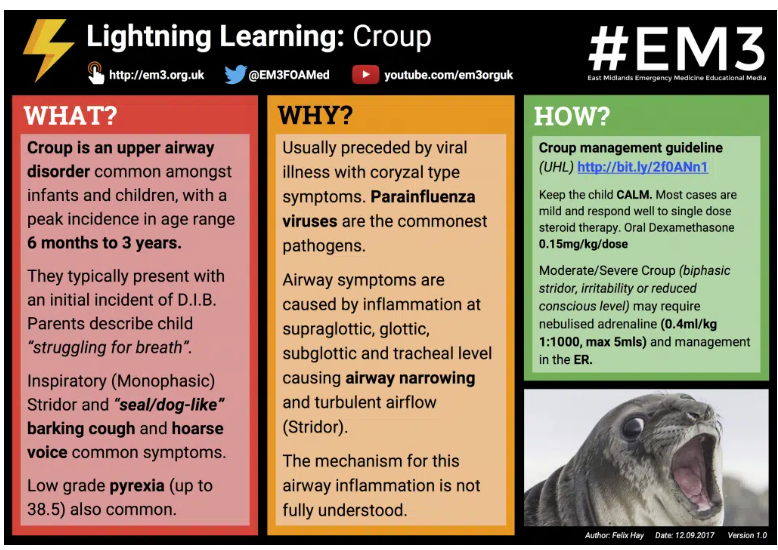
Viral laryngotracheobronchitis causing inspiratory stridor + barking cough
URTI causing mucosal inflammation anywhere bw the nose and trachea
Pathogens: Parainfluenza Virus (most common), influenza, RSV, SARS‑CoV‑2, adenovirus, measles
RF: 6m-6yrs, M>F
Sx: harsh barking cough, hoarse voice, inspiratory stridor (severe may be biphasic), sx worse at night, ↑ WOB (tracheal tug, recession)
Sx usually over 1-4d and worse at night
Red Flags: sx resp. failure (drowsiness/↓ GCS, lethargy, cyanosis, tachycardia)
Ix: Mainly a clinical dx
First Line: Clinical Exam, Westley Croup Score
Consider: FBC, CRP, UEC, CXR (rule out foreign bodies/other causes), Direct/indirect laryngoscopy
Croup Mx (R1)
Mx:
First Line: supportive mx (paracetamol, ibuprofen)
Mild: oral dexamethasone, prednisalone
Moderate/severe: nebulised adrenaline + steroids
Consider intubation if impending obstruction
Cx: Lymphadenitis, Otitis Media, Dehydration
Asthma Ix (R1)
Variable resp sx + variable airflow limitation
Reversible and paroxysmal constriction of the airways, w airway occlusion by inflammatory exudate, and late airway remodelling
RF: FHx atopy/Genetics, low birth weight, prematurity, parental smoking
Sx: Wheeze, cough, dyspnoea, chest tightness, hyperinflated chest (poorly controlled)
Indicators: recurrent sx, worse at night/early morning, triggers (exercise, cold air, allergens), FHx atopy, SABA response, post-bronchodilatory improvement on spirometry
Beware the silent chest!
Ix: Clinical Dx
First Line: Spirometry (poor control = FEV1:FVC <70%, reversible w bronchodilators)
Consider: CXR, Peak expiratory flow rate (PEFR), Bronchial provocation tests (histamine, metacholine), Exercise testing, Skin prick testing
Asthma Mx (R1)
Mx:
Ensure a written Asthma Action Plan is made
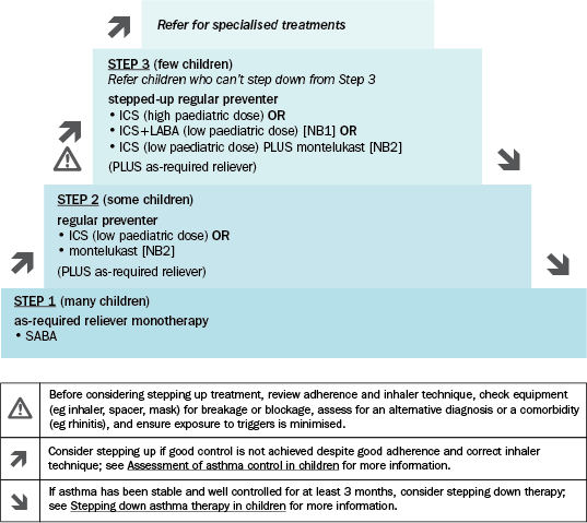
Step Up Therapy Age 1-5yrs:
SABA prn: Salbutamol, Terbutaline
Regular Preventer: ICS (fluticasone), Montelukast
Step Up Preventer: ICS high dose or ICS low dose + Montelukast
Tx of wheeze in children 1-5yrs is often not necessary
Step Up Therapy Age >6yrs:
SABA prn: Salbutamol
Regular Preventer: ICS (fluticasone, budenoside), Montelukast (+ Reliever prn)
Step Up Preventer: ICS high dose or ICS low dose + LABA or ICS low does + Montelukast (+ Reliever prn)
Severe Asthma: ipratropium (Atrovent) - SAMA, tiotropium - LAMA, mabs, prednisalone, consider deamethasone (pred. alternative)
Acute: Adolescents on combination reliever/preventer (e.g. budesonide/formoterol) should be mx w salbutamol for acute exacerbation
For first presentation, do a trial of salbutamol
Cx: asthma exacerbation
Cystic Fibrosis Ix (R1)
Autosomal recessive CFTR mutation resulting in multisystem disease
RF: Caucasian European, FHx/carrier Hx
Sx: chronic wet cough, recurrent infections, bronchiectasis, nasal polyps, digital clubbing, failure to thrive, Pancreatic insufficiency → steatorrhoea/FTT, Meconium ileus, Male infertility, neonatal jaundice
Ix:
First Line: Guthrie test (neonatal screening), Immunoreactive Trypsinogen (IRT) Heel Prick
Gold Standard: Chloride sweat test
Consider: Spirometry, CFTR genetic testing, Respiratory cultures, CT Chest
Cystic Fibrosis Mx (R1)
Mx: refer to GP, resp. paediatrician/CF specialist, dietician, physiotherapist, psychologist etc
Prioritise nutrition due to poor vitamin absorption
Acute: abx for pulmonary exacerbations
Long Term:
Airway clearance/chest physio/vest therapy
Inhaled Therapies: bronchodilator (salbutamol), Nebulised hypertonic saline, inhaled abx for chronic Pseudomonas (tobramycin, colistin)
Anti‑inflammatory therapy: long term Azithromycin (for anti‑inflammatory effect)
CFTR Modulators: depends on genotype, targets underlying protein defect → Trikafta = Elexacaftor, Tezacaftor, Ivacaftor; Orkambi = Lumacaftor, Ivacaftor; Symdeko = Tezacaftor, Ivacaftor; Kalydeco = Ivacaftor (monitor LFTs, eye/cataracts)
Pancreatic Enzyme Replacement Therapy (PERT)
Cx: airway infections (mainly Pseudomonas aeruginosa), infertility, CF-related DM (pancreatic insuff.), neonatal jaundice (cholestasis), distal intestinal obstruction syndrome (DIOS), liver disease
Bronchiolitis Ix (R1)
Viral infection of the bronchioles (smallest air passages in the lungs)
Viral LRTI in infants → small airway obstruction
Pathogens: RSV (most common), hMPV, adenovirus, influenza
RF: <12m (up to 18–24m), Winter/Spring, breast feeding for <2m, smoking, chronic lung disease (prems)
For Severe Disease: rematurity, CLD, CHD, Down syndrome, immunodeficiency
Sx: Coryza → cough → ↑ WOB, tachypnoea, low grade fever, wheeze/crackles, poor feeding, apnoea (espec. prems), nasal flaring, hyperinfl. chest, cyanosis/pallor
Sx usually over 2-5d
Ix: pulse oximetry, CXR only if atypical
First Line: bloods/urine culture (if pyrexic)
Gold Standard: Nasopharyngeal aspirate/throat swab for RSV PCR
Be sure to rule out HF!
Bronchiolitis Mx (R1)
Mx: self-limiting infection lasting 7-10d
First Line: Supportive Mx (fluids, nutrition, fever control)
Severe: Hospital Referral
Oxygen if SpO₂ < 90%
Small frequent feeds/NG hydration
No bronchodilators or steroids (don’t work)
No abx (unless secondary bacterial infection)
Note: cough may last up to 6wks
Cx: hypoxia, dehydration, fatigue, resp. failure, Bronchiolitis obliterans (permanent airway damage)

Pneumonia Ix (R1)
Infection of lung parenchyma
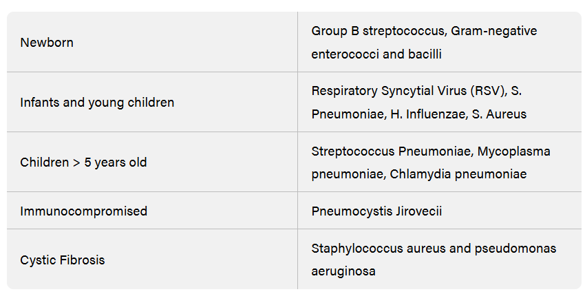
Pathogens: Viral = RSV, influ., adenovirus; Bacterial = Strep pneumoniae, Mycoplasma, Staph A, GBS, Chlam
RF: age <12yrs, immunocomp., CF, malnourishment, recent travel
Sx: fever, tachypnoea, cough, crackles (end-insp.), bronchial breathing, ↓ breath sounds, ↑ WOB, focal chest signs, dull percussion, SOB, lethargy, ↓ feeding, cyanosis
Tachypnoea = 0-5m: >60, 6-12m: >50, >12m: >40
Mild: normal mental state, mild ↑ WOB, SpO₂ ≥94%, feeding adequate
Moderate: obvious ↑ WOB, SpO₂ 90–93%, reduced oral intake
Severe: marked distress, SpO₂ <90%, apnoea, shock, need for respiratory support (HFNP/CPAP/ventilation)
Ix:
First Line/Consider: Bloods (FBE, CRP/ERC), CXR if severe/atypical, Pulse oximetry, US (effusion, empyema)
Gold Standard: nasal swab (viral PCR),Viral serology, sputum microscopy/culture
Investigate for TB/HIV if area is indicated
Pneumonia Mx (R1)
Mx: severe = resus/supportive, chest physio
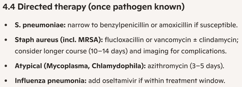
First Line: Viral = supportive, Bacterial = amoxy first line
For age >2m, viral CAP is most common → supportive
Mild/Moderate: amoxicillin
Severe: IV benzylpenicillin ± fluclox/cef/gent
Resistant/Complicated: cef ± Vanc (MRSA risk)
Atypical (post-PCR): benpen + Azithromycin
Aspiration Pneum: Amoxy-Clavulanate (mild) or Amoxy-Sulbactan (mod-severe)
Long Term: GP review 2-3d, Follow-up CXR in 4-6wks (for continued sx, collapse, consolidation, complications)
Prevention: vaccination (pneumococcal, Haem. influ.)
Cx: effusion, empyema, necrotising pneumonia, abscess
Obstructive Sleep Apnoea (R2)
Recurrent/repeated episodes of partial or complete of upper airway obstruction during sleep, disrupting ventilation and sleep quality
RF/Causes: Age 2-7yrs, Adenotonsillar hypertrophy (most common), Obesity, Craniofacial anomalies, Neuromuscular disorders
Sx: Snoring, Witnessed apnoeas (get collateral), Restless sleep, Daytime behavioural issues (learning, concentration), Morning headaches, gasping/choking, hyperextended neck positions, paradoxical breathing
Physical Sx: septal deviation, enlarged inf. turbinates, macroglossia/large tongue, RV hypert.
Ix: Polysomnography (gold standard), oximetry (O2 drops from 100% to 90-80%)
Mx: Adenotonsillectomy, CPAP (if surgery not effective), Weight Mx, Intranasal steroids (if link to allergic rhinitis)
Cx: school/behavioural issues, failure to thrive, heart strain (cor pulmonale), facial structure changes, resp. distress
Allergic Rhinitis (R2)
IgE‑mediated inflammation of nasal mucosa
Presence of >2 nasal sx: Itching, Rhinorrhoea (nasal discharge), Nasal blockage (obstruction), Sneezing
Common Allergens: house dust mite, grass/tree pollens, animal dander, moulds
RF:
Sx: Sneezing, Rhinorrhoea, Nasal congestion, Itchy nose, Allergic salute/crease, Assoc. conjunctivitis
Ix: Skin Prick Testing, Serum-Specific IgE, Lung Func.
Mx: Allergen avoidance/home environment modifications (air filter etc), Saline Douching
Pharm:
Oral non‑sedating antihistamines
Intranasal corticosteroids (INS): most effective (Mometasone furoate, fluticasone propionate)
AEs: ↓ growth, cortisol dysf. (excess steroids)
Regular dose in morning but use lowest dose
Montelukast/Leukotriene Receptor Antagonists (LTRA): less preferred than INS/Antihistamines
Rescue Therapy: Short courses of low-dose oral prednisolone/decongestants for severe sx
Allergen-Specific Immunotherapy: refractory cases
Cx: Poor sleep, Impaired school performance, Otitis media w effusion, Asthma exacerbation
Tuberculosis (R2)
Pathogens: Mycobacterium tuberculosis
RF: age <5yrs, household contacts, immunocomp., recent migrants, Aboriginal and Torres Strait Islander
Sx: Chronic cough, LOW, Night sweats, Fever, Lymphadenopathy, Failure to thrive
Sx commonly present 3m after infection
Ix: Mantoux/Tuberculin Skin Test (TST), QuantiFERON-Gold/Interferon Gamma Release Assay (IGRA), CXR, Sputum/gastric aspirates for AFB, Culture
TST and IGRA are both for Latent TB (not active)
Active TB dx via clinical, CXR, labs, drug reactivity
Mx: Standard RIPE therapy (rifampicin, isoniazid, pyrazinamide, ethambutol → CI = hepatotixicity)
Cx: TB extrapulmonary diseases (e.g. TB meningitis, Miliary TB, spinal TB)
Septic Arthritis Ix (R1)
Orthopaedic emergency involving infection of the joint space synovial) fluid and tissues
Most Common: hip and knee, also shoulder, ankle
Via haematogenous spread, adjacent osteomyelitis, direct inoculation (trauma, surgery)
Pathogens: Staphylococcus aureus (most common), GAS, Kingella kingae (children 4m-4yrs - normal pharyngeal flora), Haemophilus influenzae (unvaccinated children), GBS (neonates)
RF: age <4yrs
Sx: acute onset joint pain, non-weight bearing, fever (>38.5°C), joint held in position of comfort, warmth/swelling of superficial joints
Ix: predictive indicator = Kocher Criteria
First Line: Bloods (WCC, ESR, CRP), XRay (exclude fractures, tumours etc), US (effusion)
Gold Standard: Synovial joint fluid aspiration (cell count, gram stain, cultures/sensitivities, WCC >50 × 10⁹/L)
May consider lumbar puncture
Rule out acute rheum. fever presenting w monoarthritis
Septic Arthritis Mx (R1)
Mx: Do NOT give abx before synovial fluid is obtained
First Line: Urgent ortho referral, Surgical drainage + lavage (arthroscopy, arthrotomy), immobilise, abx (IV for 2-3wks)
Empirical Abx: 1 = Fluclox, 2 = Cephazolin, Vanc
eTG: gent/tobramycin + benpen/fluclox/vanc
Staph: IV cefazolin, fluclox ± vanc (MRSA)
MSSA: IV fluclox; MRSA: IV vanc
Strep: IV benpen, or ceft, vanc
Kingella kingae: IV cefazolin, benpen
Other: IV ceftriaxone, ciprofloxacin, amoxicillin
Septic arthritis of the hip is an emergency, needing urgent operative drainageto prevent necrosis of the femoral head
Long Term: Orthopaedic follow‑up for ≥2yrs (monitor for sequelae)
Cx: systemic sepsis, cartilage destruction, growth plate damage (limb deformity), avascular necrosis
Osteomyelitis Ix (R1)
Bacterial infection of the bone for <2wks duration
Often more subacute or insidious/slow developing than septic arthritis
Usually metaphyseal and located in lower limbs
Mostly spread haematogenously
Pathogens: Staphyloccoccus (Staph. A, MRSA), Streptococcus (S. pyogenes, S. pneumoniae), Salmonella (in sickle cell), Kingella kingae (<3yrs)
Up to 55% is culture negative
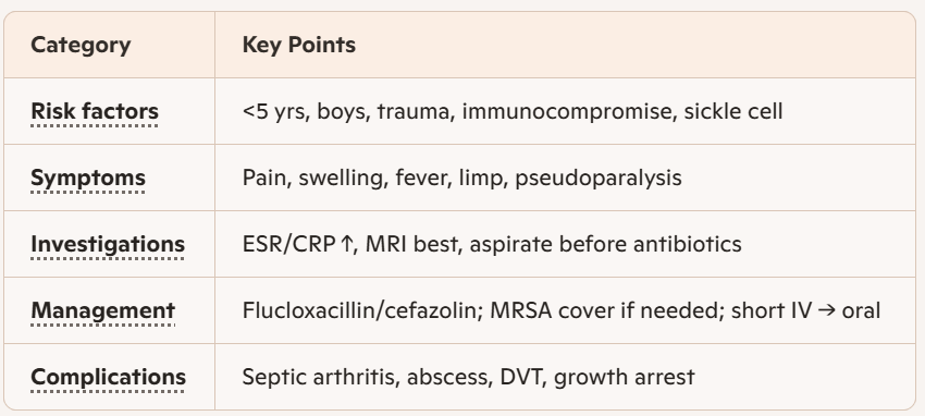
RF: age <5yrs (highest in infacts <1yr), M>F, minor trauma, immunocomp. (DM, malignancy, HIV, steroid therapy, malnutrition), prematurity, Sickle Cell Disease (commonly w salmonella inf.), indwelling lines
Sx: pain, swelling/erythema, fever, ↓ joint movement (pseudoparalysis), limp/↓ weight bearing
Infants may be afeb., irritable, refusing to use limb
Common sites: femur, tibia, long bones, intracapsular metaphyses (hip, shoulder, ankle, ellbow) → ↑ risk concurrent septic arthritis
Ix:
First Line: Bloods (ESR, CRP, WCC, Cultures), XR/CT, US, Bone Scan
Gold Standard: MRI!!! (abscess, pyomyositis, joint effusion), Cultures
Osteomyelitis Mx (R1)
Mx:
First Line: abx
Staph: IV cefazolin, fluclox ± vanc (MRSA)
MSSA: IV fluclox; MRSA: IV vanc
Strep: IV benpen, or ceft, vanc
Kingella kingae: IV cefazolin, benpen
Other: IV ceftriaxone, ciprofloxacin, amoxicillin
Long Term: surgery only if indicated (failure of medical therapy, large abscess, pelvic/long bone abscess)
Cx: concurrent septic arthritis, Subperiosteal abscess, Pyomyositis, DVT, growth arrest (deformity or limb length discrepancy), chronic osteomyelitis, organ failure
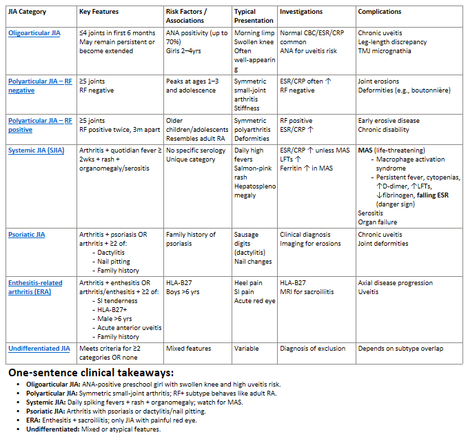
Juvenile Idiopathic Arthritis (R2)
Chronic childhood arthritis with no known cause, defined by ≥6wks of arthritis before age 16
50% achieve remission within 5yrs w early/aggressive tx
Main Subtypes:
Oligoarticular JIA: most common, ↑ risk asymp. anterior uveitis
Polyarticular JIA (RF– / RF+): affects >4 joints
Systemic JIA (SJIA): high risk MAS (macrophage activation syndrome = fever, cytopenias, ↑D‑dimer, ↑LFTs, ↓fibrinogen, falling ESR (red flag)
Characterised by: Quotidian Fever (spikes 1-2x/day, Salmon-pink rash and ↑ risk MAS
Psoriatic JIA: assoc. w psoriasis, dactylitis (‘sausage digit’) and nail pitting
Enthesitis‑Related Arthritis (ERA): inflam of tendons or ligament insertion points (e.g. Achilles)
Undifferentiated arthritis
RF: genetics/FHx (HLA-B27), FHx psoriasis
Sx: Morning stiffness ≥15 min (improves w activity), Gelling (stiffness after inactivity), Joint swelling/warmth/↓ ROM, Pain may be minimal/absent in children
Ix:
First Line: CBC/ESR/CRP (may be normal in oligoarticular JIA), ANA (uveitis), RF (RF+ polyarticular JIA), HLA‑B27 (ERA)
Gold Standard: Echocardiography (only for SJIA w suspect serositis), US/MRI (synovitis, TMJ disease, hip involvement, erosions)
Mimic Screen: Septic Arthritis = synovial fluid, neutrophils, ↓ glucose; Malignancy = bone pain, cytopenias, ↑ LDH/uric acid
Mx: early aggressive therapy, aiming for inactive disease and remission → 1= NSAIDs, Steroids; 2= Sulfasalazine, Methotrexate/DMARD; 3= Biologics (Etanercept, Adalimumab)
Oligoarticular JIA: NSAIDs (ibuprofen), Intra-articular steroids (triamcinolone hexacetonide), Methotrexate/TNF-i (extended disease: ≥3 injections/yr)
Polyarticular JIA: Methotrexate, Biologics (TNF-i = etanercept, adalimumab; IL‑6-i = tocilizumab), Prednisone
Systemic JIA: NSAIDs, Steroids, Methotrexate, Biologics (IL‑1-i = anakinra; IL‑6-i = tocilizumab)
MAS Tx = Methylpred + Anakinra + Cyclosporine
Psoriatic JIA/ERA: same as polyarticular JIA
Uveitis: topical steroids + mydriatics
If refractory: methotrexate, adalimumab, infliximab, tocilizumab
Cx: Joint Cx (cartilage loss/joint space narrowing, bone erosions, growth abnormalities/premature fusion, TMJ destruction), Eye Cx (glaucoma, cataracts, blindness), Systemic Cx (MAS, osteopenia/osteoporosis)
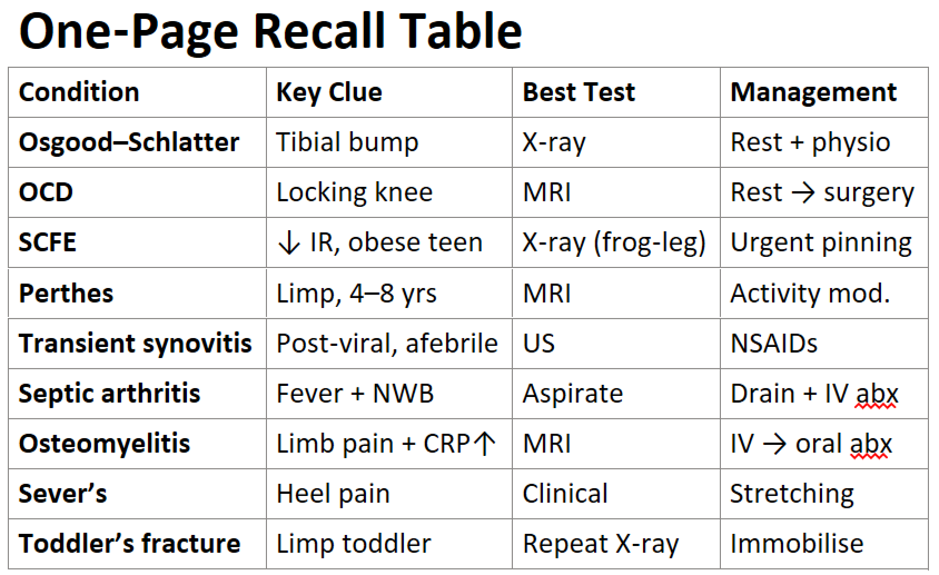
Orthopaedic Disorders in Childhood (R2)
Osgood-Schlatter Disease
Traction apophysitis of tibial tuberosity from quadriceps overuse, post growth spurt, M>F
Sx: Anterior knee pain ↑ w activity, tibial tubercle swelling/bump
X‑ray: soft‑tissue swelling, tubercle fragments
Osteochondritis Dissecans
Focal aseptic necrosis of subchondral bone → detachment of bone-cartilage fragment from repetitive trauma/mechanical stress
Sx: Activity‑related knee pain w locking/catching, antalgic gait, Preserved ROM
X‑ray: subchondral fragment ± radiolucency
Unstable SCFE: unable to weight‑bear → ↑ AVN risk
Slipped Capital Femoral Epiphysis (SCFE)
Posteroinferior displacement of femoral head at growth plate
Sx: Hip/groin/thigh/knee pain, limb, ↓ int. rotation/abd., Drehmann sign (ext. rotation during hip flexion)
AP pelvis + frog‑leg lateral X‑ray
Perthes Disease (Legg-Calvé-Perthes)
Idiopathic AVN of the femoral head
RF: M <10yrs
Sx: ↓ hip abd. + int. rotation, limp, hip/knee pain
X‑ray: may be normal early → MRI/bone scan
Cx: femoral head deform., early OA
Transient Synovitis
Benign, self‑limiting post‑viral hip inflam/effusion
Sx: hip pain (most common cause in children), afebrile; Hip in ext. rotation, abd., slight flexion
Overuse Apophysitis Conditions
Osgood–Schlatter: tibial tuberosity
Sever’s disease: calcaneal apophysis
Sinding–Larsen–Johansson: inferior patella
Sx: Activity‑related pain at tendon insertion
Toddler’s Fracture
Spiral fracture of tibia in ambulant toddlers
Sx: Limp/refusal to walk, minimal trauma
X‑ray: may be normal → repeat or oblique views
Muscular Disorders Mimicking Orthopaedic Disease
Duchenne muscular dystrophy
Spinal muscular atrophy
Present w limp, gait abnormalities, delayed motor milestones
Febrile Convulsions Ix (R1)
Common paediatric emergency associated with high fever and seizures in children b/w 6m-5yrs
Simple Febrile Seizure: usually generalised, last <15min, do not recur within 24hrs
Complex Febrile Seizures: focal onset, last >15min, or recur within 24hrs
RF: age b/w 6m-5yrs, genetics, high fever (>40°C), Viral Infection (HHV-6, influenza), recent immunisation (MMR, DTaP), Recent Inf (Otitis Media, UTI, Gastroent, LRTI)
Sx: seizures and fever (>38°C) in the absence of CNS infection, metabolic abnormalities or Hx afebrile seizures
Usually Tonic-Clonic Seizure (generalised, rolling eyes, tongue biting, incontinence, shaking/twitch)
Ix: Identify cause of fever
First Line: Clinical Exam (Simple Febrile Seizures do not require diagnostic workup)
Other Screening: Urinalysis/Urine Culture, Bloods (CBC, CRP, UEC, BGL), Imaging (US, XR - find source of infection),
Workup for Complex Seizures: Lumbar Puncture (excl. meningitis/enceph), CT/MRI, EEG
Exclude herpes encephalitis in Complex Seizures!

Febrile Convulsions Mx (R1)
Mx: identify and address cause of the fever!
Most febrile seizures are spontaneous and do not require treatment
Abortive Therapy (Seizures Persisting >5min or Complex Febrile Seizure): IV Benzodiazepines (1 = Lorazepam, 2 = Diazepam, Midazolam)
Antipyretic Therapy (post-febrile seizure): NSAIDs, Acetaminophen
Long-Term/Prophylaxis: Not needed due to being caused by a rise in temperatures (fever)
Cx: epilepsy (rare - 10% risk), Febrile status epilepticus
Febrile status epilepticus: subgroup of complex febrile seizure where the seizure duration exceeds 30min, or there are multiple seizures lasting a total of 30min w/out recovery b/w each one

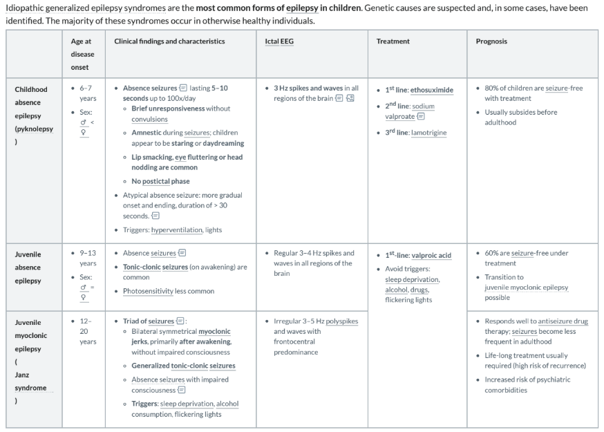
Epilepsy Syndromes - Idiopathic Generalised (R1)
Congenital or Idiopathic; Most common epilepsy form
Epilepsies are stereotyped, intermittent disturbances of behavior or movement resulting from abnormal electrical discharges within the brain
Causes: Infections, Head Trauma, Metabolic Disorders
Childhood Absence Seizure:
RF: 6-7yrs, M>F; Triggers: hyperventilation, lights
Sx: Absence Seizures 5-30sec (up to 100x/d) = brief unresponsiveness, amnestic (staring/day-dreaming), Lip smacking/Eye fluttering/Head nod
Sudden stop of motion ± minor facial motions
No postictal phase
Atypical Absence Seizure: more gradual onset and ending, duration >30sec
EEG: 3Hz sym. spikes and waves (all brain areas) → ‘wallpaper’
Juvenile Absence Seizure:
RF: 9-13yrs, M>F
Sx: Absence/Tonic-Clonic Seizures (T-C on awakening), Photosensitivity (uncommon)
EEG: Regular 3-4Hz spikes/waves (all brain areas)
Juvenile Myoclonic Seizure (Janz Syndrome):
RF: 12-20yrs; Triggers: sleep dep., ETOH, lights
Sx (Seizure Triad): Myoclonic Jerks (bilat. sym., after awakening, no impaired consc.), Generalised Tonic-Clonic Seizures, Absence Seizures (w impaired consciousness)
EEG: 3-5Hz polyspikes and waves w frontotemporal predominance
Ix:
First Line/Consider: Bloods (UEC, BGL - rule out metabolic abnormalities), Tox Screen, Blood Cultures, Lumbar Puncture (febrile pts)
Gold Standard: EEG, Seizure Hx
Mx:
Childhood Absence: 1 = Ethosuxemide, 2 = Sodium Valproate, 3 = Lamotrigine
Juvenile Abs/Myo: Valproic Acid, Avoid Triggers
Cx: psychiatric comorbidities, seizure recurrence
Epilepsy Syndromes - Symptomatic/Cryptogenic Generalised (R1)
Congenital or Idiopathic. Sx = structural/metabolic abnormalities; Crypto = unknown eitiology
Causes: Infections, Head Trauma, Metabolic Disorders
Infantile Spasms (West Syndrome):
RF: 3-7m, M>F
Causes: perinatal inf., hypoxic-ischemic injury, PKU/Tuberous Sclerosis
Sx: Sudden Sym./Synchronous Spasms (clusters of 5-10), Jerking Flexion/Extension (‘jackknife movements', ‘fling arms’) of neck/torso/limbs
"Crunch-like" movement of the head, knee flexion, appearing frightened, Moro-like reflex
Spasms appear in clusters
Followed by tonic phase
EEG: hypsarrhythmia (high Delta waves w irreg. multifocal spikes + slow waves)
Lennox-Gastaut Syndrome:
RF: 3-5yrs
Causes: structural brain abnormalities, cryptogenic
Sx: myoclonic/tonic/atonic/absence seizures, developmental delays, frequent Status Epilepticus periods (seizure >5min)
EEG: multifocal sharp/slow wave, slow spike-wave
Ix:
First Line/Consider: Bloods (UEC, BGL - rule out metabolic abnormalities), Tox Screen, Blood Cultures, Lumbar Puncture (febrile pts)
Gold Standard: EEG, Seizure Hx
Mx: do not respond well to antiseizure drugs
West Synd: ACTH, Prednisalone, Vigabatrin
L-G Synd: try Anticonvulsants (Valproic Acid, Clobazam, Lamotrigine, Felbamate), Ketogenic Diet, Vagus Nerve Stimulation, Surgery
Cx: developmental delays, motor/cognitive impairments

Epilepsy - Other Forms (R1)
Pediatric vs. Adult Epilepsy: Children differ from adults in seizure behavior, drug metabolism, and specific medication side effects
Broad Categories:
Focal Fits: Originating from one specific part of the brain
Generalized Fits: Originating from both hemispheres simultaneously
Causes: include genetic causes, structural abnormalities, metabolic conditions, unknown causes (increasingly identified as genetic)
Complex Cases: Conditions like Tuberous Sclerosis (one of the phakomatoses) involve masses within the brain that cause focal fits. May present as generalized epileptic encephalopathy
Focal Discognitive Seizures:
Sx: Partial interruption of consciousness starting in one part of the brain and potentially spreading
Occur less frequently than absence seizures but last longer
Focal Clues:
Contralateral signs: Head/eye deviation to the side opposite of the seizure focus
Ipsilateral signs: Automatisms (semi-purposeful, repetitive, stereotyped movements) → eye blinking, nose wiping, picking at clothes
Benign Rolandic Epilepsy:
Benign Focal Epilepsy of Childhood/Benign Childhood Epilepsy w Centrotemporal Spikes
RF: middle childhood
Sx: Focal fits involving the face and mouth because discharges originate in the Rolandic area, some secondary generalisation
Most seizures occur overnight
EEG: Classic centrotemporal spikes, Spikes often flip from the left to the right side
In the EEG layout, odd numbers correspond to the left hemisphere, even to the right, and "Z" to the midline
Ensure to differentiate epilepsy from other conditions like night terrors, tick disorders, ritualistic behaviors, simple staring, or pseudo-seizures

Cerebral Palsy Ix (R1)
Non-progressing disorders affecting muscle tone and development of movement/posture. Caused by brain damage/ABI in utero or up to age 3 (hypoxia etc)
Types: Spastic (most common), Ataxic, Dyskinetic
Spastic: injury to motor cortex → ↑ Muscle tone
Ataxic: cerebellar injury → ↓ balance/coordination
Dyskinetic: injury to basal ganglia → involuntary movements, twisting postures
Can have a mixture of spastic/dyskinetic
Functionality can be classified by GMFCS Levels 1-5
RF: prematurity, perinatal cx (chorioamnionitis, TORCH, birth trauma, intracerebral haemorrhage, asphyxia), Postnatal inf. (meningitis/sepsis), placental abruption, Teratogens (warfarin, sodium valproate), Multiparity, thyroid disease, IUGR
Sx: failure to meet milestones, joint contractures, seizure disorder, intellectual disorder, ADHD
Spastic: spastic paresis (>1 limb), scissor gait, persistent primitive reflexes (pos. babinski), ↑DTR, ↑ Muscle tone, scoliosis, impaired hearing/vision
Non-Spastic: dyarthria, dyskinetic/abnormal invol. movements (worsen w stress), Ataxic movements (intention tremor, lack of balance/coordination)
Ix:
First Line: Clinical Exam/Hx
Consider: cranial US, MRI (show causative lesions - haemorrhage, brain malformations)

Cerebral Palsy Mx (R1)
Mx: focus on treating contractures (bracing, antisposmodics, physical therapy, surgery), social therapy (speech therapy, social support)
Non-Pharm: Physical Therapy (prevent muscle contractures), OT (motor skill development), Speech Path, Orthotic Devices (braces, splints, casts), Assistive Devices (wheelchairs, walkers), Education Support, Nutrition Support (for dysphagia), Social/Psych Support
Pharm: Antispasmodics for tone (Botulinum, Baclofen, Gabapentin, Dantrolene, Benzodiazepines), Anticonvulsants, Anticholinergics (for rigidity, sialorrhea)
Surgery: ortho surgery (scoliosis, relieve contractures, spasm neurosurg mx/Selective Dorsal Rhizotomy)
Cx: seizure disorders, intellectual disability, impaired sensation/perception, chronic pain, MSK cx
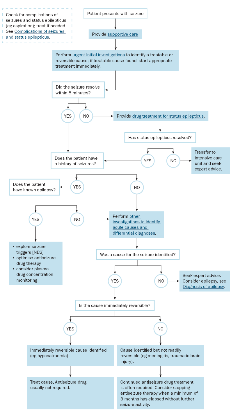
Status Epilepticus (R2)
Seizure lasting >5min or repeated seizures w/out full recovery to normal conscious level between episodes
Neurological emergency
RF: epilepsy, infection, FHx, Intracranial inf., severe metabolic disturbance (hypoglycaemia, E- dist.), neurological damage, neurosurgery
Sx: Seizure lasting >5min/repeated seizures
Ix: EEG, BGL, UEC, Calcium/Magnesium, Antiepileptic drug (AED) levels, ECG, Neuroimaging
Mx: Resus, Seizure Termination (1 = Midazolam, Diazepam, Lorazepam, 2 = Phenytoin, Phenobarbitone, Levetiracetam, Sodium Valproate)
Cx: seizure becoming refractory (risk ↑ w ↑ seizure duration) → high neurological morbidity and mortality
Migraine (R2)
Condition causing a headache w throbbing pain and other sx (N/V, dizziness, sensitivity to light/sound/smell)
Childhood migraines are more likely to be bilateral than adult migraines
RF/Triggers: FHx, Fatigue, OCP, Certain Foods, Weather, Menstruation, Stress
Sx: Headache (Recurring/Relapsing pattern, Dull/Throbbing, worse on sides), paleness, photophobia (light), phonophobia (sound), N/V, dizziness
Rare Syndromes: Aura w/out headache, hemiplegic migraine, basilar symptoms (ataxia), ophthalmological migraine, Alice in Wonderland synd. (altered perception of size and movement)
Childhood Periodic Syndromes: cyclical vomiting and abdominal migraine (recurrent abdominal pain and upset as a migraine prodrome)
Headache Patterns:
Acute/Rapid Escalation: Suggestive of acute raised intracranial pressure
Recurring/Relapsing: Suggestive of a migrainous pattern
Chronic Daily: Suggestive of intracranial hypertension, tension-type headaches (described as a "band" at the front), or analgesic-overuse headaches
Analgesic Withdrawal Headaches: Caused by frequent use of medications like Panadol or Nurofen
Ix: Clinical Exam/Hx (headache pattern)
Mx: NSAIDs (Ibuprofen → for early use), Triptans (Rizatriptan → for severe attacks), Avoid Trigger, Nutraceuticals (Riboflavin, Coenzyme Q10), Cold Packs/Supportive
Cx: missed school, anxiety, depression, status migrainosus (unbroken attacks lasting over 72hrs), medication overuse headaches (frequent pain relief use)

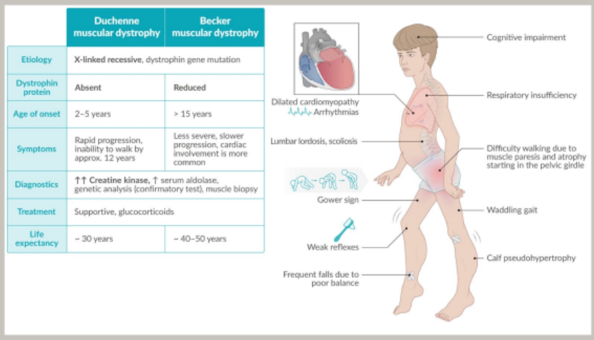
Duchenne Muscular Dystrophy (R3)
X-linked recessive disease affecting MSK system (most severe form of muscular dystrophy) causing progressive muscle paresis and atrophy
Disease onset usually at age 2-3yrs; Life exp. 30yrs
Rapidly progressing (ambulatory inability by age 12)
RF: Males only, age 2-5yrs, genetic/FHx
Sx: progressive muscle paresis/atrophy (starts proximal LLs/pelvic girdle, extends upwards), ↓ reflexes, ↓ cogn, waddling gait (Duchenne limp) w bilat. Trendelenberg sign, Gower Maneuver (‘walking up’ body), Calf pseudohypertrophy, scoliosis, dilated cardiomyopathy
Ix: Creatine Kinase (↑↑), Serum Aldolase (↑), Genetic analysis (dystrophin gene mutation), Muscle Biopsy (absent dystrophin protein)
Mx: Supportive (physiotherapy, psychology, Ventilation, assistive devices/wheelchair), Glucocorticosteroids (Prednisalone), Eteplirsen (Antisense Oligonucleotide)
Cx: cardiac/resp failure
Spinal Muscular Atrophy (R3)
Autosomal Recessive Motor Neuron Disease caused by apoptosis of lower motor neurons
Typically presents during infancy/early childhood w progressive weakness
Disease severity related to age of onset
RF: M>F
Sx: progressive weakness, hypotonia, muscle atrophy, hyporeflexia/areflexia, bulbar weakness
Ix: genetic testing, muscle biopsy (hypertrophied motor neurons)
Mx: Nusinersen (halts SMA progression), Onasemnogen Abeparvovec (potentially curative gene therapy), Supportive therapy (prevent resp/ortho cx)
Cx: Type 1 SMA (Werdnig-Hoffmann disease) = death within first 2yrs (resp muscle paralysis/asp/ pneum); delayed motor milestones, joint contractures, spinal deformities
Guillain-Barré Syndrome (R3)
Acute immunological demyelinating polyneuropathy where antibodies attack peripheral nerves, causing temporary conduction failure and often triggering muscle weakness/paraesthesia starting in the feet/legs and spreading upward
May be triggered by infection (eg Gastroenteritis, Campylobacter enteritis, Mycoplasma pneumoniae, EBV, CMV, HIV) or vaccination (eg COVID, influenza)
RF: immunocomp., age <10yrs
Sx: rapidly progressing ascending weakness, pain and sensory disturbance (often w/out sensory signs) → starts peripherally w impaired/lost tendon reflexes; autonomic instability (BP, cardiac arrhythmia, urinary retention), ophthalmoplegia, ataxia, areflexia
Ix: CSF (↑ protein → sign of demyelination, ↓ cellular response), Nerve Conduction Study (slowed conduction), Spinal MRI w Contrast (‘spider’ nerve roots)
Ensure HIV testing
Mx: IV immunoglobulin, plasma exchange, supportive therapy (resp + swallowing function)
May require ICU transfer
Cx: resp. failure, DVT/Embolism
Gastroenteritis Ix (R1)
Acute diarrhoeal illness, usually infectious, w passage of loose/watery stools, V, fever, abdo pain
Pathogen: Norovirus (most common, spread in schools/daycares), Rotavirus, Salmonella, Shigella, Campylobacter jejuni, E. coli (bloody diarrhoea), C. dif, Giardia (parasite)
RF: childcare facilities/schools, immunocomp., sickle cell disease
Sx: sudden onset watery diarrhoea ± V, acute watery/bloody diarrhoea (dysentery), V, mild fever (>40°C), tachypnoea, malnutrition, abdo pain/dist/guard
Ix:
First Line: stool microscopy/culture, bloods (UEC, glucose, renal function/eGFR)
Gastroenteritis Mx (R1)
Mx: supportive mx
First Line: Oral Rehydration Therapy (ORT), IV fluids, early feeding, Antidiarrhoeals (loperamide), Abx, Ondansetron
Pt should not return to school/nursery until 48hrs since last ep. diarrhoea/V
Cx: dehydration/shock, Haemolytic Uraemic Synd./HUS (acute renal failure, haemolytic anaemia), Toxic megacolon, Acquired/secondary lactose intolerance
Red Flags: bloody diarrhoea, bilious V, severe abdo pain, shock, neuro sx, persistent diarrhoea >14d
Gastro-Oesophageal Reflux/GER Ix (R1)
GER = Physiological Reflux
Invol. passage of gastric contents into the oesophagus
Normal regurgitation or "positing" in babies. Does not require treatment if the baby is thriving and well
Note: GORD = Pathological Reflux w secondary consequences (sig. irritability/pain after feeding, LOW etc) → only GORD when leads to problems
80% of infants experience reflux due to several anatomical factors: liquid diet, lying flat, shorter esophagus, and ↓ Angle of His
RF: infants <3m, prematurity, neurodisability (CP), repaired oesophageal atresia, congenital diaphragmatic hernia, chronic lung disease, hiatus hernia
Sx: FTT, significant irritability, evolving food aversion, assoc. resp. problems (related to aspiration), "Happy spitter", dysphagia, frequent regurg/V, excess hiccups
Note: Not all vomiting babies have GOR
Ix:
First Line: growth assessment
Gold Standard: Clinical Exam
Consider: Barium Swallow (contrast), Scopes (Upper GI Endoscopy w Biopsies), Oesophageal pH Monitoring (Reflux index >7% = abnormal, 30% = GORD) - Good for acid exposure measurements
Gastro-Oesophageal Reflux/GER Mx (R1)
Mx: Conservative - Most sx resolve by 12-14m of age
Positioning (holding upright after feeds, cot head elevation), Smaller/Frequent feeds
Thickened anti-reflux (AR) formulas/Alginates (e.g., Gaviscon)
Acid suppression: Acid Suppression/H2-Receptor Antagonist (Ranitidine), PPIs (Omeprazole, Nexium)
Cx: failure to thrive/malnutrituon, anaemia, esophagitis, aspiration pneumonia, Recurrent acute otitis media
Coeliac Disease (R2)
Gluten-sensitive autoimmune disease of the small intestine requiring lifelong exclusion of wheat, barley, and rye (not an allergy)
Immunological responses to an environmental (gliadin) and genetic factors (HLA-DQ2/DQ8)
Immune system overreacts to gluten, damaging villi lining small intestine → malabsorption
RF: northern european descent, T1D, Thyroid Disease, Down/Turner Syndrome, Other Autoimmune Diseases
Sx: fts of Malabsorption (FTT/LOW, loose stool, steatorrhea, anorexia, abdo pain/distention, muscle waste), Anaemia, GIT Sx, Delayed Puberty, Arthritis, Peripheral Neuropathy, Dermatitis Herpetiformis
Ix: Small Bowel Biopsy!, Anti-Tissue Transglutaminase (tTG-IgA - most sensitive blood test), Anti-endomysial IgA (most specific)
Biopsy Triad: Subtotal villus atrophy (small, stumpy, or absent villi), Crypt hyperplasia (biopsy appears full of crypts), ↑ intraepithelial lymphocytes
Histology = crypt hyperplasia + villous atrophy
Mx: gluten free diet, dietary supplementation, iron
Cx: anaemia, osteoporosis, malignancy (Enteropathy-associated T-cell lymphoma/EATL, Non-Hodgkin Lymphoma/NHL), Fertility Issues, Refractory Coeliac Disease (sx persists despite diet)
Infantile Colic (R2)
Excessive/inconsolable crying in first few months of life
May be caused by ↑ gas production in gut, abnormal GIT motility, gut inflammation
RF: infants <6wks, Parental anxiety/depression, Family tension, Inadequate parent-infant interaction
Sx: Recurrent and prolonged periods of infant crying, fussing or irritability with non-obvious causes and cannot be prevented or resolved by caregivers; Crying usually in late afternoon/evening, fist clenching
Ix: diagnosis of exclusion, Rome IV diagnostic criteria, growth monitoring
Mx: Self-limiting (usually resolves within 5m of age), Infant Comfort (rocking, ‘white noise’, warm bath)
DDx: Intussusception or volvulus intestinal obstruction, pyloric stenosis, Incarcerated/strangulated hernia, sepsis, trauma/non-accidental injury, Hydrocephalus, hunger/dehydration, constipation, GER, food allergy
Red Flags: fever, poor feeding, bilious/projectile V, bloody stool, lethargy
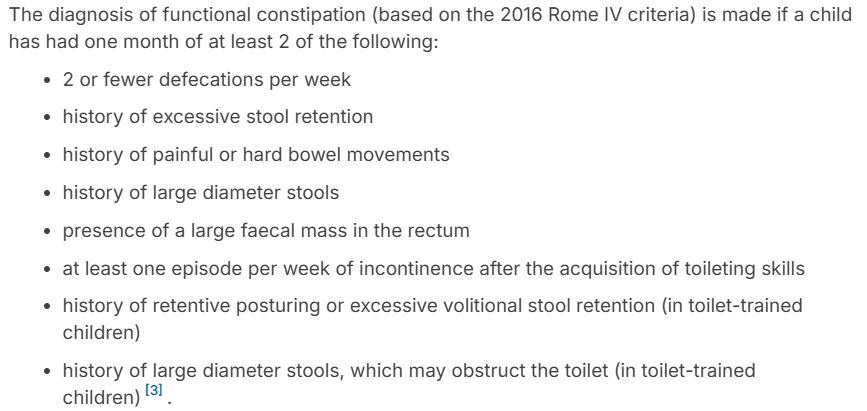
Functional Constipation (R2)
Infrequent passage of hard, painful stools without underlying organic cause
Most common cause of acute abdo pain in children
RF:
Sx: infrequent stools, large/hard stools, abdo pain/dist, painful defecation, stool withholding, palpable faecal mass, faecal incontinence/overflow
Ix: Rome IV criteria, Clinical Exam
Abdo XR has no role in dx of FC in children
Mx: Polyethylene glycol (PEG), Lactulose, Long-term stool softener, Regular toilet sitting, High fiber diet, Behaviour modification
Neonates: Poloxamer
Infants >1m: Lactulose, Macrogol (Movicol Jr), Poloxamer
Children >1yr: Lactulose, Macrogol (Movicol Jr, OsmoLax), Paraffin, Docusate, Poloxamer
Refractory >1yr (not improving): Bisacodyl (oral or rectally), Senna, Sodium Picosulfate
Cx: Fecal impaction → pelvic floor damage, haemorrhoids, Rectal prolapse, Anal Fissures
Red Flags (consider organic cause): FTT, delayed meconium, neuro sx, abnormal anus, blood w/o fissure
Inflammatory Bowel Disease (R3)
UC: Continuous inflammation limited to the large bowel, typically pw bloody diarrhoea
Crohn's: from the mouth to anus. Includes transmural inflam., granulomas, skip lesions (areas of normality and abnormality)
Often has systemic features like LOW, growth failure, anaemia (tiredness)
RF: age >6yrs, FHx, Abx use, Processed Foods
Sx: abdo pain, LOW, FTT, diarrhoea (UC = bloody), urgency, tenesmus (UC), arthritis (CD), Uveitis, Erythema nodosum, Delayed puberty
Ix: Fecal Calprotectin (protein marker indicates inflam cause of bloody diarrheoa), Bloods (FBC, CRP/ESR, Iron Study), Scope (Gastroscopy/Colonoscopy)
Do a stool sample to rule out inf. (esp. in UC)
Mx: Sulfasalazine (DMARD), Corticosteroids (Prednisalone/Hydrocortisone), Methotrexate (Immunomod), Azathioprine (Immunomod), Infliximab (Biologic)
Cx: FTT, delayed puberty, osteoporosis, anaemia, toxic megacolon, GIT damage (strictures, fistulas, abscess)
Cow’s Milk Protein Allergy (R3)
Immune-mediated allergic response to naturally-occurring milk proteins casein and whey
Classifications: IgE-mediated, Non-IgE-mediated, Mixed
IgE = Type I hypersens.; Non-IgE = T-cell Activity
RF: PMHx/FHx Atopy
Sx: sx worse after formula introduction, blood in stool, failure to thrive, skin reactions (pruritus, erythema, urticaria), Facial Angiooedema, N/V, diar/const, colicky abdo pain, GER, infantile colic, fatigue, U/LRT sx
Ix: IgE Ab bloods/RAST test, Clinical Exam, FBE/other bloods (iron-def anaemia etc), Growth charts
Mx: avoidance of cow’s milk (incl. mother), Extensively hydrolysed or amino acid formula, Dietetic support
Cx: malabsorption, reduced intake → ↓ growth, anaphylaxis
Biliary Atresia (R3)
Progressive fibro-obliterative disease of extrahepatic bile ducts causing neonatal cholestasis
Obst. progresses from extrahep. biliary tree to intrahep. ducts → jaundice, hepatomegaly
RF: genetics/FHx (HLA-B12), ?viral infection
Sx: prolonged neonatal jaundice (>2wks), pale stool, dark urine, hepatomegaly, poor weight gain, ascites, hepatomegaly
Suspect in all infants w jaundice ± pale stools after age 14d, or conj. bilirubin ≥17.1micromoles/L
Ix: Bilirubin, LFTs, US (hidden gallbladder), HIDA scan, PT/INR (INR >1.5, PT >14s = Coagulopathy), Liver Biopsy
Conjugated Bilirubin: ≥17.1 micromoles/L
LFTs: ↑↑↑ GGT, ↑ ALT, ↑ ALP, ↑ AST,
Mx: Surgery (liver transplant; before 6-8wks old → Kasai portoenterostomy), Vitamin Sups, Nutritional support
Cx: growth failure, portal HTN, cholangitis, ascites
Type 1 Diabetes Ix (R1)
Insulin deficiency caused by autoimmune destruction of pancreatic beta cells (insulin producers)
T1DM = ‘starvation in the midst of plenty’
Blood glucose levels are ↑ as it cannot be used for metabolism/storage due to an absolute deficiency of insulin (due to autoimmune destruction of pancreatic beta cells)
If a child presents w high blood glucose (BG >25) ± sx, it is a medical emergency until proven otherwise!
RF: FHx (DM, Autoimmunes)
Sx: polyuria, polydipsia, LOW, lethargy, noct. enuresis
DKA: N/V/dehydration, abdo pain, tachypnoea, Kussmaul breathing, Ketotic breath (‘fruity’), ↓ GCS
Ix: be sure to Ix/rule out sepsis/infection!
First Line: Blood gas (pH <7.3, Bicarb <15mmol/L = DKA), Urine Dipstick (high ketones = red flag!), Bloods (UEC, FBE), ECG
Gold Standard: Blood Glucose (usually >11mmol/L in DKA), HbA1c
Comorbidity Screening: Coeliac disease, Thyroid disease, ?Adrenal/Addison’s disease (if sx)
Type 1 Diabetes Mx (R1)
Mx: continuous glucose monitors, insulin pumps
Acute (DKA): Monitor fluids, potassium, sodium and glucose (SPIDER); Potassium Mx/replacement
Long-Term: Insulin, Dietary Mx/Nutritionist
Basal-Bolus Inj: basal = long-acting, bolus = rapid
Continuous Subcut Inf (CSII): pump
Mixed Insulin Regimen: Rapid/Short + Intermediate
Mix: NovoMix, Humalog Mix
Rapid-Acting: NovoRapid, Humalog
Ultra-rapid (Adults): FIASP (not yet approved for paeds)
Cx: retinopathy, neuropathy, nephropathy, delayed growth/puberty
DKA: cerebral oedema (avoid rapid fluid resus and dramatic E-/glucose shifts), Hypokalaemia, Aspiration Pneumonia, Hypoglycaemia
Obesity Ix (R1)
BMI: Overweight = 85-95th percentile, Obese = ≥95th percentile, Severe Obesity = Class II or III obesity
Healthy = 5th-85th percentile
RF: maternal obesity. excess gestational weight gain, maternal smoking, excess calorie intake, physical inactivity/excess screen time, short sleep, FHx
Sx: ↑ BMI, ↓ exercise tolerance
Ix:
Consider: BP, Sleep Study (if OSA sx), HbA1c/Fasting glucose, ALT/AST (NAFLD screen)
Gold Standard: BMI percentile, Lipid profile
Consider Secondary Cause if: poor linear growth, developmental delay, dysmorphic fts (e.g. medications, hypothyroidism, Cushing’s, Growth Hormone def.)
Obesity Mx (R1)
Mx:
First Line: lifestyle (diet, physical activity)
Long-Term: Pharm (Orlistat, Metformin, GLP-1 agonist), Bariatric Surgery
Cx: Cardiometabolic (HTN, dyslipidaemia, insulin resistance, T2DM, metabolic syndrome, NAFLD), OSA, PCOS, ortho issues, psychosocial distress
Growth Hormone Deficiency (R2)
Insufficient secretion of growth hormone from the anterior pituitary leading to impaired linear growth
Growth hormone (GH) also has effects on bone mineral density, body composition, metabolic profile, and QoL
May be linked to pituitary hormone deficiencies
RF: FHx, pituitary hormone deficiencies, CNS tumours/cysts, radiotherapy
Sx: short stature, ↓ growth velocity, delayed bone age, ↑ central adiposity, immature facial features
Ix: Growth Assessment (anthropometry, growth velocity, mid-parental height calculation, bone age evaluation), Bone Age XR, IGF-1/IGFBP-3, GH stimulation testing, Pituitary MRI (if indicated)
Values of insulin-like growth factor 1 (IGF1) and its binding protein (IGFBP3) of >2 standard deviations
Mx: Recombinan GH therapy (rhGH), Ongoing growth monitoring, Monitor pubertal progression
Cx: stunted/delayed growth/puberty, osteoporosis, GH-linked T2DM, CAD
Thyroid Disorders (R2)
Hyperthyroidism, Hypothyroidism
RF: FHx, PMHx Autoimmune, ↑ iodine intake, smoking, female, prematurity, low birth weight (+ twins)
Causes: Hyper = Graves; Hypo = Thyroid Dysgenesis (congenital), Hashimoto’s, Iodine Def (acquired)
Sx: Weight changes, Temp. intolerance, anxiety, lethargy hyper/hypoactivity, ↓ school performance, goitre, tachy/bradycardia, tremor, constipation
Thyroid issues can masquerade as pubertal or growth disorders
Ix: Pediatric Thyroid Function Tests (TFTs), TRAbs, Anti-TPO, Anti-Tg Ab/TgAb, US
Both TSH and Free T4 must be ordered together to identify pituitary-related primary issues that a TSH-only screen might miss
Abs indicate autoimmune thyroid disease
Hyper = ↓ TSH, ↑ T4; Hypo = ↑ TSH, ↓ T4
Mx:
Hyperthyroidism: carbimazole, BB, surgery
Hypothyroidism: levothyroxine
Cx: ↓ school performance, growth changes, thyroid storm, Thyroid eye disease, mpaired neurocognitive development and growth

Protein-Energy Malnutrition (R2)
Undernutrition caused by inadequate calorie intake, absorption or increased metabolic demand resulting in poor growth
RF: Inadequate Intake (neglect, poor feeding habits, improper formula prep, feeding disorders), Malabsorption (coeliac disease, CF, IBD, food allergy), ↑ Calorie Requirements (CHD, chronic lung disease, hyperthyroidism, malignancy)
Sx: Weight-for-height z-score <−2, Declining growth percentiles, Reduced growth velocity
Ix: No specific Ix, FBC, UEC, Coeliac Screen, CRP/ESR
Mx: Behavioural (structured meals, eliminate grazing), Nutritional (calorie enrichment, formula fortification, supplements)
Cx: Frequent infections, hypoglycaemia, hypothermia, shock, HF, developmental delays
Vitamin Deficiencies (R2)
Main Deficiencies: Vitamin D, Iron, Vitamin B12/Folate, Fat-Soluble Vitamins (A,D,E,K)
RF: obesity, limited sun exposure (Vit D), CF, cholestatic liver disease, Malabsorption (fat-solubles)
Sx:
Vit D: Rickets, bone pain, fractures
Iron: Pallor, fatigue
Vit B12/Folate: macrocytic anaemia, neuro sx
Ix: 25-hydroxyvitamin D, Calcium, Phosphate, ALP, PTH, FBE
Mx: Vitamin supplements (D etc), Calcium optimisation
Cx: rickets, soft skull bones, bowed legs, calcium-induced seizures, scurvy, bleed/bruising issues, neuro issues, megaloblastic anemia
Ambiguous Genitalia (R3)
Genital appearance does not allow clear assignment of male or female sex at birth
RF: Genetics (46XY, 46XX, 45XO, mosaicism), Congenital Adrenal Hyperplasia (enzyme blocks adrenal glands = ↓ hydrocortisone/aldosterone, ↑ androgens)
Sx/Exam: is there abnormal or internal anatomy?
Phallus: more consistent with penis or clitoris? Penile length <2.5 cm in a phenotypic male
Labioscrotal Folds: Flat (labia), rugose (scrotum)
Gonads: Palpable gonads? (Only testes are palpable in this region)
Perineum: One or two openings? (F = urethral and vaginal; M = single urethral opening primarily at the glans)
Ix: Karyotype (46XY, 46XX, 45XO, mosaicism) Electrolytes, 17-hydroxyprogesterone, Testosterone, Cortisol, Pelvic/abdominal US
Mx: Urgent endocrine assessment, Family counselling, Mx adrenal crisis (if present)
Cx: infertility, short stature, gender identity disorder, gonadal malignancy, adrenal crises
UTI Ix (R1)
Infection of the urinary tract. Mostly caused by organisms of the GIT
Lower UTI = bladder (cystitis), urethra; Upper UTI = renal pelvis, kidneys (pyelonephritis)
Pathogens: E. coli! (75%), Klebsiella, Staphylococcus saprophyticus, Proteus, Pseufomonas
RF: age <1yr, F>M (but <3m = M), PMHx, Voiding Dysfunction, Vesicoureteral Reflux, Sexual Abuse, Spinal Abnomalities, Constipation, Immunosuppression
Sx: fever (>38°C), abdo/loin pain, acute dysuria or frequency, V, not tolerating oral feeds, failure to thrive, unsettled/irritable, lethargy
Ix: All infants w unexplained temp >38oC should have urine sent for microscopy and culture within 24hrs
First Line: Urine Dipstick (leuks, nitrites, blood/protein), Bloods (FBE, UEC, CRP/ESR), US KUB, CT KUB
Gold Standard: Urine Sample (clean catch, urine collection pads)
Clean Catch Urine (CCU) or In-Out Cath preferred
MSU for older children w bladder control
Suprapubic aspirate is considered the gold standard urine collection method but is invasive
Consider US KUB, DMSA, MCUG if atypical or recurrent
UTI Mx (R1)
Mx:
First Line: oral Abx (Trimethoprim, Nitrofurantoin, cephalosporin/Cefalexin, Amoxicillin), Supportive Mx
First Line (Monash): Cephalosporins or Augmentin; Gent is good but ↑ risk of deafness
Neonates: treat as acute pyelonephritis = Amoxy + Clavulanate (Augmentin), Cefalexin, Cef, Gent
Acute Pyelo in Children: IV Gent, Tobramycin
Cx: Renal scarring/damage, HTN, Renal insufficiency or failure (CKD)
Vesico-Ureteric Reflux Ix (R1)
Retrograde flow of urine from the bladder back into the ureters and kidneys (abnormal backwash of urine from the bladder to the upper urinary tract)
Kidney development depends on the ureteric bud arising from the primitive cloaca. Abnormalities in timing or position lead to hypoplasia, dysplasia, cysts, or obstruction
Types:
Primary Reflux: Due to embryological bud development in the wrong position
Secondary Reflux: Often caused by high bladder pressures (e.g., "holding on" to avoid bathroom breaks)
Grades I-V: I/II = non-dilating (resolve spont.), III-V = dilation, V = intrarenal reflux, severe distention of the entire collecting system
RF: genetics
Sx: usually asmp.; sx of UTI, kidney inflam., bladder dysfunction
Ix:
First Line: US KUB (for screening renal size/swelling, does not exclude reflux)
Gold Standard: Micturating Cystourethrogram/MCUG (dx test, contrast into bladder vizualises reflux)
Consider: DMSA Scan (Dimercaptosuccinic acid identifies renal scars and calculates functional "meat" of each kidney), Radionuclide Cystogram/RNC scan (radioactive material in bladder), Cytoscopy/scope

Vesico-Ureteric Reflux Mx (R1)
Mx: aim to minimise infections
First Line: Observation for low-grade reflux (often resolves by age 5)
Long-Term: long-term Abx (up to 5yrs), Surgery
Surgeries:
Cohen Reimplantation: Mobilizing and re-stitching the ureter across the bladder wall
Deflux/Collagen Injection: Minimally invasive, injects material into base of the ureter
Circumcision: In boys with significant reflux, circumcision can drop infection rates
Cx: UTI, kidney scarring/damage, CKD, HTN, impaired kidney growth
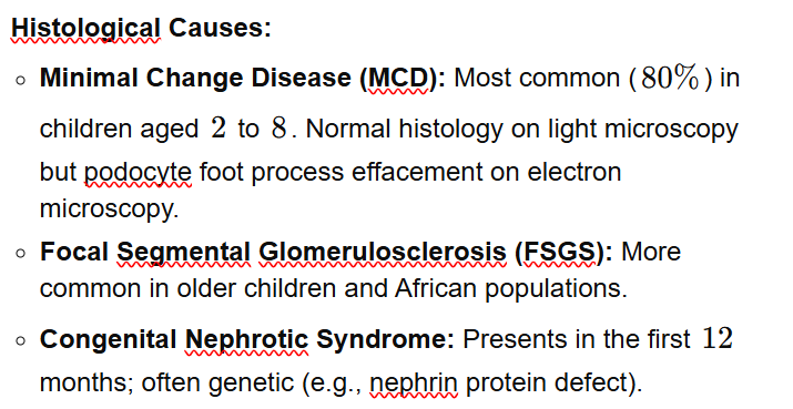
Nephrotic Syndrome Ix (R1)
Glomerular disorder presenting as a triad of: Generalised Oedema, Proteinuria (>200mg/mmol) and Hypoalbuminaemia (<25g/L)
Causes damaged glomerulus to leak proteins (podocytes flatten and leak)
Types: Minimal Change Disease (most common), Congenital Nephrotic Syndromes, Focal Segmental Glomerulosclerosis, Mesangiocapillary glomerulonephritis
RF: Asian ethnicity, M>F, age <4yrs, Varicella Zoster infection (Chicken pox)
Sx: Triad = oedema, proteinuria, hypoalbuminaemia
Generalised oedema: Periorbital (often mistaken for allergy initially), peripheral, ascites, pleural effusions, scrotal/vulval edema
Ix:
First Line: Urine Dipstick (↑↑ protein: 3+/4+), Urine Protein:Creatinine Ratio (>100mg/mmol), UEC, FBE, Serum Albumin (>25g/dm), Varicella Zoster Serology
Atypical Features: Renal Biopsy, Complement Levels, Hepatitis Serology, Anti-streptolysin O Titre (ASOT), Autoimmune Investigations (ANA, ANCA, anti-dsDNA)
Consider Renal Biopsy if Atypical: impaired renal function, frank haematuria, age <1yr/>12yrs, HTN, steroid resistance
Nephrotic Syndrome Mx (R1)
Mx:
First Line: High dose Steroids (Prednisalone), Diuretics if Fluid Overloaded, VZV immunoglobulin/IV Acyclovir if VZV positive
Will usually respond quickly but most will relapse
Long-Term: Low Salt Diet (↓ oedema), Prophylactic Abx (↓ immunoglobulins = ↑ inf. risk), Relapsing Syndromes = low-dose maintenance Steroid Therapy or Immunomod. Drugs (Steroid Sparing Agents = Rituximab, Levamisole, Cyclophsphamide; Calcineurin inhibitors = Ciclosporin, Tacrolimus)
>1 Relapse = Frequently Relapsing Nephrotic Syndrome or Steroid-Resistant Nephrotic Syndrome (SRNS)
Cx: thrombosis/PE, infection → Spontaneous Bacterial Peritonitis (Strep. pneum.)/cellulitis, fluid overload, CKD
Glomerulonephritis (R2)
Glomerular injury w inflammation
Haematuria, Proteinuria ± HTN, Oedema and Renal Impairment
Types/Causes:
Acute Post-Streptococcal (Post-Infectious) GN
Causes: GAS (most common), EBV, CMV, Hep B, endocarditis infection
Features: acute nephritic synd., RPGN, Low C3, Normal C4
Biopsy: ‘starry sky’
IgA Nephropathy (Berger Disease)
Causes: mesangial IgA complex deposits
Features: Synpharyngitic macroscopic haematuria (haematuria during respiratory infections), Acute/Chronic nephritis, Nephrotic syndrome, RPGN (less common)
Biopsy: Mesangial IgA deposition ± C3
Henoch-Schönlein Purpura (HSP) Nephritis/IgA Vasculitis
Causes: Renal disease w same pathological features as IgA nephropathy
Features: haematuria, proteinuria, nephritic syndrome, nephrotic syndrome
Biopsy: Similar to IgA nep. w mesangial IgA deposition
Membranoproliferative GN (MPGN)
Causes: Immune Complex-Mediated = Hep B/C, Autoimmune disease (Lupus etc); Complement-Mediated = C3 nephropathy, Dense Deposit Disease (DDD)
Features: Acute/Chronic GN, Nephrotic syndrome, RPGN, Low C3
Biopsy: "Tram-track" double-contoured capillary walls, Subendothelial/mesangial deposits
Lupus Nephritis
Causes: Immune complex deposition in systemic lupus erythematosus (SLE)
Features: Acute/Chronic GN, Asymp. urinary abnormalities, Nephrotic synd, RPGN
Biopsy: "Full house" staining (IgG, IgA, IgM, C3, C1q)
ANCA-Assoc. GN (Pauci-Immune Vasculitis)
Causes: Granulomatosis w Polyangiitis (GPA), Microscopic Polyangiitis (MPA), Eosinophilic Granulomatosis w Polyangiitis (Churg-Strauss), Renal-limited vasculitis
Features: RPGN, Haematuria, Proteinuria, Acute Nephritis
Biopsy: Pauci-immune necrotising GN, Crescents, Minimal immune deposits
Anti-GBM Disease (Goodpasture Syndrome)
Causes: Abs target type IV collagen in glomerular and alveolar basement membranes
Features: RPGN, Pulmonary haemorrhage
Biopsy: Crescentic GN, Linear IgG staining along the GBM
Membranous Nephropathy
Although often presenting as a nephrotic synd rather than nephritic, it is included among important glomerular diseases
Causes: Primary (often assoc. w PLA2R Abs), Secondary to SLE, Hep B, malignancy
Biopsy: Diffuse GBM thickening, Spike and dome appearance, Granular IgG and C3 along GBM, Subepithelial deposits
RF: recent inf., autoimmune disease, genetics
Sx: macroscopic haematuria, oedema, HTN, proteinuria, renal impairment
Ix: Urine Dipstick, Renal Biopsy, Light Microscopy Histology (does not define disease)
Mx: Supportive therapy (BP control, ACEi/ARB, fluid/salt management), Immunosuppression (steroids, cyclophosphamide, MMF, rituximab), Disease-specific therapy (plasma exchange, complement inhibition, treating infection)
Cx: AKI, CKD, HTN, Fluid Overload/Oedema, Nephrotic Syndrome, Pulmonary Haemorrhage, Thromboembolism

Enuresis (R2)
Bed-wetting
Monosymptomatic Enuresis (MSE): enuresis w/out any other LUTS or Hx of bladder dysfunction
Divided into primary and secondary enuresis
Primary: sustained night-time dryness has never been achieved
Secondary: bedwetting after being completely dry overnight for >6m (new-onset sx after a min. 6m period of night-time dryness)
Non-Monosymptomatic: night-time wetting and at least one daytime lower urinary tract sx
RF: age <4yrs (1 in 3 children at this age), FHx, DM, social environment (vulnerability/trauma), ↑ fluid intake
Daytime bladder control/coordination usually occurs by age 4yrs
Night-time bladder control is not expected until age 5-7yrs
Sx: bed-wetting, nocturia, ?daytime LUTs (urgency, ↑/↓ voiding freq., incontinence, dysuria)
Identify pattern of enuresis episodes (including frequency, timing, volume)
If acute onset (d-wks), consider systemic illness
Secondary/PHx dryness for >6m = consider possible medical, emotional, or physical triggers
Ix: Bladder/Bowel diary, consider dipstick/microscopy if red flags (DM/polydipsia), bloods if faltering growth
Mx: If enuresis is infrequent ± not distressing to the child/parents, tx is not indicated
Education/encourage regular fluids and toileting throughout the day
Bedwetting Alarms (Pad and Bell) → 6-8wks
Desmopressin (Minirin): short-term improvement, indicated when alarm therapy is unsuccessful/CI
Cx: shame/embarrassment/social anxieties
Otitis Media Ix (R1)
Middle ear inflammation and effusion
Acute Otitis Media: effusion is infected (usually a self-limiting viral infection)
Bact. Pathogens: Strep. pneum, Haem. influ, Moraxella catarrhalis
Otitis Media w Effusion: effusion is not infected
RF: age <3, Aboriginal/Torres Strait Islander, low socio-ec, Child Care, Allergic Rhinitis, Struct. Abnormalities
Protective Factors: breastfeeding, avoid supine bottle feeding
Sx: inflammatory sx (pain, bulging/immobile tympanic membrane) + middle ear effusion, otorrhoea (indicates tymp. membrane perforation/effusion), fever, irritability, poor feeding, ear rubbing/tugging
Ix:
First Line: Clinical Exam/Otoscope
Otitis Media w Effusion: loss of lucency of tymp. membrane + immobile, grey-white or blue fluid


Otitis Media Mx (R1)
Mx: usually self-limiting, sx may persist up to 7d
First Line: pain relief (paracetamol/ibuprofen)
Abx only for high risk groups (cochlear implants, immunocomp, remote comms, age <2) = Amoxy
Long-Term: grommets (recurrent AOM), Myringotomy
Cx: sepsis/shock, hearing/speech/learning delays, tympanic membrane perforation, chronic ear discharge, mastoiditis, facial nerve palsy, brain abscess

URTI/Tonsillitis/Sinusitis Ix (R1)
Tonsillitis: inflammation of the palatine tonsils from either a bacterial or viral infection (mostly viral)
May inflame other areas of the mouth → tonsillopharyngitis (pharynx involvement), adenotonsillitis (adenoids involved)
Pathogens: Adenovirus, EBV, GAS (Strep. Pyogenes)
RF: smoke exposure
Sx: odynophagia, fever, ↓ oral intake, halitosis, new onset snoring/apneic eps, SOB, red/inflamed tonsils, white exudate (pus) spots on tonsils, Cervical lymphadenopathy
Large clearly visible tonsils are not always the result of infection (tonsils largest at age 4-8)
Sx b/w 5-7d: >7d indicate glandular fever
Ix:
First Line: Bloods (FBC, LFTs, UEC, CRP/ESR)
Gold Standard: Throat swab, RAT
Throat swabs have limited differential value, may detect S. pyogenes
URTI/Tonsillitis/Sinusitis Mx (R1)
Mx:
First Line: supportive, analgesia, steroids (prednisalone, dexamethasone)
Abx for GAS: benpen, amoxicillin
Long-Term: surgery (Tonsillectomy - use SIGN criteria)
Cx: peritonsillar abscess/Quinsy, spread to deep neck spaces (retropharyngeal, parapharyngeal) → abscess, recurrent tonsillitis, Post-Strep Cx (glomeruloneph, RF)
DDx: Quinsy/peritonsillar abscess (unilat. w swelling most prominent superior to the tonsil), Pharyngitis (sore throat/dysphagia in the absence of tonsillar inflam.), Glandular Fever (specific viral cause of tonsillitis w longer sx duration and assoc. neck swelling/abdo pain)

Conjunctivitis (R2)
Inflammation of the conjunctiva from viral (most common), bacterial and allergic causes
Conjunctiva: clear membrane covering the white part of the eye and the inside of the eyelids
Pathogens: Adenovirus, Staph. A, Strep. pneum, Haem. influ, Neis. gon/C. trachomatis (neonates)
RF: age <5, contaminated exposure (contact, fluids, water/swimming pools)
Sx: red/’pink’ eye, erythema behind eyes, swollen eyelids, excessive tears/production, yellow-green discharge ± crusting, photophobia, ‘gritty’ sensation, itchiness, eye rubbing, coryzal sx
Unilat/purulent = bacterial
Unilat/watery = viral
Bilat/watery = allergic
Gonococcal Conj. (N. gonorrhoeae): severe, green discharge, ulceration, risk of blindness/corneal perf.
Beware Chlamydial Conj (C. trachomatis)
Ix: Clinical Exam, PCR (viral), Culture (bacterial), Consider Allergy testing (allergic conj.)
Mx: Isolate/Keep Home!, Supportive mx
First Line = chloramphenicol, framycetin (drops)
Viral: conservative mx
Allergic: antihistamine
Gonococcal: ceftriaxone, cefotaxime
Chlamydial: azithromycin
Will remain infectious until discharge resolves
Cx: Corneal Infiltration/Ulceration, chronic conjunctivitis, Orbital Cellulitis/tissue spread, Ophthalmia Neonatorum in neonates (from N gon/C. trach → rapid corneal perforation and blindness)
Cervical Adenitis (R2)
Swelling and inflammation of lymph nodes in the neck, typically from viral (most common) or bacterial infections
Abnormal enlargement of LNs in the head and neck usually >1 cm
Pathogens: Staph. A, Strep. pyogenes, EBV, CMV, Rubella, Mycobacterium tuberculosis, Toxoplasmosis gondii
RF: recent travel (tuberculosis exp.), animal exposure (cats), ↓ vaccination
Sx: neck lump, restricted neck movement, LOW, fever, night sweats, bruising, palpable Lymphadenopathy, Features of deep tisse head/neck infection (trismus, muffled voice)
Ix: Clinical Exam, neck US if suspected abscess, CT, CRP/ESR, serology, CXR (if suspected malig.), Biopsy, Tuberculin skin test/Quantiferon Gold (if TB suspected)
Mx: often self-limiting, abx (Cefalexin, Cefazolin), MCS + incision/drainage (if abscess)
Cx: abscess

Orbital Cellulitis (R2)
Dangerous infection inside the eye socket, usually spread from the ethmoid sinus through the thin lamina papyracea (infection posterior to the orbital septum)
Different from preseptal cellulitis, which is limited to tissues anterior to the septum
Pathogens: Strep, Staph. A, Anaerobes
RF: sinusitis, trauma, insect bites, skin/eyelid infections
Sx: Red eye, painful eye movements (inability to move the eye full-range), abscess near the optic nerve, opthalmoplegia, proptosis, ↓ vision, RAPD (Relative Afferent Pupillary Defect), Fever/Systemic sx
Buzzwords: Sinusitis + painful eye movements = think orbital cellulitis
Ix: CT Orbit w Contrast
Mx: surgical drainage, urgent opthal referral, IV abx (ceftriaxone ± metronidazole if sinusitis)
Cx: optic neuritis, optic nerve ischeamia, intracranial cx (meningitis, epidural/subdural abscess, cavernous sinus thrombosis, cerebral asbcess), blindness
Strabismus (R2)
Misalignment of the eyes due to abnormal ocular muscle control
Crossed eyes or squinting eyes
Esotropia: Eyes turning inward (convergent)
Accommodative: most common, long-sightedness (hyperopia) → convergence, required corrective glasses to straighten eyes
Congenital: large inward angle, normal spectacle numbers (no glasses), causes dense ambylopia, surgery shifts muscles
Sixth Nerve Palsy: Esotropia + Diplopia (double vision) → red flag for brain tumour!
Exotropia: Eyes turning outward (divergent)
Intermittent: common, eye turns out from fatigue/loss of focus, 6/6 vision, cosmetic surgery
Strabismus: crossed eyes/"squint"
RF: FHx, prematurity, low birth weight, maternal smoking
Sx: diplopia, eye misalignment, abnormal eye movements, visual confusion, intermittent eye closing
Ix: Clinical Exam (neurological and ophthalmic exams), Simultaneous Prism and Cover Test (SPCT)
Mx: surgery, corrective glasses/patch
Cx: ambylopia (lazy eye → early detection of strabismus is important for prevention!)

Retinoblastoma (R3)
Primary malignant tumor of the retina involving the autosomal dominant RB gene (tumor suppressor gene)
Most common intraocular malignancy of childhood
Potentially fatal (spreads to brain)
RF: genetics
Sx: Leukocoria (white pupil reflection → often noticed in flash photography). Strabismus, Ambylopia, painful eye, red eye, poor vision
A leukocoria is a retinoblastoma until proven otherwise!
Ix: US, MRI, Eye exam under anaesthesia (by opthal), Red Reflex/Slit-Lamp
Mx: Enucleation (removal of affected eye), Chemo, Radiotherapy, Immunotherapy, Frequent screening under anesthesia for at-risk families
Cx: blindness, mets, retinal detachment, glaucoma
Cataract (R3)
Clouding of the crystalline lens causing visual impairment. Congenital or Acquired
Congenital: congenital rubella, genetics, metabolic disorders
Acquired: Juvenile Idiopathic Arthritis (JIA)-associated uveitis, Trauma, Steroid Exposure
RF: steroid exposure, rubella, Down Syndrome, FHx
Sx: leukocoria, poor visual development, strabismus, ↓ fixation in infants, constant glare
Ix: Clinical Exam/Opthalmoscope, Slit-Lamp, B-Scan US
Mx: early surgical removal (prevent ambylopia), Intra-Ocular Lens (IOL → lens replacement)
Mx is dependent on cataract density
Cx: amblyopia (blocked visual input → lazy eye)
Management of the Seriously Ill Child (R1)
Fever:
DDx: Viral infections (most common), bacterial (otitis media, tonsillitis, pneumonia, UTI, meningitis, cellulitis, osteomyelitis), and rarely, malignancy or autoimmune disease
Age-Driven Protocols:
Neonates (<2wks): Highly susceptible to sepsis. Require a "full septic workup" (Lumbar puncture, blood culture, urine test) and empiric antibiotics regardless of appearance
Infants (1-3m): "Limited septic workup." A well-looking infant might get blood and urine tests but potentially no antibiotics; a sick-looking infant receives a full workup including Lumbar puncture and abx
Children (>3m): Selective testing based on whether the child is "sick" vs. "well" and the presence of localized signs
Special Considerations:
Immunosuppressed/Chemotherapy: Fever in a patient on chemotherapy is treated as priority as neutropenic sepsis. Requires immediate blood cultures, empiric abx, and admission until cultures are negative
Returned Travelers: Must screen for Malaria (blood film/ICT test), Dengue fever (serology), Typhoid (Salmonella typhi bacteremia via blood culture), and Hep A
Acute Abdo Pain/Appendicitis:
Appendicitis indicators: Constant worsening pain (rather than intermittent/colicky), pain shifting to the right lower quadrant, and severe localized tenderness
DDx in Adolescent Females: Ovarian torsion or severe ovulation pain (requires US for differentiation)
Respiratory Emergencies:
Croup (Laryngotracheobronchitis):
Inflammation of the larynx, trachea, and proximal bronchi resulting in stridor
Sx: Tracheal tug, subcostal/intercostal indrawing, and a "seesawing" motion of chest/abdomen
Mx: Oral steroids (Dexamethasone, Prednisolone) to reduce swelling. Nebulized Adrenaline in severe cases as a rapid topical vasoconstrictor
Bronchiolitis:
Common in infants >12m
Characterized by diffuse crackles and wheeze
Mx: Supportive care only. No specific pharmacological treatment. Includes oxygen therapy and hydration (nasogastric or IV) if the infant is too breathless to feed
Asthma:
Severity graded as mild, moderate, or severe/life-threatening
Mx: Inhaled bronchodilators (‘hour of power‘), oral/IV steroids. Severe cases may require BiPAP or PICU admission
Education: Essential components include spacer technique and a written asthma management plan
Anaphylaxis/Allergic Reactions
Food Allergy (Immune-Mediated): Reactions stimulated by the immune system
IgE-Mediated: immediate onset (within minutes to 1-2hrs → typically <1hr). Caused by the IgE Ab responding to a specific food
Process: Sensitisation/First Exposure → Re-Exposure and Effector Phase → Reaction
Non-IgE Mediated: Mediated by aberrant T-cell lymphocytes or innate immunity. Sx are delayed (occur >1-2hrs, up to days/wks later)
Ix: Skin Prick Test (SPT), Serum Specific IgE (ELISA), Oral Food Challenge (OFC)
Anaphylaxis: Acute onset of hypotension, bronchospasm, or upper airway obstruction OR skin features (urticaria, angioedema) combined w respiratory, cardiovascular, or severe GI sx
Mx:
Remove the allergen, DRSABCD
First Line Mx: Adrenaline
Dose: 0.2-0.3ml (based on weight/age), repeated every 5min if needed
EpiPen Jr: 150mcg, weight <20kg
EpiPen: 300mcg, weight >20kg
Note: Steroids and antihistamines are not primary treatments for acute anaphylaxis and have no proven effect on the immediate reaction
Action Plans:
Green Plan: mild-moderate allergic reactions
Red Plan: for anaphylaxis (includes EpiPen instructions)
Common Fractures (R1)
Common Fractures: Wrist, Forearm (Buckle, Transverse), Supracondylar (elbow), Clavicle, Ankle
Supracondylar Fracture: Type 3 (fully displaced) is an orthopedic emergency as it can compromise the brachial artery, becoming a limb-threatening injury. Usually requires pin fixation and immediate surgery
Analgesia Mx Options:
Physical: Splinting, ice, elevation
Pharmacological: Paracetamol, Ibuprofen, Oxycodone
Intranasal Fentanyl: Proved equivalent in efficacy to IV Morphine for severe pain from fractures or burns
Ketamine: A dissociative anesthetic used for procedural sedation (e.g., straightening a deformed limb or dressing severe burns)
Meningitis Ix (R1)
Inflammation of the meninges surrounding the brain and spinal cord
May be bacterial, viral (aseptic), fungal, or tuberculous
Bacterial meningitis is a medical emergency requiring immediate treatment
Pathogens: Pneumococcus, Meningococcus/Neisseria meningitidis, Haem influenzae type B; Neonates = E. coli, GBS, Listeria monocytogenes (rare)
RF: age <5yrs, immunocomp/unvaccinated, asplenia (no spleen), maternal GBS, cochlear implant/med device
Sx: Fever, maculopapular non-blanching rash, neck stiffness, irritability, bulging anterior fontanelle, N/V, photophobia, lethargy, seizures, poor feeding, headache, Kernig/Brudzinski signs
Note: neck stiffness sign is unreliable in age <18m
Neonates/children <2m often present with nonspecific signs of sepsis rather than the classical signs of meningitis
Ix:
First Line: bloods (CRP/ESR, CBC, UEC)
Gold Standard: CSF/Lumbar Puncture
Bacterial CSF: Cloudy/turbid, polymorph (neutrophil) predominance (except Listeria → lymphocyte predominant), ↑ protein, ↓ glucose
Viral CSF: Clear, lymphocyte predominance (polymorphs may appear early), normal protein, normal glucose
Tuberculosis CSF: opalescent CSF, ↑↑↑ lymphocytes, ↑↑ protein, ↓↓ glucose
Consider: neuroimaging if signs of ↑ intracranial pressure or focal neurological deficits
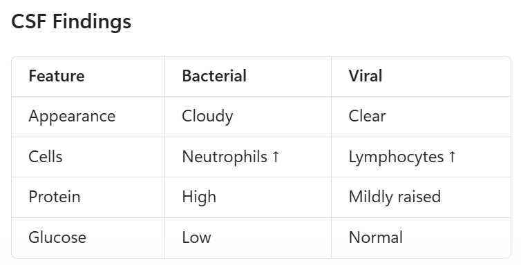
Meningitis Mx (R1)
Mx: Fever w purpura should be treated as meningococcal disease until proven otherwise
First Line: Immediate IV Abx!, Supportive Care/ICU, Seizure Mx, Consider Dexamethasone
Abx: Gent/Tobramycin + Benpen/Amoxy/Amp
Empirical >2m: Cef/Cefotaxime + Dexamethasone ± Vanc (S. pneum/Cocci)
Hos-Acq N/<2yrs: Cefotaxime/Ceft + Vanc
Com-Acq N/<2yrs: Gent/Tobr + Cef + Amoxy
Ensure CSF has been done prior to Abx Mx
Cx:
Acute: Convulsions (often suggesting late presentation), ↑ intracranial pressure, Syndrome of Inappropriate Antidiuretic Hormone (SIADH), cerebral oedema
Long-term: Sensorineural hearing loss (most common measurable cx), ongoing convulsions/epilepsy, motor deficits, neurological sequelae
Sepsis/Septicaemia Ix (R1)
Sepsis: life-threatening organ dysfunction caused by a dysregulated host response to infection
Septicaemia: bloodstream infection
Pathogens: N. meningitidis, Strep. pneum, S. pyo, GBS (neonates), E. coli, Staph infections (Staph. A), HSV (N)
RF: age <3yrs, immunocomp, preterm, indwelling cath, recent trauma/infection
Sx: fever/hypothermia, tachycardia, irritability, poor feeding, lethargy, Hypotension, ↓ GCS, ↓ urine output, mottled skin, petechiae/purpura, shock, ↓ cap. refill
Ix:
First Line: Blood Culture, FBE, CRP/ESR, Urinalysis, Blood Gas/Lactate, Consider Lumbar Puncture
Gold Standard: Culture/Sensitivity Testing

Sepsis/Septicaemia Mx (R1)
Mx:
First Line: Resus, Early IV Abx, Vassopressors (if in shock), Control/Adress Source
Abx: Gent/Tobra, Cefo/Ceft, Vanc
Neonates: Cefotaxime + Benpen
Com-Acq (>2m): Gent/Tobra + Cefo/Ceft + Vanc
Hosp-Acq (>2m): Piptaz + Vanc
Hosp-Acq (N): Gent/Tobra + Fluclox/Vanc
Suspected HSV Encephalitis: add Aciclovir
Suspected Meningitis: add Dexamethasone
Toxic Shock Synd: Clindamycin + IVIg
Cx: DIC, Multi-organ failure, death, limb loss (meningococcaemia), Toxic Shock Syndrome
Toxic Shock Syndrome: toxin-producing Strep. pyogenes infection causing hypotension + 2 of: kidney impairment, coagulopathy, resp. distress hyperbilirubinaemia, rash/soft tissue necrosis
Immunisation (R1)
Inactivated Vaccines:
Contain dead organisms or subunits
Require more doses for an immune response
Adverse events occur early
Includes: Inactivated Polio (IPV), Subunit (Pertussis, Influenza), Toxoid (Diphtheria, Tetanus), Conjugate (Pneumococcal)
Live Attenuated Vaccines:
Contain weakened whole organisms
Often require fewer doses and multiply in recipient
Adverse events often delayed (e.g., MMR fever/rash at 7-10d)
Recipient may be infectious to others (e.g., Oral Polio)
Includes: MMR, Varicella, Rotavirus, BCG (Bacillus Calmette-Gurin)
Australian Schedule (Example Highlights):
Birth: Hepatitis B
2, 4, 6 Months: Hexavalent (DTP, Polio, Hep B, Hib), Pneumococcal, Rotavirus
12 Months: MMR, MenACWY, Pneumococcal
18 Months: MMRV, DTP, Hib (Haem. influ)
Pregnancy: Funded Pertussis and Influenza vaccines to provide passive protection for the baby in the first 6m
Annual Influenza: from 6m
Additional vaccines for Aboriginal and Torres Strait Islander children and special-risk groups
Kawasaki Disease Ix (R1)
Acute systemic medium-vessel vasculitis of childhood
Mucocutaneous Lymph Node Syndrome
Rare but critical disease that affects coronary arteries → leads to coronary artery aneurysms if left untreated
RF: age <5yrs (6m-4yrs), Asian ancestry, M>F
Sx: Diagnostic criteria, irritability, sterile pyuria, aseptic meningitis, arthritis, abdo pain, uveitis, diarrhoea
Diagnostic Criteria: Fever for >5d PLUS 4 of: Bilateral non-purulent conjunctivitis, oral/mucous membrane changes (e.g., strawberry tongue, lip cracking), extremity changes (swelling/erythema, later desquamation), polymorphous rash, cervical lymphadenopathy (>1.5cm → typically unilat)
Warm CREAM: Warm = Fever 4-5d, Conjuncitvitis, Rash, Erythema/Edema of Extremeties, Adenopathy (cervical LN), Mucosa/Mucus Membranes
Ix:
First Line: CRP/ESR, FBE (↑ Neutrophils, Anaemia, Thrombocytosis, Leukocytosis), UEC/LFTs, ECG, Urinalysis, consider Septic Workup (CSF, bloods etc)
Gold Standard: Clinical Exam (Dx Criteria), Echocardiogram (coronary artery lesions)
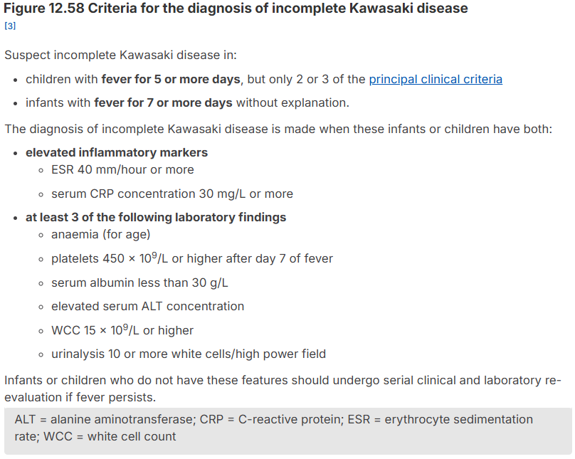
Kawasaki Disease Mx (R1)
Mx:
First Line: Aspirin!, IV Immunoglobulins (IVIG → ↓ aneurysm risk and inflammation/if severe)
Consider: corticosteroids (Methylprednisalone, Prednisalone) or biologics
Long-Term: Echocardiogram follow-up
Cx: coronary artery aneurysms/damage
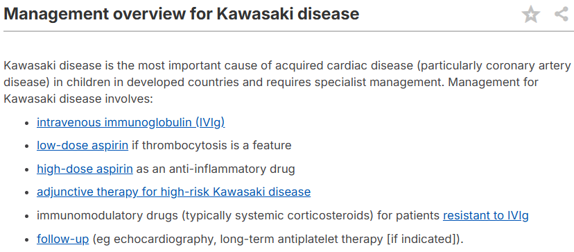
Childhood Viral Infections (R2)
Measles (Rubeola):
Clinical triad: Cough, Coryza, Conjunctivitis
Plus: Koplik spots, High fever, Descending maculopapular rash
Cx: Pneumonia, Encephalitis, Subacute sclerosing panencephalitis (SSPE)
Prevention: MMR vaccination
Rubella:
Sx: Mild fever, Tender posterior cervical/postauricular lymph nodes, Pink facial rash spreading to trunk
Cx: Major concern is congenital rubella syndrome in pregnancy
Roseola (HHV-6):
Sx:
High fever for 3-5 days
Fever resolves then rash appears
Rash begins on trunk and spreads outward
Cx: Common cause of febrile seizures
Parvovirus B19 (Fifth Disease):
Sx: "Slapped-cheek" rash, Lacy reticular rash on limbs/trunk
Cx: Aplastic crisis, Fetal hydrops
Hand Foot and Mouth Disease:
Pathogens: Coxsackie virus, enterovirus
Sx: Oral ulcers, Vesicles on hands, feet and buttocks
Mx: Usually self-limiting
Cx (rare): Meningitis, Encephalitis
Varicella (Chickenpox):
Sx: Generalised pruritic vesicular rash, Lesions in different stages
Mx: Usually self-limiting, give immunoglobulins to neonates/infants to prevent systemic disease
Cx: Secondary bacterial infection, Encephalitis, Pneumonia
Preventable by vaccination
Viral Hepatitis: Hep A, B, C (R3)
Hepatitis A:
Transmission: Faecal-oral route, Contaminated food/water, Household spread
Sx: Children often asymp, Fever, Malaise, Jaundice, N
Mx: Supportive only
Prevention: Vaccination recommended for risk groups (incorporated in some Aboriginal and Torres Strait Islander childhood programs)
Hepatitis B:
Transmission: Vertical (mother-to-child), Blood exposure, Sexual transmission
Sx: Usually asymp. in childhood
Cx: Infants are at high risk of chronic infection and later Cx (Cirrhosis, Hepatocellular carcinoma)
Prevention: Birth-dose Hep B vaccine within 24hrs, Routine infant immunisation schedule
Hepatitis C:
Transmission: Vertical, Blood-borne exposure
Sx: Usually asymp in childhood
Cx: Chronic hepatitis, Cirrhosis, Hepatocellular carcinoma
Mx: Direct-acting antiviral agents in eligible children
Currently no available vaccine
Dengue (R3)
Mosquito-borne flavivirus infection transmitted by Aedes mosquitoes
RF: tropical/subtropical regions (Southeast Asia, Pacific Islands Northern Australia)
Sx:
Classic Dengue: Sudden high fever, Severe headache, Retro-orbital pain, Myalgia ("break-bone fever"), Arthralgia, Rash
Warning Signs:, Persistent vomiting, Abdo pain, Mucosal bleeding, Lethargy, Hepatomegaly
Severe Dengue: Plasma leakage, Shock, Severe bleeding, Organ dysfunction
Ix: FBC (thrombocytopenia), Haematocrit rise, Dengue NS1 antigen/PCR, Serology
Mx: Supportive Mx, Careful fluid Mx, Monitor for shock
Avoid NSAIDs and aspirin due to bleeding risk
Use Prevention Measures: mosquito bite avoidance, vector control, selected vaccine in specific endemic areas
Cx: dengue shock syndrome/DSS (Rapid fluid loss from blood vessels causes dangerously low blood pressure and poor blood flow), plasma leakage, severe bleeding/hemorrhage, fluid accumulation (e.g. pleural effusions), single/multi-organ dysfunction (acute liver failure, myocarditis, encephalopathy, AKI)
Iron Deficiency Anaemia Ix (R1)
Inadequate iron stores for haemoglobin synthesis result in nutritional deficiency and anaemia
Most common cause of anaemia in childhood
RF: age >12m + teens, F>M (menstruating), prematurity, LBW, excess intake of cow’s milk (>500ml/d), breastfeeding w/o iron supplements, poor dietary iron intake, adolescent menstruation, vegetarian diet, low socioeconomic status
Sx: often asymp., fatigue, pallor, irritability, ↓ exercise tolerance, ↓ concentration, Pica, ↓ school performance, behavioural/developmental delay
Ix: screen for more sinister causes of anaemia!
First Line: FBE (↓ Hb, ↓ MCV = microcytic, ↑ RDW)
Gold Standard: Iron Studies (↓ Ferritin = most sensitive marker, ↓ serum iron, ↑ transferrin/TIBC)
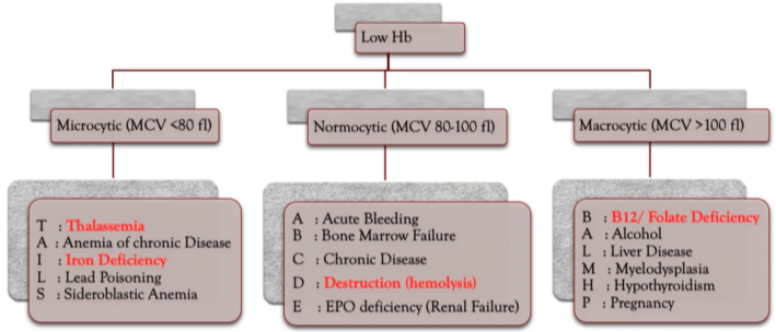
Iron Deficiency Anaemia Mx (R1)
Mx: treat underlying cause!
First Line: Oral Iron (ferrous sulfate), dietary counselling
Long-Term: Continue iron therapy for ~3m after Hb normalises to replenish stores
Cx: Neurodevelopmental impairment, Learning difficulties, Cognitive/behavioural deficits
Acute Lymphoblastic Leukaemia/ALL Ix (R1)
Malignant clonal proliferation of lymphoid precursor cells within the bone marrow
Most common (25%) childhood cancer. B cell ALL is most common form. Good prog. in ages <11yrs
Leukaemic Blasts infiltrate bone marrow, blood, liver, LNs, spleen, CNS, testes → bone marrow failure
RF: age 2-5yrs, M>F, genetic abnormalities (Trisomy 21)
Buzzwords: primary school age (rarer and worse prognosis in older ages >11yrs)
Sx: bone pain (hyperproliferation causes pressure in bone marrow → pain) + pallor/anaemia, petichial bruising (thrombocytopenia), systemic/B sx (fever, sweats, LOW)
Bone Marrow Failure: pallor (anaemia), fatigue, bruising, petechiae, mucosal bleed, recurrent inf.
Infiltrative Sx: bone pain, limp, lymphadenopathy, hepatosplenomegaly, enlarged testicles, CNS sx
Deficiency in Main 3 Cell Lines = anaemia (lethargy, pallor), thrombocytopaenia (easy bruising/bleeding), leukopaenia (fevers/infections)
Ix:
First Line: FBE (anaemia, thrombocytopenia, abnormal WCC), Peripheral Smear (blasts), CXR (mediastinal mass)
Gold Standard: Bone marrow aspirate/biopsy (>25% lymphoblasts), Flow Cytometry (identify markers)
Leukoerythroblastic Picture: The presence of nucleated red cells indicates a primary bone marrow pathology (bone marrow failure or infiltration)
Consider: cytogenetics (risk stratification), lumbar punc.
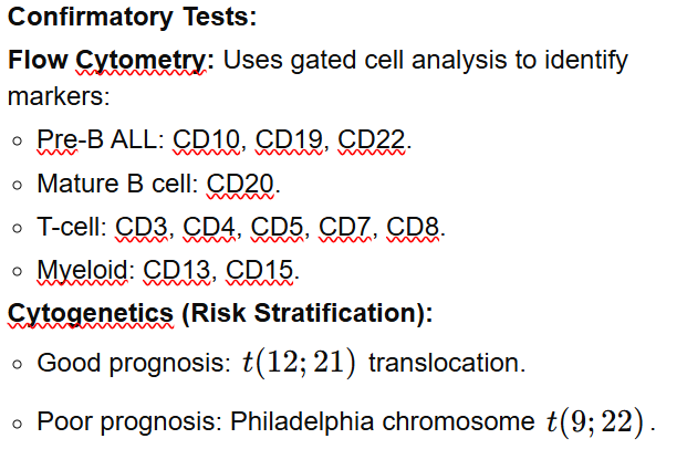
Acute Lymphoblastic Leukaemia/ALL Mx (R1)
Mx: steroids, chemo, radiation therapy, CNS prophylaxis, stem cell transplantation (selected pts), blood products
Note: do not give steroids until biopsy has confirmed ALL as these would affect accuracy of diagnostic testing
Prognosis: Current cure rates exceed 85-95% in developed countries for moderate disease (worse in severe disease/older pts)
Cx: Infertility, avascular necrosis, peripheral neuropathy, anxiety
ITP/Immune Thrombocytopaenic Purprura Ix (R1)
Autoimmune destruction of platelets causing isolated thrombocytopenia, often following viral infection
Isolated low platelet count of <100 x 109/L in an otherwise well child with an otherwise normal FBE and film
Most common acquired bleeding disorder in children
Persistent ITP = 3-12m; Chronic ITP = >12m
RF: age 2-7yrs, viral inf., recent live virus immunisation (e.g. MMR), FHx autoimmune, recent med changes
Sx: otherwise well child with: petichiae, purpura, easy bruising, epistaxis, gingival bleeding
Importantly no: hepatosplenomegaly, lymphadenopathy, constitutional sx (otherwise consider leukaemia)
Typically sudden onset sx
Ix: dx of exclusion!
First Line: Peripheral Smear (normal RBCs/WBCs, may have large platelets)
Gold Standard: FBE (isolated thrombocytopenia = ↓ platelets)
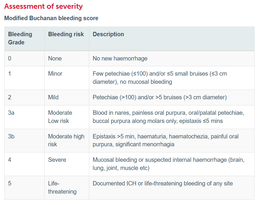
ITP/Immune Thrombocytopaenic Purprura Mx (R1)
Mx: Decision to treat a child is based on clinical fts, not platelet count → majority do not require mx
Goal of mx is to stop active bleeding
First Line: Observational/Conservative Mx
Significant Bleeding: 1 = Corticosteroids (Prednisalone), 2 = IVIG, Anti-D immunoglobulin
Epistaxis: oral tranexamic acid
Life Threatening: thrombopoietin receptor agonists (Romiplostin, Eltrombopag)
Prognosis: ~80% recover within 6-12m, s mall proportion develop chronic ITP
Cx: Chronic ITP, Risk of Intracranial Haemorrhage is very low (<1%)

Lymphoma (R2)
Malignant proliferation of lymphoid tissue
Types:
Hodgkin Lymphoma: more common in adolescents, good prognosis (>90% survival)
Non-Hodgkin Lymphoma: more common in younger children, aggressive but highly treatable
RF: M>F, older aged children, EBV infection, immunosuppression
Sx: lymphadenopathy (persistent, firm, non-tender, rubbery, enlarging), B Sx (fever, night sweats, LOW), fatigue, pruritus, hepatosplenomegaly, cough/dyspnoea or airway obstruction (from mediastinal mass or SVC obstruction), supraclavicular nodes (SC lymphad. is pathological until proven otherwise!)
Buzzwords: (left) Indolent Neck Mass (H-L)
Ix: Excisional LN/Large Core Biopsy = gold standard (for flow cytometry), FBE, ↑ LDH, ESR/CRP, CXR (hilum/mediastinal prominence), CT/PET scan
Note: Needle biopsy is discouraged
Mx: chemotherapy, radiotherapy, immunotherapy
Consider stenting if SVC compromises airway
Hydration ± Allopurinol/Rasburicase for tumour lysis syndrome
Cx: tumour lysis syndrome (in lymphomas w rapid cell turnover → ↑ P3-, K+, Ca2+ → kidney damage), alopecia, neutropenia, sub-fertility
