Chapter 11: Fundamentals of the Nervous System and Nervous Tissue
 Call Kai
 Learn
 Practice Test
 Spaced Repetition
 Match
 Flashcards
 Knowt Play
1/58

Earn XP
Description and Tags
Exam 4
59 Terms
Nervous system
Master controlling and communicating system of the body
regulates and coordinated all body activities
cells communicate via electrical and chemical signals:
rapid and specific
usually results in immediate responses
Functions of nervous system
sensory input: sensory receptors monitor changes in internal/external environments
integration: process and interpret sensory input
motor output: activates effector organs (muscles and glands) -→ response to stimuli
Central nervous system (CNS)
the brain and spinal cord
interprets sensory input and dictates motor output based on reflexes
integration and control center
Peripheral nervous system (PNS)
the nerves and ganglia outside of CNS
cranial nerves(12)
spinal nerves(31)
ganglia
acts as a communication highway, connecting your brain and spinal cord to your limbs, skin, and internal organs
Spinal nerves (31)
impulses to and from spinal cord
Cranial nerves (12)
impulses to and from brain
Sensory division (afferent) of PNS
somatic and visceral sensory nerve fibers
conducts impulses from receptors towards CNS
almost all neurons are unipolar
cell bodies are located in ganglia outside of CNS
Somatic sensory fibers
Impulses from skin, skeletal muscles, and joints to CNS
Visceral sensory fibers
impulses from visceral organs to CNS
Motor division (efferent) of PNS
motor nerve fibers
conducts impulses away from the CNS to effectors (muscles and glands)
multipolar neurons
most cell bodies are located in CNS (except some autonomic neurons)
Somatic nervous system of motor division
somatic motor (VOLUNTARY)
conducts impulses from the CNS to skeletal muscles
Autonomic nervous system (ANS) of motor division
visceral motor (INVOLUNTARY)
conducts impulses from CNS to cardiac muscle, smooth muscle, and glands
Sympathetic division of ANS
mobilizes body system during activity
fight or flight
speed
stress
Parasympathetic division of ANS
conserves energy
promotes house-keeping functions during rest
rest and digest
Neuron (nerve cell)
excitable cells that transmit electrical signals
extreme longevity
amitotic-lose ability to divide; cant be replaced
high metabolic rate: requires continuous supply of O2 and glucose
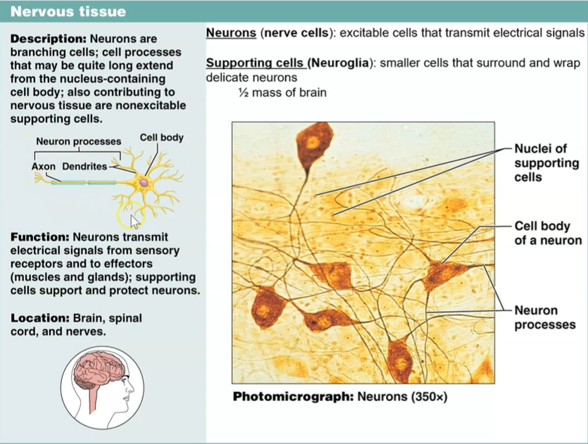
Neuroglia/glial cells (supporting cells)
smaller cells that surround and wrap delicate neurons
dark staining nuclei
outnumber neurons in CNS 10:1
6 types of glial cells: 4 in CNS and 2 in PNS
functions:
provide supportive scaffolding four neurons
segregate and insulate neurons
guide young neurons to their proper connections
promote health and growth of neurons

Astrocytes (CNS)
most abundant, versatile, highly branched glial cells
radiating processes cling to neurons and their synaptic endings, and cover capillaries
satellite cells of PNS
functions:
support and brace neurons
anchor neurons to their nutrient supplies (capillaries); supply nutrients themselves to neurons
guide migration of young neurons
aid synapse formation
monitor exchange and permeability of capillaries
“mop up” leaked K+; recapture/recycle released neurotransmitters
connected by gap junctions, can respond to nearby nerve impulses

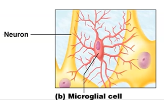
Microglia cells (CNS)
immune defense
small, oval cells with spiny processes →immune
processes touch nearby neurons
follow chemotaxis → damages cell send chemicals
migrate towards damaged neurons and phagocytize microorganisms and neural debris
important because cells of immune system are denied access to CNS
Ependymal cells (CNS)
aid in production of CSF
Line the central cavities of brain and spinal cord
often ciliated →move CSF; nourishes neurons
range from squamous to columnar

Oligodendrocytes (CNS)
branches cells that form insulating myelin sheaths around CNS nerve fibers

Schwann cells (PNS)
surround fibers forming myelin sheaths of PNS

Satellite cells (PNS)
surround neuron cell bodies in PNS

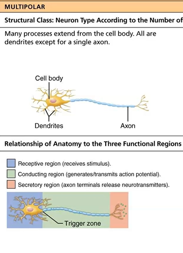
Dendrites
Carry message into body of neuron; receptive regions
Can contain 100s of short, tapering, diffusely branches processes
Large surface area to receive signals from other neurons
incoming messages are usually not action potentials
they are graded potentials: short distance signals
Axon hillock
propagation of action potential
Axon
impulse-generating and conducting region; carry info away
generates AP along axolemma to axon terminal
some axons are 3-4 feet long
long axons are called nerve fibers
conducting region of neuron
axon collateral: a side branch that splits off from the main shaft of an axon
axons usually branch profusely at their end: terminus
10,000+ terminal branches
distal endings= axon terminals→region that secretes neurotransmitters into extracellular space
Anterograde movement: movement away from cell body: mitochondria, cytoskeletal elements, renew axon
Retrograde movement: movement towards cell body: organelles to be recycled/destroyed in cell body
Myelin sheath
whitish, fatty segmented sheath found around axons
not continuous
functions:
protect axon
electrically insulate fibers from one another
increase speed of nerve impulse transmission
axons can be myelinated or unmyelinated
Myelination in PNS
formed from Schwann cells
cytoplasm is squeezed from between the membrane layers
few proteins in membranes (good insulators)
encloses axon w/ plasma membrane
has concentric layers of membrane that make up myelin sheath

Myelination in CNS
formed by oligodendrocytes
each cell can wrap up to 60 axons at once
myelin sheath gap in present
white matter: regions of brain and spinal cord w/ dense collections of myelinated fibers
gray matter: mostly neuron cell bodies and nonmyelinated fibers
Nodes of Ranvier
gaps between adjacent Schwann cells
axon collaterals can emerge
nerve cell body
usual organelles but NO centrioles (amitotic)
biosynthetic center of neuron
synthesizes proteins, membranes, chemicals
rough ER (Nissl bodies)
spherical nucleus with nucleolus
most neuron cell bodies are in CNS where they are protected by skull and vertebral column
contains axon hillock
Neuron processes
armlike extensions from cell body
CNS contains neuron cell bodies and their processes
PNS contains only neuron processes
Two types of processes
dendrites
axons
Nuclei
Cluster of neuron cell bodies in CNS
Ganglia
Cluster of neuron cell bodies in PNS
Tract
Bundle of axons in CNS
Nerve
Bundle of Axons in PNS
Multipolar neurons
3+ processes (1 axon, 2+ dendrites)
most common and major neuron type in CNS
interneurons
motor
Interneurons
lie between motor and sensory neurons in neural pathways
confined to CNS
multipolar
99% of body’s neurons are interneurons
Bipolar neurons
2 processes (1 axon, 1 dendrite)
1 axon and 1 dendrite extend from opposite sides of the cell body
rare (ex: retina, olfactory)
sensory

Unipolar
one T-like process (2 axons)
peripheral and central processes
mainly in ganglia of PNS
Sensory

Leakage (nongated) channel
always open but selective to the ion they let in
Chemically gated/ligand gated channel
open with binding of a specific chemical
Voltage gated channel
open and close in response to changes in membrane potential
Mechanically gated channels
open and close in response to physical deformation of receptors (i.e. touch or pressure receptors)
Resting membrane potential
the potential difference (-70mV) across the membrane of a resting neuron
ionic differences are the consequence of:
differential permeability of the plasma membrane to Na+ and K+
differences in ionic composition of ICF and ECF
At rest:
membrane is slightly permeable to Na+
membrane is 75x more permeable to K+
membrane is freely permeable to Cl-, which balances the Na+ and K+ charge
Generating the resting membrane potential
differences in plasma membrane permeability
more K+ diffuses out than Na+ diffuses in
as result, the inside of cell is more negative
establishes resting membrane potential
sodium potassium pump (Na+/K+ ATPase) stabilizes RMP
maintains gradients for Na+ and K+
3 Na+ are pumped out: 2 K+ are pumped in
Graded potential
incoming short distance signals
decrease in intensity with distance
magnitude varies directly with the strength of the stimulus
triggered by change in environment (stimulus) that causes gated ion channels to open
essential for initiating action potentials
a graded potential can become an action potential but a action potential cannot become a graded potential
receptor potential
postsynaptic potential
Receptor potential
a local, graded change in the membrane potential of a sensory receptor cell caused by sensory transduction (the conversion of an environmental stimulus—such as light, sound, pressure, or temperature—into an electrical signal)
Postsynaptic potential
a temporary, graded change in the electric polarization of a postsynaptic neuron's membrane caused by the binding of neurotransmitters to receptors. These electrical changes propagate passively toward the axon hillock, where they determine whether the neuron fires an action potential
Action potential
long distance signals of axons
only occur in excitable membranes (neurons and muscle cells)
a brief reversal of membrane potential with a total amplitude of 100mV
action potentials do not decrease in strength over distance
in neurons, the AP is generated in the axon
a stimulus changes the permeability of the neurons membrane by opening specific voltage gated ion channels on the axon

depolarization
reduction in membrane potential; inside of membrane becomes less negative (moves closer to 0 mV) than resting
ex: -70mV → 45 mV
Repolarization
membrane returns to its resting membrane potential
ex: 45mV → -70mV
Hyperpolarization
inside of the membrane becomes more negative (moves further from 0mV) than resting
ex: -70mV → -80mV
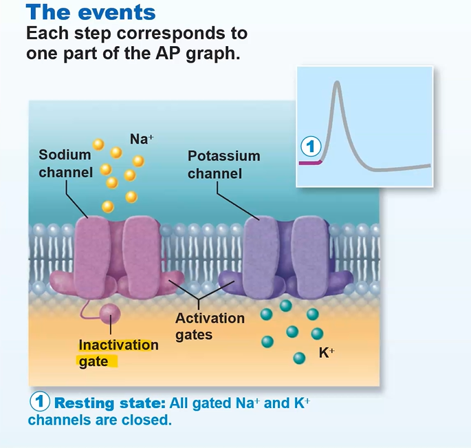
Action potential: 1. Resting state
Na+ and K+ channels are closed
Leakage accounts for small movements of Na+ and K+
Each Na+ channel has two voltage-regulated gates:
Activation gates- closed at resting state; open at depolarization
Inactivation gates- open at resting state; prevents more Na+ enter
maintaining -70mV
depolarization opens and then inactivates Na+ channels
both must be open for Na+ to enter
only one must be closed to close Na+ channels
Action potential: 2. depolarization phase
Na+ permeability increases
Na+ gates are opened; K+ gates are closed
Threshold- a critical level of depolarization (-55mV to -50mV)
at threshold, depolarization becomes self-generating an progresses by positive feedback → all Na+ channels open

Action potential: 3. Repolarization phase
Slow Na+ inactivation gates close
A) Membrane permeability to Na+ declines to resting levels
AP spike stops rising
B) As sodium gates close, slow voltage-sensitive K+ gates open
K+ exits the cell and internal negativity of the resting neuron is restored (-70 mV)

Action potential: 4. Hyperpolarization
K+ gates remain open, causing an excessive efflux of K+
the neuron is insensitive to stimulus and depolarization
Na+ channels also begin to reset
