X-Ray Crystallography for Protein Structure Determination
 Call Kai
 Learn
 Practice Test
 Spaced Repetition
 Match
 Flashcards
 Knowt Play
1/45

There's no tags or description
Looks like no tags are added yet.
Name | Mastery | Learn | Test | Matching | Spaced | Call with Kai | Chat |
|---|
No analytics yet
Send a link to your students to track their progress
46 Terms
Protein Structure Determination
When we talk about structure in this lecture we are talking about tertiary and quaternary structures
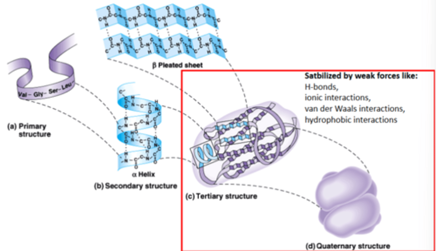
Why do you want to determine protein structure? What does it help us do?
• Understand precise mechanism of protein function
-Predict and engineer protein function
-Depict regulatory conformational changes
• Understand mechanism of action of drugs
What are the 3 ways to determine protein structure?
1. Experimental determination (X-ray crystallography, NMR, Cryo EM)
2. Comparative modeling (based on homology of your protein of interest compared to another protein in nature with a known structure)
3. Ab initio (de novo) prediction-computer prediction of structure
-CryoEM-used for larger proteins
What are the 2 experimental approaches to determine protein structure?
1. X-ray crystallography
2. NMR
X-Ray Crystallography
-What is it used to determine? What percentage?
-What 2 things does it require?
-How sensitive is it/what can it detect? What sized proteins?
-What was the earliest protein structure found?
- Was used to determine 80% of known protein structures
- Requires high protein concentration
- Requires crystals
- Able to trace amino acid side chains
- Earliest structure solved using this method was myoglobin
- Can find structure of any protein regardless of its size
NMR
-What size proteins can it detect?
-How does it work?
- Magnetic field applied to proteins in solution
- Does not require crystallization
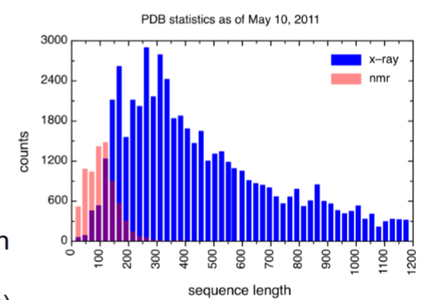
- The largest structures you can find with this method are 350 amino acids (40 kD)-if your protein is bigger than this you can't find the structure using NMR
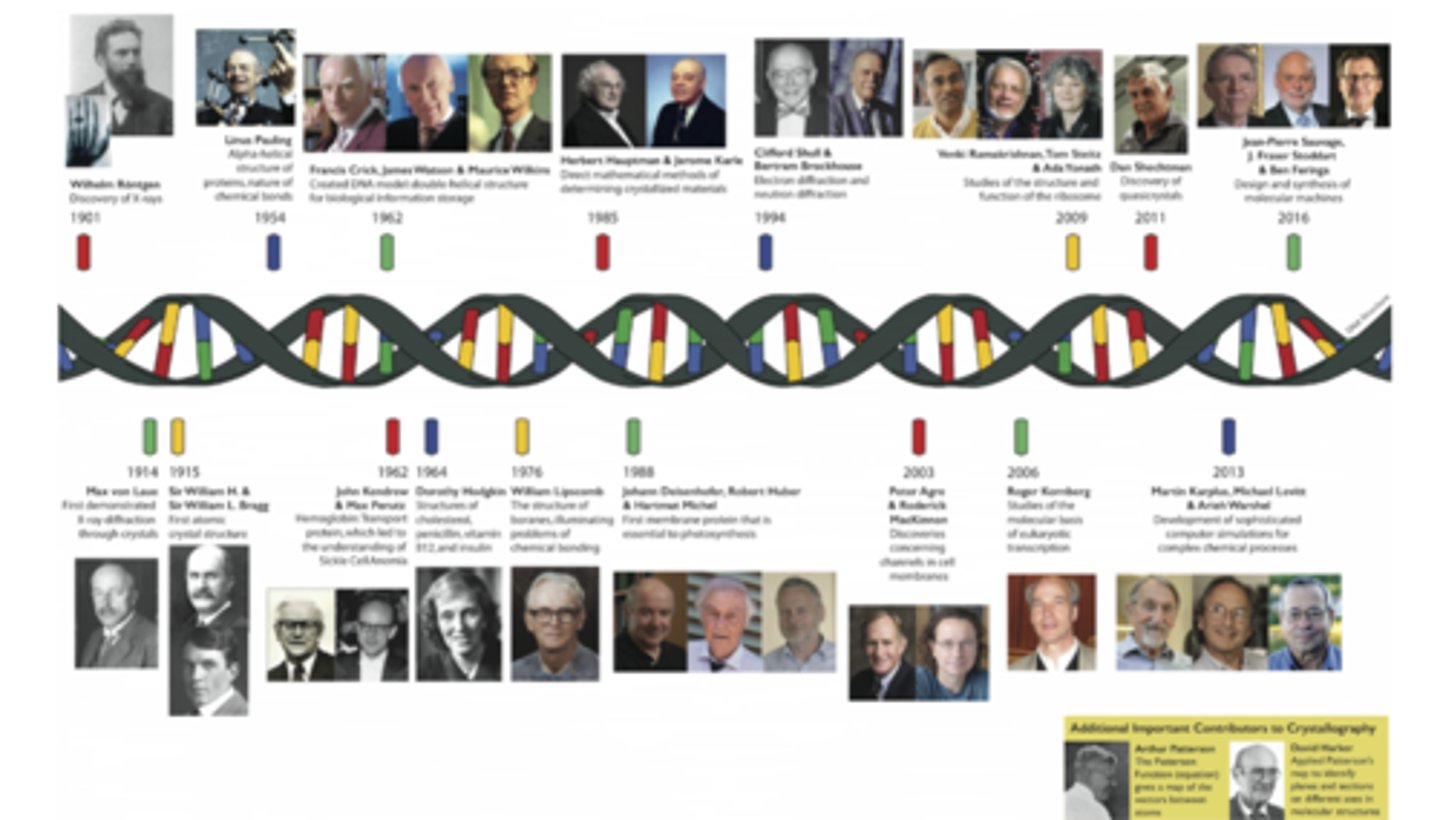
Nobel Prizes Awarded to Crystallographers
-How many were awarded nobel prizes?
30 were awarded nobel prizes
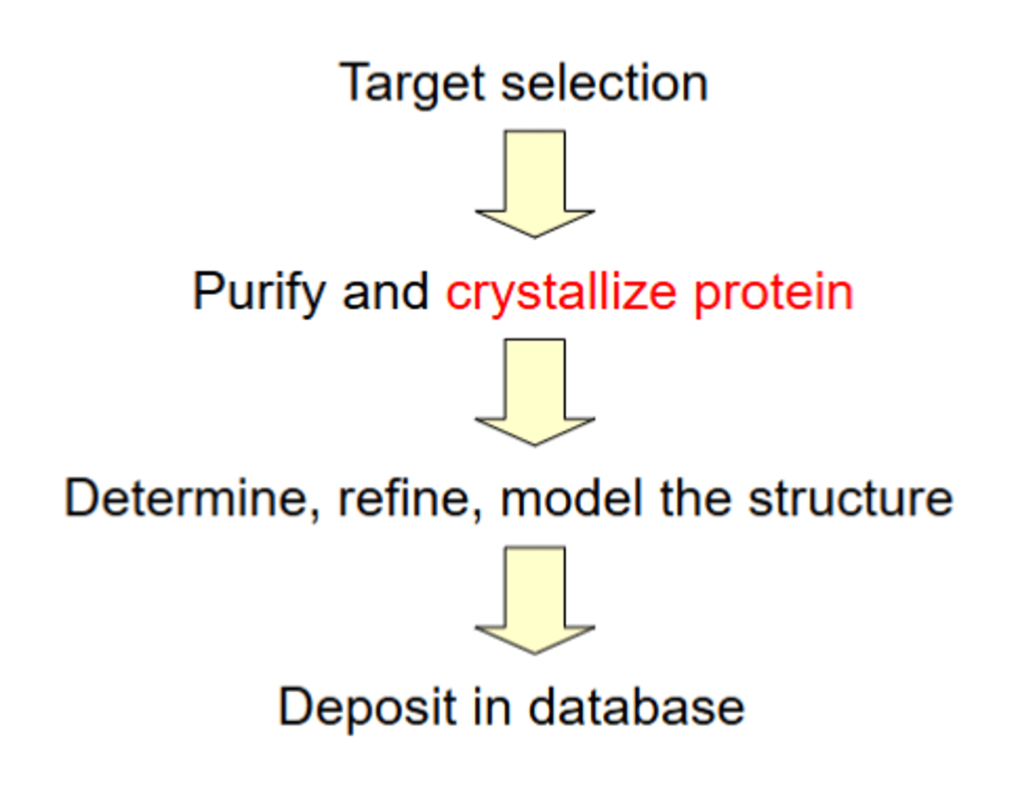
4 Broad Steps in Obtaining Protein Structure
1. Target selection
2. Purify and crystallize protein
3. Determine, refine, model the structure using x ray crystallography
4. Deposit in database
Protein Crystallization Steps
-What are the steps?
-How much protein is needed? Should it be soluble or insoluble?
-How pure should the protein be?
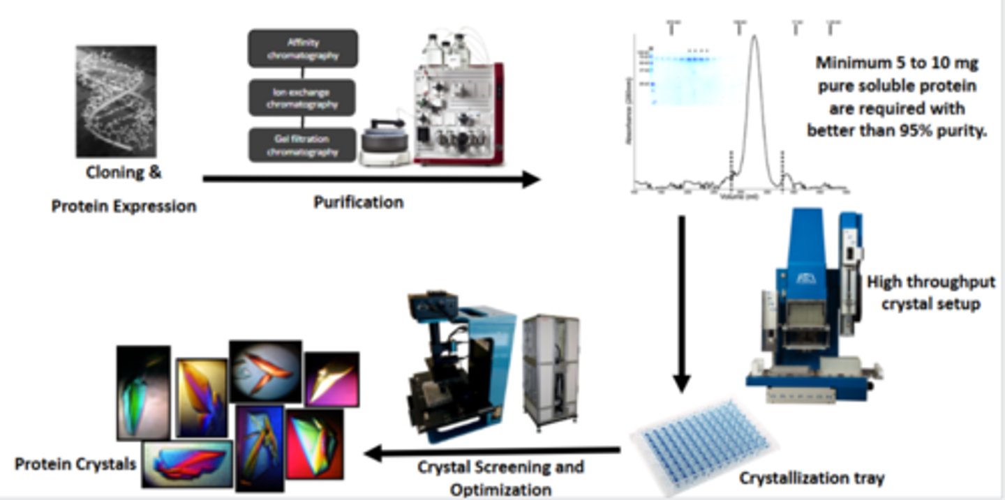
-Minimum 5 to 10 mg pure soluble protein are required with better than 95% purity
Steps:
1. Cloning the gene that encodes the protein of interest
-Put gene of interest in plasmid vector for expression of protein of interest
2. Purify protein of interest
-Minimum 5 to 10 mg pure soluble protein are required with better than 95% purity
3. Crystallization: Mix purified protein with multiple different buffer solutions to make protein crystals (picture on bottom left) in tray
4. Incubate protein crystals at constant temperature
-Because of evaporation of water, the proteins start coming together to form crystals
-There are robots/machines to help you do this whole process
5. Detection of protein crystals by computer that can scan for drops of protein crystals or microscope
Protein Crystallization
-Why do we need to make protein crystals?
-Can we use diffraction data from single molecules? What happens in X-ray crystallization with a single molecule? Can the rays be detected?
-What is the solution to this? What is measured instead? How does it work? How are the rays detected?
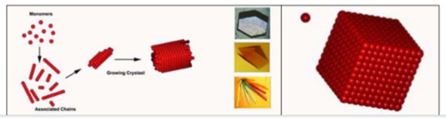
-We need to make protein crystals because a single molecule is a very weak scatterer of X-rays. Most of the X-rays will pass through the molecule without being diffracted. The diffracted rays are too weak to be detected.
-Solution: Analyzing diffraction from crystals instead of single molecules. A crystal is a three-dimensional array of ordered molecules held together by noncovalent interactions. Under certain circumstances, macromolecules (protein, DNA, RNA) can form crystals. Signals from multiple molecules (~10^14) in crystals reinforce each other. The resulting diffracted rays are strong enough to be detected so you can determine protein structure.
-Need to make proteins arranged in tandem arrays/packed in a uniform, symmetric way in crystals
-Crystals are needed to determine protein structure
Crystallization
-What 3 things does the success of crystallization depend on? Which cause more success?
-What does a crystal look like?
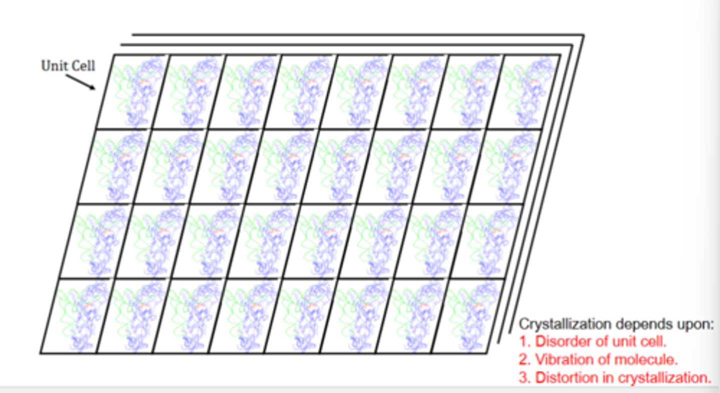
Crystallization depends upon:
1. Disorder of unit cell-more disorder=less symmetric unit cells-->low success of crystallization
-Want low disorder for better crystallization
2. Vibration of molecule-if proteins or other molecules like buffers are vibrating/moving around, its hard for them to pack into a crystal
-Want low vibration for better crystallization
3. Distortion in crystallization
-Distortion can cause crystallization to not work
-Crystal has many unit cells with proteins in it and is 3D (picture)
Protein Crystals
-What are the structures of the ideal, real, and protein crystals?
-What would cause imperfections?
-The Ideal Crystal-the ordered disposition of molecules such that there exists a regular repetition of a pattern in 3-D space, where this repetition extends over a distance equal to or greater than thousands of molecular dimensions (molecule is repeated in a perfect symmetric fashion)
-The Real Crystal-a crystal with less than perfect periodicity, imperfections are often caused by impurities and the effects of non-zero temperatures (nonideal temperatures for crystallization) ex. salt crystals
-The Protein Crystal-the crystal contains a high amount of solvent, meaning that some molecules present are not in the crystalline state, but in the liquid state, creating disorder (aren't perfectly symmetric)
What are 2 pieces of evidence that show the solution and crystal structure are similar?
-What percentage of protein crystals is solvent?
This shows that the structure from x-ray crystallography can be trusted/are the real structures:
1- NMR and X-ray crystallography have been used to determine the structure of the same molecule. The
two methods produce similar models.
2-Many macromolecules are still functional in the crystalline state. Most protein crystals contain 50-70% solvent.
Protein Phase Diagram
-What is it? What are the axes and parts of the diagram?
-What happens at each of the 4 zones of the diagram?
-What 2 zones are crystals found in?
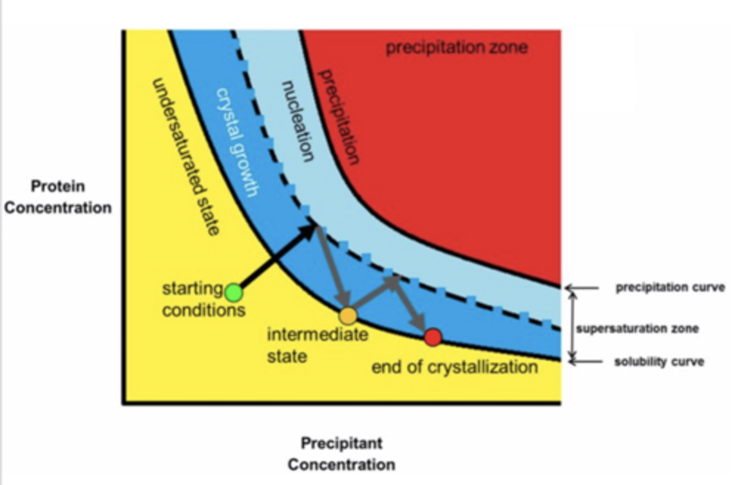
-The protein phase diagram is a map which represents the state of a protein (e.g., solid and liquid) as a function of the ambient conditions (e.g., temperature and concentration)
-The graph represents what happens when the protein is mixed with buffers and crystallizes
-Graph in pic: x axis-precipitant concentration-the precipitant is one of the components of buffers you add that facilitates evaporation of solvent from protein and buffer mixture which leads to protein packing/precipitation-->protein crystals. Y axis: concentration of protein.
-Crystals are semi-solid structures (have some liquid in them and are somewhat solid) and are found in crystal growth and nucleation zones
1. Undersaturated state-protein is liquid (no crystals)
2. Zone 1 - Crystal growth /metastable zone
-The solution may not nucleate for a long time but this zone will sustain growth. It is frequently necessary to add a seed crystal.
3. Zone 2 - Nucleation zone
-Nucleation is a phenomenon whereby a "nucleus", such as a dust particle, a tiny seed crystal, or commonly in protein crystallography, a small protein aggregate, starts a crystallization process (crystal starts to form around nucleus as more molecules join)
-Protein goes through crystal growth then into the border between crystal growth and nucleation to form nuclei, then goes back into crystal growth for the crystals to grow/get larger
4. Zone 3 - Precipitation zone
-Proteins do not nucleate but precipitate out of solution
-Want to stay in crystal growth and nucleation zones, not precipitation zone-any protein or precipitant concentrations above or below these zones will precipitate your protein
-This is why we evaporate water very slowly so we don't hit precipitation zone
What are 7 factors that affect crystallization? How do they affect it?
1) Purity of proteins-less pure=won't work
2) Protein concentration-too high or low can cause precipitation of protein
3) Starting conditions (makeup of the protein solution/buffer composition)
4) Precipitating agent (precipitant)-precipitant on x axis in previous flashcard
5) Temperature
6) pH
7) Additives: Detergents, reducing agents, substrates, co-factors, etc.
Hanging/Sitting Drop Vapor Diffusion
-What are the steps? What substances are in the 2 different solutions?
-How does it work?
-What is the mother liquor?
Most popular method among protein crystallographers
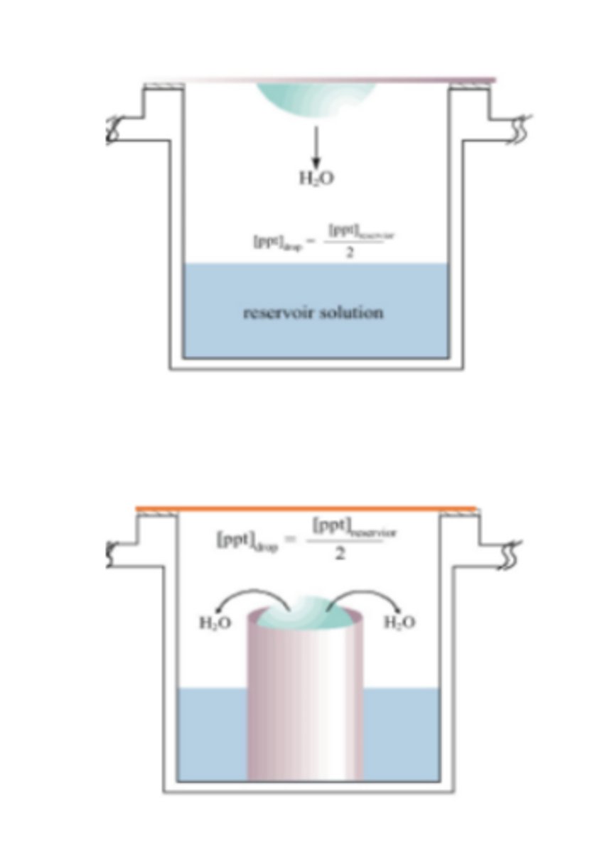
1. Crystal screen buffer/mother liquor is the well solution (0.5-1 mL), mix this with protein and you make a drop with it
-Mother liquor-the solution in which the crystal exists-this is often not the same as the original crystallization screening solution, but is instead the solution that exists after some degree of vapor diffusion, equilibration through dialysis, or evaporation
2. Drop (on siliconized glass cover slip) is 1/2 protein solution, 1/2 crystal screen buffer (0.5-4 uL). So, the concentration of precipitant in the drop is 1/2 the concentration in the well
-Hanging drop method-drop is hanging upside down from top
-Sitting drop method-drop is sitting on a pedestal in a closed chamber
3. Cover slip is inverted over the top of the well an sealed with vacuum grease (airtight) (hanging drop method)
-Put buffer solution in rest of the chamber and close the top
4. The precipitant concentration in the drop will equilibrate with the precipitant concentration in the well via vapor diffusion
-Water evaporates from your drop very slowly and then the drop ends up in the same buffer that you filled the chamber with (this buffer contains precipitant)
-Is a very slow process-protein and precipitant concentration increase very slowly over time (is good because you don't want a sudden increase in concentration which could lead to precipitation)
-Protein eventually forms crystals
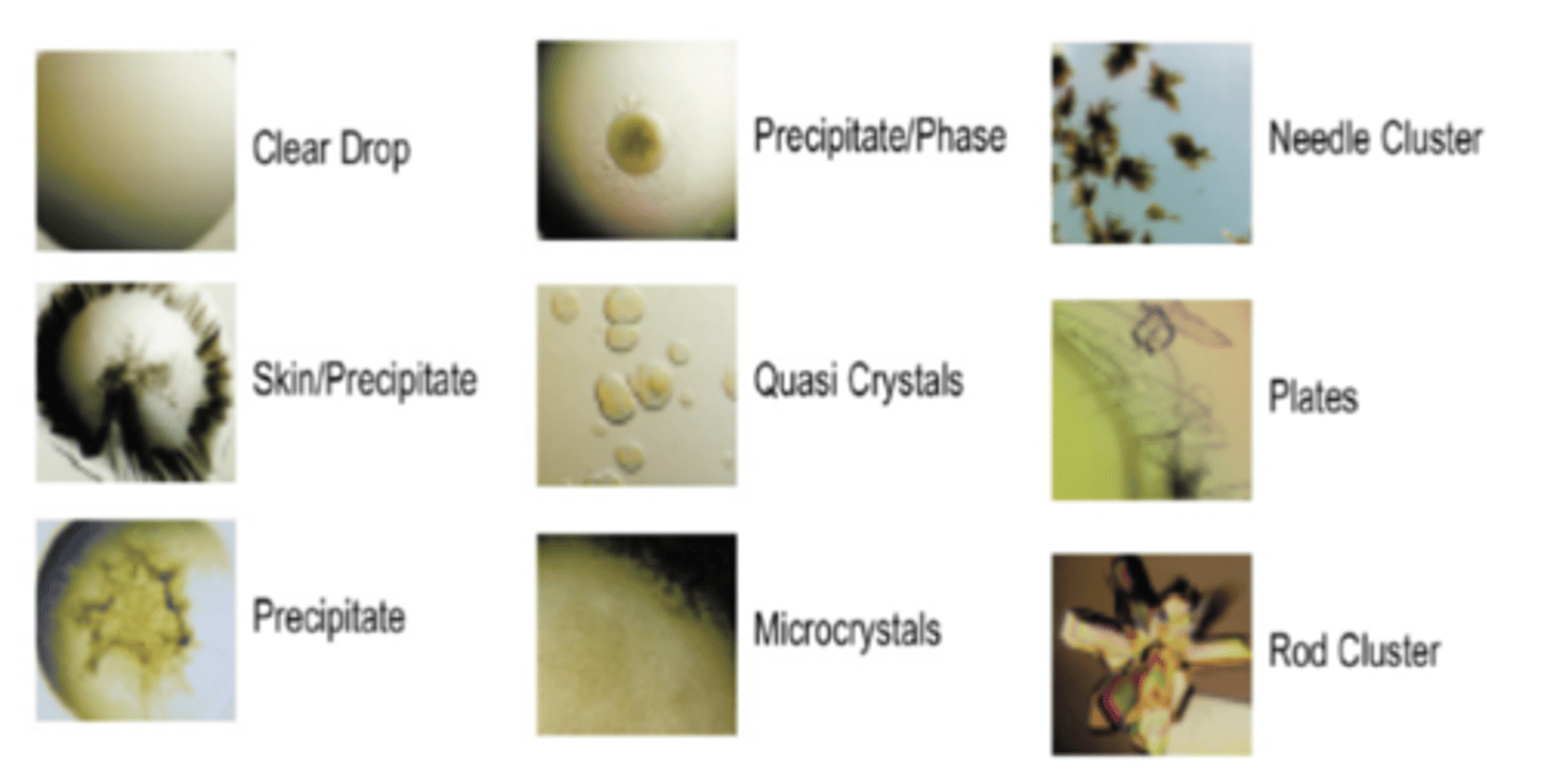
Interpreting Results of the Crystallization Experiment
-What do different results look like and what do they mean?
-Plates=good crystals
-Precipitate/skin and needle clusters=not good
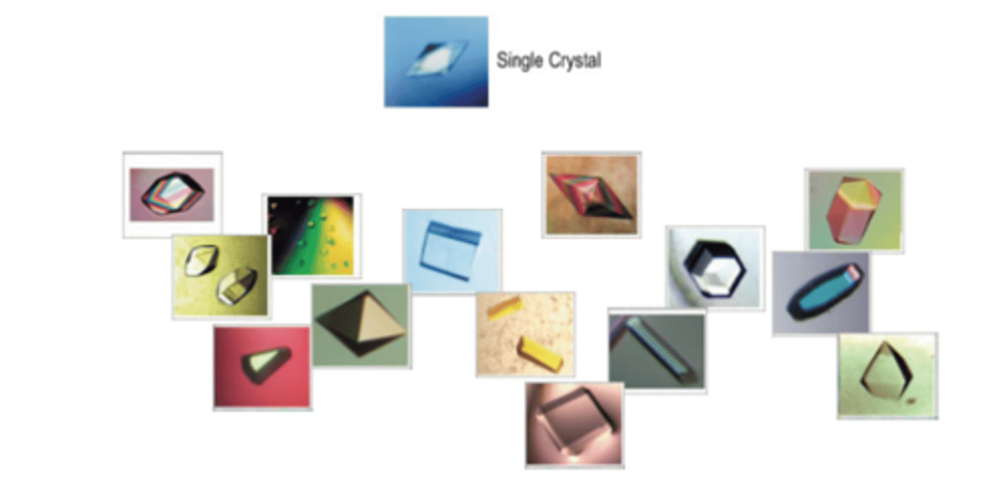
What are the sizes and shapes of crystals?
-What is the typical size of a crystal?
-What do you want the crystal to look like?
-Crystals come in all shapes and sizes
-The typical size of a crystal is 0.3 nm x 0.3 nm x 0.1 nm
-Want crystal to have linear edges and be symmetrical
-This is what your crystals should look like
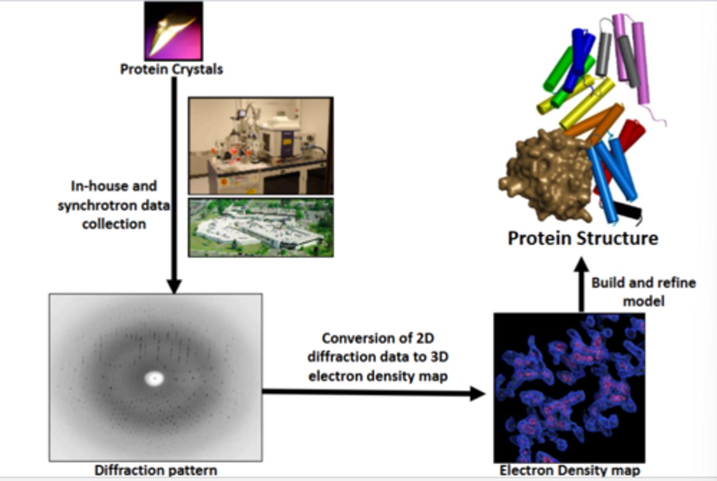
What are the steps performed in x-ray diffraction?
1. Shine x-rays at protein crystals
-Can use in house small source of x-rays (small amount of x-rays generated and quality of data is limited) or use national facility source of x rays (makes x-rays with a very high intensity which gives you very good quality data)
-Electrons around crystals scatter the x-rays
2. Collect scattering patterns using a detector
3. See diffraction pattern picture (white dot surrounded by black dots, white dot is x rays being blocked by a blocker) (diffraction pattern picture)
-Most of the x rays go through the crystal, some are scattered and hit detector and form image
-Each spot on picture is an array of x-rays that was detected at different spots
4. Make 3D data (electron density map picture)
-Is electron density clouds of the atoms in the crystal
5. Find protein structure (protein structure picture)
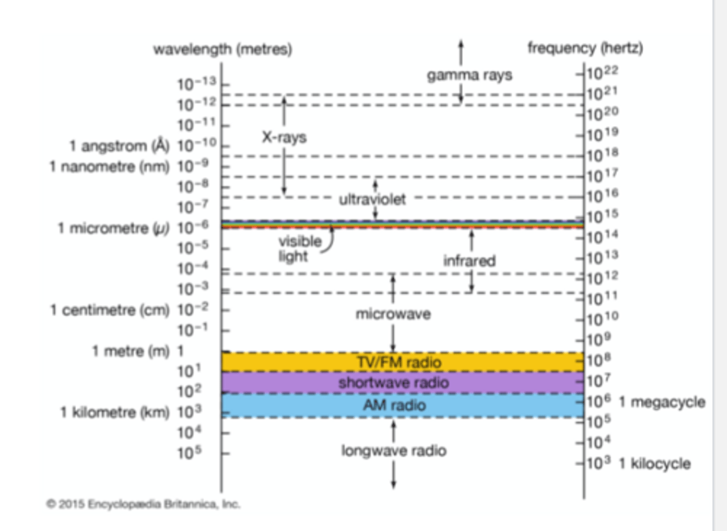
Why are x-rays used?
-What should the wavelength of light be for the object to be visible under magnification?
-Can visible light be used to image proteins? Why?
-What light wavelength is used for imaging proteins? How does this method work?
-In order for an object to be visible under magnification, the wavelength of the light must be, roughly speaking, no larger than the object
-Visible light (400-700 nm) cannot produce an image of protein molecules, in which bonded atoms are about 1.5Å apart (0.15 nm). Electromagnetic radiation of this wavelength (0.15 nm) falls into the X-ray range, is why we use x rays
X-ray crystallography principle:
-That X-rays are diffracted by crystal
-Direct detection of atom position in crystal based off of diffraction pattern
-If you just shined x-rays on proteins in a tube, the light would be scattered in many different directions and wouldn't give you information about the structure of the protein
-You have to get the protein in an ordered structure (crystal) for the x-rays to be scattered in a way that shows you the structure of the protein
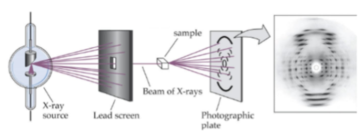
X Ray Diffraction Experiment
-How does it work? What happens?
-X rays penetrate crystal, get scattered and are captured as diffraction pattern on a detector
-Rotate crystal and repeat process and record XRDs pattern
-There is a screen to allow only a fine stream of x-rays to come out onto the protein crystal sample
-X rays are diffracted in different directions onto a photographic plate or detector-->diffraction pattern picture
-Where the dots are compared to center and intensity of each dot is used to determine protein structure based on diffraction pattern picture
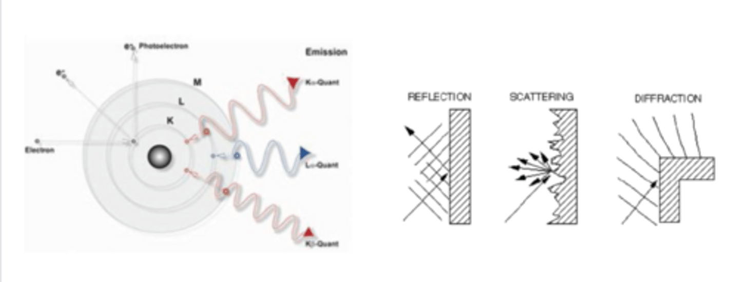
X-Ray Generation and Diffraction
-Left side: generation of x-rays
-Right side: diffraction of x-rays
Generation of X-rays:
-Knocking off an electron from an atom (like copper)
-In source you hit a highly intense ray of electrons onto the atoms of copper, the beam of electrons hit the copper inner orbital electrons and kick them out of their orbits
-When these electrons are kicked out, the other electrons try to occupy the orbits of the inner electrons that were lost. To do this the electrons jump from outer to inner orbitals of the atom which generates x-rays.
-Reflection-Rays are reflected back at 90 degrees because they are being hit at a uniform linear surface
-Scattering-since your surface is irregular, the ray you hit at the surface can be scattered back in all different directions
-Diffraction-similar to scattering, but the surface is semi-uniform so you can predict what angle your rays will come back out at, this is what happens in x ray crystallography
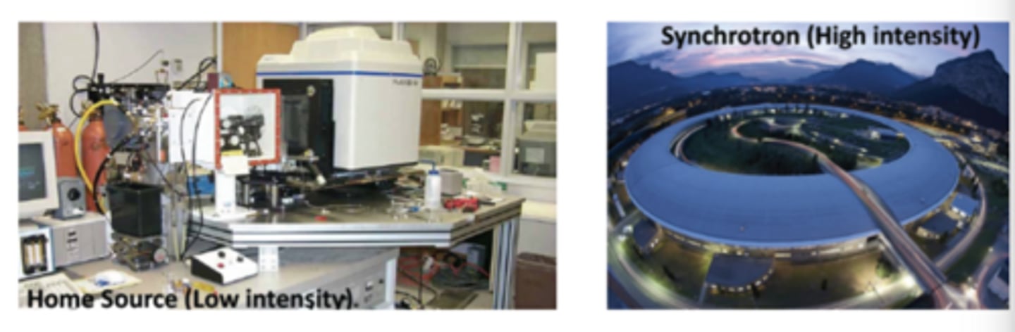
X-Ray Sources and Intensity
-What sources of X rays have high and low intensity?
-In house-small source of x-rays (small amount of x-rays generated and quality of data is limited), uses copper ions like in previous flashcard
-Synchrotron-source of x rays (makes x-rays with a very high intensity which gives you very good quality data), facility is in a ring structure
Data Collection
-Why do you rotate the crystal?
-What data is collected? How can this be used to determine protein structure?
-Shine x-rays at crystal and rotate crystal to expose different planes in the unit cells to deflect the x-rays onto a detector (rotate crystal to get structure of whole thing)
-As you rotate your crystal many pictures are taken of the spots detected, the spots change as you rotate your crystal
-Can use location and intensity of spots to figure out where electrons/atoms are in your crystal
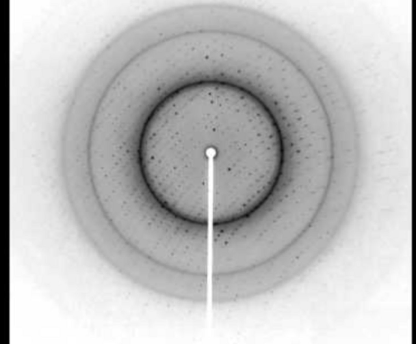
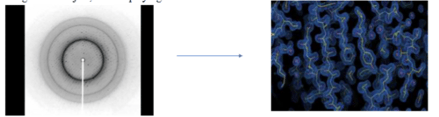
2D Diffraction Data to 3D Structure
-Can X rays be focused by lenses? What is the solution to this? How does it work?
-What is the refractive index of x-rays in all materials?
-What do you need to get structure of protein?
-Unlike visible light, X-rays cannot be focused by lenses. The refractive index of X-rays in all materials is very close to 1.0.
-Solution: Use a computer to simulate an image-reconstructing lens. In short, the computer plays the part of the objective lens, computing the image of the object, then displaying it on a screen.
-X-ray diffraction pattern provides amplitude of X-rays, but to obtain electron density map of crystal, phase information of X-rays is also needed.
-Need location and intensity of spots as well as phase information to make electron density cloud picture-->find protein structure
-Phase information-phase of x-rays used in the experiment
-Phase information (AI description): the precise position of the wave's peak relative to other waves—is lost (Phase information in X-ray crystallography refers to the missing angular data () needed to calculate the 3D electron density of a crystal structure, as experiments only measure the intensity (amplitude) of diffracted waves)
-Need to know if the phase of the waves made from diffraction off of the crystal. If 2 waves are constructive they are going up and down at the same time. If they are not constructive they go up and down at different times. The difference between the peak of the waves/difference between the waves is called phase.
The Phase Problem
-What are the 3 methods that can be used to provide phase information?
Phase information can be obtained using three methods (not detailed in this lecture):
1. Molecular Replacement (MR)
2. Multiple Isomorphous Replacement (MIR)
3. Single- or Multiple- wavelength Anomalous Diffraction (SAD or MAD)
Applications of Structural Analysis
-What 3 things do protein structures help determine?
Structures help to:
1. Understand precise mechanism of protein function
2. Depict regulatory conformational changes
3. Understand mechanism of action of drugs
What is the primary purpose of X-ray crystallography in protein studies?
a. To determine protein solubility
b. To determine protein structure in 3D
c. To analyze protein sequences
d. To study protein dynamics
To determine protein structure in 3D
Which structures are analyzed using X-ray crystallography?
a. Primary structures only
b. Tertiary structures only
c. Tertiary and quaternary structures
d. Both primary and secondary structures
Tertiary and quaternary structures
What stabilizes tertiary and quaternary protein structures?
a. Ionic interactions and hydrogen bonds
b. Covalent bonds exclusively
c. Disulfide bonds only
d. Van der Waals forces alone
Ionic interactions and hydrogen bonds
Why is it essential to understand a protein's 3D structure?
a. It helps in identifying amino acid sequences
b. It determines the solubility of proteins
c. It helps predict protein function and mechanism
It enables DNA sequencing
It helps predict protein function and mechanism
What is a prerequisite for X-ray crystallography?
a. Low concentration of proteins
b. High purity and high concentration of proteins
c. DNA sequencing data
d. Amino acid profiling
High purity and high concentration of proteins
Why are crystals required for X-ray diffraction studies?
a. Single molecules scatter X-rays strongly
b. Crystals amplify diffraction signals
c. Single molecules are weak X-ray scatterers
d. X-rays cannot penetrate liquid samples
Single molecules are weak X-ray scatterers
What is the role of precipitant in crystallization experiments?
a. To increase protein solubility
b. To degrade proteins in solution
c. To facilitate evaporation of solvent and crystal formation
d. To stabilize protein tertiary structure
To facilitate evaporation of solvent and crystal formation
What type of molecule arrangement is found in protein crystals?
a. Random arrangement of molecules
b. Ionic lattice structure
c. Ordered grid-like structure
d. Semi-solid amorphous state
Ordered grid-like structure
Which method does not require crystallization for structure determination?
a. X-ray crystallography
b. Cryo-EM imaging
c. Nuclear Magnetic Resonance (NMR)
d. Mass spectrometry
Nuclear Magnetic Resonance (NMR)
What is the limitation of NMR compared to X-ray crystallography?
a. Requires crystallization of proteins
b. Lacks atomic resolution for small proteins
c. Cannot resolve proteins larger than ~40 kDa
d. Limited to DNA studies
Cannot resolve proteins larger than ~40 kDa
What percentage of Protein Data Bank entries were determined using X-ray crystallography?
a. 20%
b. 50%
c. 70%
d. 80%
80%
Why do flexible proteins pose challenges for crystallization?
a. They degrade rapidly in solution
b. Flexible regions interfere with diffraction signals
c. They hinder packing into ordered arrays
d. Vibration affects their solubility
They hinder packing into ordered arrays
What is the typical solvent content in protein crystals?
a. 10%
b. 30%
c. 50%
d. 50-70%
50-70%
What does the phase diagram of protein crystallization depict?
a. Protein folding dynamics
b. Salt concentration effects on purity
c. Protein states as a function of concentration and precipitant levels
d. Tertiary structure formation
Protein states as a function of concentration and precipitant levels
What happens during nucleation in crystallization experiments?
a. Proteins dissolve into solution
b. Nuclei form randomly without order
c. Nuclei grow into precipitates over time
d. Nuclei act as starting points for crystal growth
Nuclei act as starting points for crystal growth
Which experimental setup is commonly used for protein crystallization trials?
a. Open-air incubation method
b. Hanging drop method only
c. Sitting drop method only
d. Both hanging drop and sitting drop methods
Both hanging drop and sitting drop methods
How are x-rays generated for diffraction experiments?
a. Through high-temperature heating of copper atoms
b. By focusing visible light on metal surfaces
c. By cooling copper to absolute zero temperatures
d. By knocking electrons out of copper atoms with high-energy electrons
By knocking electrons out of copper atoms with high-energy electrons
What does the intensity of diffraction spots indicate?
a. Protein purity levels
b. Molecular weight distribution within crystals
c. Salt concentration effects on diffraction efficiency
d. Electron density within unit cells
Electron density within unit cells
Which Nobel Prize-winning discovery utilized X-ray crystallography?
a. DNA sequencing
b. RNA transcription mechanisms
c. Cryo-EM imaging advancements
d. Atomic-resolution ribosome structure determination
Atomic-resolution ribosome structure determination