Anterior Segment and Anterior Uveal Tract
 Call Kai
 Learn
 Practice Test
 Spaced Repetition
 Match
 Flashcards
 Knowt Play
1/170

There's no tags or description
Looks like no tags are added yet.
171 Terms
What structures of importance are located in the anterior chamber of the eye?
Cornea, Drainage angle, and anterior face of iris
What structures of importance are located in the posterior chamber of the eye?
Ciliary body, lens zonules, anterior face of lens, interior face of iris
What structure produces the aqueous humor?
Ciliary body
How is globe shape maintained?
Steady balance of aqueous production and drainage
What structure is responsible for draining aqueous humor?
Drainage angle
Why is there ACAID (Anterior Chamber Acquired Immune Deviation)?
Classic T helper cell response in eye can lead to ocular tissue damage
How is the blood-aqueous barrier formed?
Tight junctions between ciliary body epithelial cells, an epithelial cell basement membrane, stroma, and a tight junction between vascular endothelial cells
What congenital diseases may impact the iris?
Heterchroma, coloboma, aniridia and hypoplasia, and persistent pupillary membrane
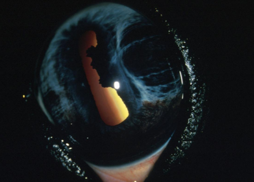
What congenital condition is seen here?
Iris hypoplasia
How can perisitant pupillary membranes occur in the eye?
Mesenchymal tissue forming the vascular suppy to the developing lens is formed abnormally and leads to incomplete regression
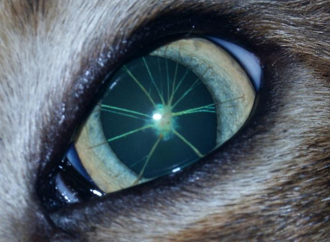
What condition is seen here?
Persistent pupillary membrane
What structures can a persistent pupilalry membrane impact?
Iris, lens, and cornea

What condition is seen here?
Persistent pupilalry membrane
What degenerative iris changes can occur in our patients?
Senile iris atrophy and uveal cysts
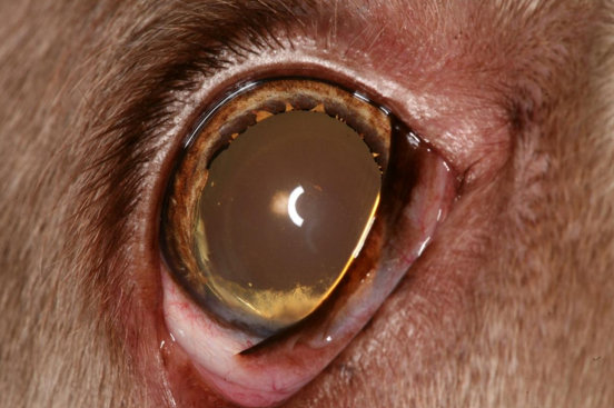
What condition is seen here?
Iris atrophy (note transparence at pupil margin)

What condition is seen here?
Iris cyst

What condition is seen here?
Ventral iris cyst

What condition is seen here?
Multiple iris cysts
What is uveitis?
Inflammation of the uveal tract
What structures may be impacted by uveitis?
Iris, ciliary body, and/or the choroid
What is the etiopathogenesis (etiology and pathogenesis) of uveitis?
Can be primary or secondary to lens, scleral or corneal disease (neoplasia, infection, Immune mediated, etc)
How do the ocular tissues modulate the level of inflamamtion during inflammatory incidences? (Not sure if this is written right…)
Blood-aqueous barrier, anti-oxidants in aqueous humor, ACAIDs, and a lack of intrinsic lymphatic system
What may initiate inflammation in the uvea?
Tissue injury, disruption of the blood-aqueous barrier, and/or release of inflammatory mediators
What clinical signs may indicate uveitis?
Aqueous flare, fibrin, hyphaema, hypopyon, miosis (pupil constriction/spasm), reduced intraocular pressure, limbal neovascularization and corneal edema, iris hyperpigmentation, keratic precipitates, iris swelling, synechiae, reduced vision
If we see a serous exudate in the uvea, we would call that
An aqueous flare
If we see a fibrinous exudate in the uvea, we would call that
Fibrin clot
If we see a sanguinous exudate in the uvea, we would call that
Hyphema
If we see a purulent exudate in the uvea, we would call that
Hypopyon

What is seen here?
Aqueous flare

What is seen here?
Hypopyon
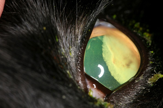
What is seen here?
Hypopyon and cellular flare

What is seen here?
Fibrin and hemorrhage

What is seen here?
Hemorrhage/Hyphema

What is shown here?
Hyphema
What changes do we expect to see in uveitis?
Pupil miosis, iris rubeosis, and corneal neovascualrization
What may occur in a subacute case of uveitis that has been localized by the immune cells?
PMN and mononuclear phagocytes are present and injury is resolved
What may occur in a subacute case of uveitis that has been uncontrolled by the immune cells?
Leukocytes throughout, blood vessel proliferation and fibroblast reactions
May resolve or become chronic
Why may we get chronic uveitis?
Initiating factor not eliminated or immune mediated disease established

What is shown here?
Iris neovascularization with keratic precipitates

What is shown here?
Iris lymphoid follicles
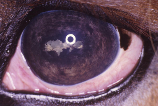
What is shown here?
Posterior synechiae and cataract

What is shown here?
Synechiae and iris rests
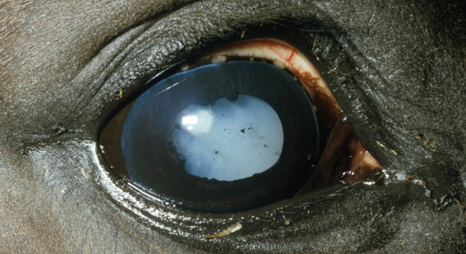
What is shown here?
Iris hyperpigmentation, synechiae, iris rests, and cataract
What are potential specific causes of uveitis?
Lens induced, hyperlipidemia, trauma
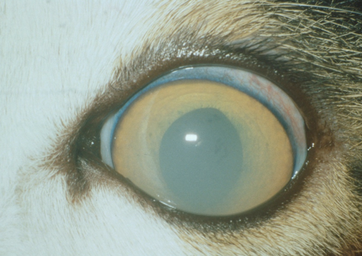
What is shown here?
Hyperlipidemia

What is shown here?
Migrating foreign body
What diseases/infections in canines can lead to uveitis?
Infectious hepatitis, Brucellosis, Ehrlichiosis, Uveodermatological syndrome, Leishmaniasis, Toxoplasmosis

What is shown here?
Pigmentary glaucoma
What potenital diseases can lead to feline uveitis?
FIP, bartonella henselae, Toxoplasma, FIV, FHV, FeLV
What potenital diseases can lead to equine uveitis?
Leptospira, EHV, EVA
What potenital diseases can lead to farm animal uveitis?
Bovine malignant catarrhal fever, IBR, CSF (Classical swine fever)
What types of neoplasia may be seen in the iris?
Melanoma/melanocytomas, adenoma/adenocarcinomas, and lymphomas

What is shown here?
Benign melanocytoma

What is shown here?
Malignant melanoma

What is shown here?
Iris melanoma

What is shown here?
Lymphosarcoma
What is glaucoma?
A collection of neuro-destructive diseases that cause the death of the neural cell alyers within the retina and optic nerve head. Typically accompanied by a raised intraocualr pressure
How are aqueous pathways/production regulated?
Difusion of solutes down a concentration gradient, active secretion using ATP against a concentration gradient, and ultrafiltration as a result of a hydrostatic force

What is shown with the yellow arrows?
The direction of aqueous humor movement/drainage
What may contribute to variations in intraocualr pressure?
Diurnal variation, age, blood pressure, drugs, ocular inflammation, position and restraining of animal, instrumentation used for measurement
What intraocualr pressure do most species have as physiologically normal?
15-25 mmHg
What is the pathology of glaucoma? (fancy words)
Buphthalmia leading to Phthsis bulbi
What is Buphthalmia?
An increase in globe size
How does Buphthalmia occur?
Stretching of ocualr tunics (associated with ulceration, fractures of Descemet’s membrane, and lens luxations)
When may acute buphthalmia resolve?
If underlying glaucoma is relieved
What is phthsis bulbi?
A reduction in globe size
How does Phthsis bulbi occur?
Pressure induced atrophy of the siliary processes leading to decreased aqueous production

What is seen in both of these eyes?
Haab’s striae
When do Haab’s striae occur?
As a result of breaks in Descemet’s membrane
How is the cornea affected by the pathology of glaucoma?
Edema as a result of increased intraocular pressure → stretching and fractures of Descemet’s membrane during globe nelargement → Neovascularization to accomodate increased size and damage sustained
How is the sclera affected by the pathology of glaucoma?
Thinning and stretching due to globe size increase
How is the iris affected by the pathology of glaucoma?
Mydriasis(paralysis of pupil) ± imparied neural or blood supply to sphincter muscle ± lack of sensory input due to retinal ganglion dysfunction
Eventual atrophy
How is the ciliary body affected by the pathology of glaucoma?
Advanced atrophy will halt aqueous production → hypotony
How is the choroid and tapetum affected by the pathology of glaucoma?
Vascular perfusion decreases → poor autoregualtion and poorer perfusion
__ fundus may be less affected initially, but eventual __ thinning
How is the lens affected by the pathology of glaucoma?
Can result in cataract formation and __ luxations
How is the vitreous affected by the pathology of glaucoma?
Liquifaction occurs leading to lens luxations and interference of outflow pathways
How is the retina and optic nerve head affected by the pathology of glaucoma?
Axonal degeneration and atrophy accompanied by shearing forces and decreased axoplasmic flow (What even does any of this mean)
How can you assess the drainage angle of the eye?
Using gonioscopy
What is special about goinoscopy?
Use of a mirrored or convex lense to image the angle beneath the edge of the cornea at the limbus
How can we classify primary glaucomas?
Open/normal angle or Narrow/closed angle
How can we classify secondary glaucomas?
Blockage along the aqueous pathway (Lens luxation, iris bombe, drainage angle)
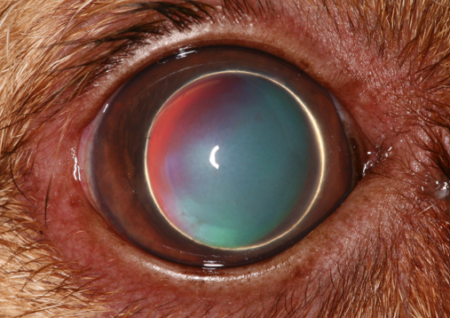
What is seen here?
Anterior lens luxation
What diseases may affect the lens?
Cataracts, luxation, penetration/rupture, Secondary affects on the globe
Where is the basement membrane of the lens located?
On the surface of the structure, facing the intertior of the structure (“backwards” from the rest of epithelial layers)
Why are diseases affecting the lens of importance?
Sight threatening, painful, and potential indicator of other disease
What congenital diseases may be seen in the lens?
Aphakia, Microphakia, Lenticonus/lentiglobus, coloboma, or cataracts
What is microphthalmia?
A failure of full sized lens development, or due to reduced intraocular pressure, or from other lens effects

What is seen here?
Microphakia
What is a lens coloboma?
Normal lens, apart from a small area where zonules fail to form so the lens is not stretched accross the whole region it should

What is seen here?
Lens coloboma

What is seen here?
Mesodermal remnants
What vitreal deficits may affect the lens?
Persistent hyaloid artery, persistent hyperpalstic primary vitreous, or persistent hyperplastic tunica vasculosa lentis
Any can progress to dysplasia due to failrue to regress
What is the definition of a cataract?
An opacification of the normally optically clear lens or its capsule
What features should be known to properly classify a cataract?
Position, age at onset, etiology, maturity, and progression
From external to internal, what are the regions of the lens?
Capsule, subcapsule, cortex, nucleus
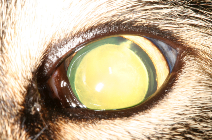
What is seen here?
A large, dense, extensive nuclear cataract with cortical extensions at the 2 o clock position

What is seen here?
Juvenile cataract
What are potential etiologies of a cataract?
Hereditary, traumatic, toxic/dietary, senile, infectious, secondary to other ocular disease

What is seen here?
Cataract (secondary to capsular rupture)
What classification would you give a cataract impacting a very small area (<15%)?
Incipient