WOOOOOO
 Call Kai
 Learn
 Practice Test
 Spaced Repetition
 Match
 Flashcards
 Knowt Play
1/95

There's no tags or description
Looks like no tags are added yet.
96 Terms
Normal RBCs

Microcytic and crenated RBCs

Yeast.The presence of budding forms aid in dis-
tinguishing from RBCs

Air bubble

KOVA-stained squamous epithelial cells and oil
droplets

Dysmorphic RBCs

RBCs and one WBC

WBCs (multilobed nucleoli)

WBC clump

Hansel stained eosinophils

WBCs with acetic acid nuclear enhancement

Phenazopyridine-stained sediment showing squa-
mous epithelial cells and phenazopyridine crystals formed fol-
lowing refrigeration

KOVA stained SECs

Squamous epithelial cells identifiable under low
power

Clump of squamous epithelial cells

Clump of squamous epithelial cells with folded
forms

KOVA-stained spherical transitional epithelial cells

Caudate transitional epithelial cells

Syncytia of transitional epithelial cells

RTE cell. Columnar proximal convoluted tubule
cell with granules and attached fat globules

RTE cells. Oval distal convoluted tubule cells

RTE cells, cuboidal from the collecting duct

Fragment of RTE cells under phase microscopy

Prussian blue–stained hemosiderin granules.

Oval fat body

Sudan III-stained oval fat body

Oval fat body under bright-field and polarized
microscopy

KOVA-stained bacteria and WBC

Yeast showing mycelial forms

spermatozoa

mucus threads

Hyaline cast under low power with amorphous
granules and mucus

Hyaline cast and amorphous urates attached to
mucus pseudocast.

Hyaline cast

hyaline cast under phase microscopy

convoluted hyaline cast

Hyaline cast containing occasional granules

rbc cast (hypochromic and dysmorphic free rbcs)

KOVA-stained RBC cast under phase microscopy

Disintegrating RBC cast (free rbc present)

Cast containing hemoglobin pigment. A com-
parison of RBCs and yeast also can be made

granular dirty brown cast

disintegrating wbc cast

KOVA stained wbc cast

WBC clump! matrix absent

RTE cell cast

KOVA stained RTE cell cast

KOVA-stained RTE cell cast under phase
microscopy

RTE cast with bilirubin stained cells

Fatty cast showing adherence of fat droplets to
cast matrix

fatty cast

fatty cast under phase microscopy

Finely granular cast and uric acid crystals

Granular cast formed at a tubular bend

Granular disintegrating cast

Coarsely granular cast, squamous epithelial cell,
and mucus

Granular cast degenerating into waxy cast

KOVA-stained waxy casts ( 100x).

KOVA-stained waxy casts ( 200x).

KOVA-stained waxy cast ( 400x)

KOVA-stained broad waxy cast ( 400)

Broad granular cast becoming waxy

Broad bile-stained waxy cast

amorphous urates

uric acid crystals

uric acid crystal clumps

Uric acid crystals under polarized light x100

Uric acid crystals under polarized light (400x)

Classic dihydrate calcium oxalate crystals

Classic dehydrate calcium oxalate crystals under
phase microscopy
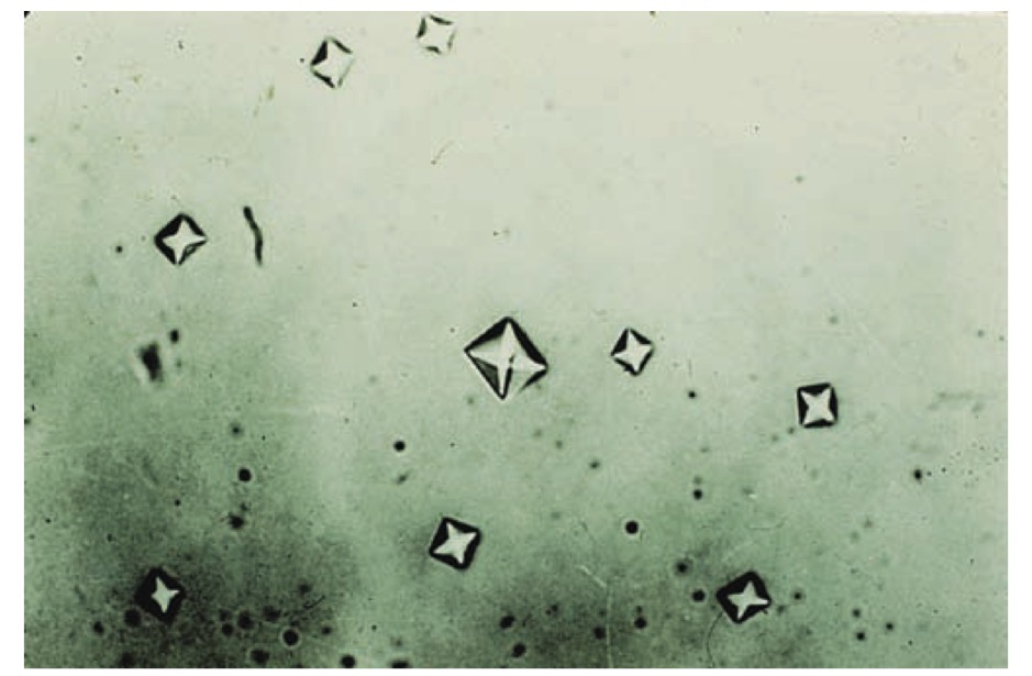
Monohydrate calcium oxalate crystals

Amorphous phosphates

Triple phosphate crystals

“Coffin lid” and other forms of triple phosphate
crystals

Triple phosphate crystals and amorphous phos-
phates

calcium carbonate crystals

Ammonium biurate crystals

cystine crystals

clump of cystine crystals

cholesterol crystals

cholesterol crystals under polarized light

tyrosine crystals in fine needle clumps

Tyrosine crystals in rosette forms

Leucine crystals

bilirubin crystals (bright yellow)

sulfa crystals in rosette form

Sulfa crystals,WBCs, and bacteria seen in UTI

Ampicillin crystals, nonrefrigerated

Ampicillin crystals following refrigeration

starch granules (dimple centered)

Fecal material and oil artifacts

pollen grain (concentric circles)

Fiber resembling a cast

Diaper fiber resembling a cast

Fiber under polarized light

Vegetable fiber resembling waxy cast
