Chapter 1 and 2 - CHM136
 Call Kai
 Learn
Learn Practice Test
 Spaced Repetition
 Match
Match Flashcards
 Knowt Play
1/52

There's no tags or description
Looks like no tags are added yet.
Name | Mastery | Learn | Test | Matching | Spaced | Call with Kai | Chat |
|---|
No analytics yet
Send a link to your students to track their progress
53 Terms
Infrared spectroscopy
determines the functional groups of a molecule
cm-1 - wavenumber
energy is proportional to 1/wavelength of the oscillation
bonds in a molecule absorb photons of light from the IR region of the spectrum , causes change in bond vibrations
bond in diff fg’s vibrate at different characteristics therefore the wavenumber of the absorbed light reveals the molecules fg’s
bonds
in molecules are constanlty vibratins around a certain equilibirum distance - bond length
Molecular vibrations
constantly stretching or bending motions
s-1

Stretching modes of vibration
vibration either lengthen or shorten bonds to distances slightly longer or shorter than their most stable bond lengths
relative to the equilibrium bond length - constanlty gets shorter or longer or to the equillibrium
Bending modes of vibrations
increase and decrease the angles between atoms relative to the equillibirum bond angles in the molecule
each vibration occurs at slightly different frequency s-1
which molecuels undergo more stretching and bending vibrations
molecules w/ more atoms
Number of vibrations
3n-6

Vibrating bodns create change in dipoles
creates oscillating electric field
these functional groups of the elctric field absorbs light between 4000 and 600 cm-1
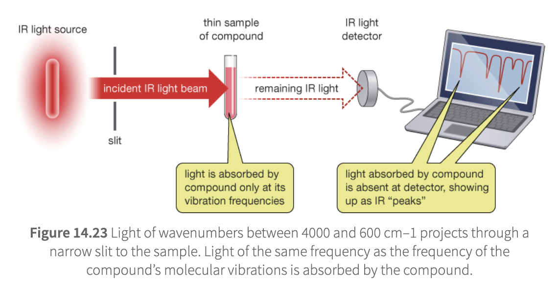
IR spectroscopy
when IR passes through a sample of th ecompound the photons w/ frequencies that match those of molecular vibrations are absorbed
those not absorbed read a detector and are converted to spectrum

IR spectrum

Hydrogen region
3600-2700 cm-1
OH , NH, CH (sp3, sp2, sp)
bonds with hydrogen vibrate at largest frequencies - due to lightness of h atoms
terminal alkyne most high
aldehydes ahve two medium peaks
Amines or amides
narrower than alcohol peaks and broader than alkyne
primary amines and amides produce 2 peaks
secondary amines and amides produce only 1 peak
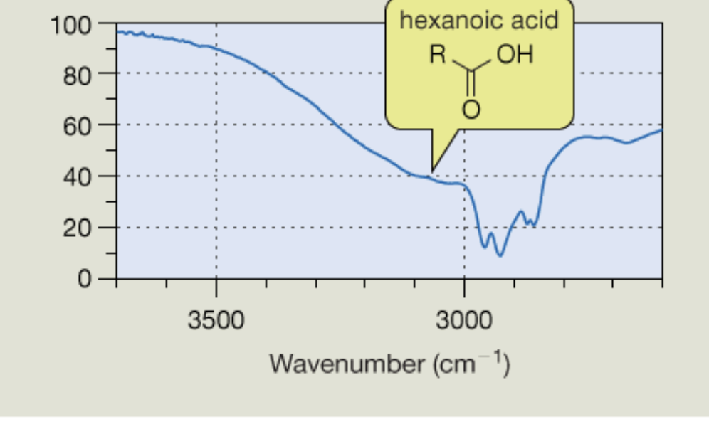
Carboxylic acid peaks
Triple bond region
alkynes and nitriles (more intense)
Terminal alkyne
induces a greater change in dipole therefore producing stronger signal
Finger print region
1500 - 500 cm-1
Spin
all nuclei posses this quantum mechanic property
spin state number is determined by atomic number and mass
H1 and c13
only 2 possible spin states
+1/2 or -1/2
Spins of 0
nuclei w/ even atomic mass and numbers
odd nuclei
can occupy several spin states

nuceli responding to magnetic fields
yield significant information about the structure of the molecule containing the nuclei
magenetic moments of nuclei in an external magnetic field (B0) align either parallel to the external field (alpha lower energy) or antiparallelt to it (beta, higher energy)
alpha more stable because lower energy
delta E
difference in energy between alpha and beta states - is small
depends on the magnetic field
stronger magnetic field results in larger delta E
Spin flip
transition from nuclei between apha and beta states
Resonance frequency
frequency at which a spin flip occurs
this happens when EM radiation of a frequency corresponding to deltaE - to the nuclei - the alpha state are excited and move to higher energy beta state
while the beta state moves to the alpha state
this equalizes the population of a and beta states and produces detectable magnetic field that contains a frequency component for each hydrogen type
Free induction decay
after excitation - the magnetic field weakens as the systme relaxes and goes back to equilibirum
signal detection provides info on teh type of each hydrogen
different chemical envrionments in a molecule cause variations int he magnetic field around the nuclei
H2 resonance
CDcl3 - d is deuterium h2
tuned to select a type of nucleus
signals corresponding to the sample are much stronger than those of the solvent
TMS
used as ppm 0
Integration
the calculation of the area under the peak - that states how many hydrogens are present in this chemical environment
Chemical shift
Lenz’s law
the principle that an induced current always generates a magnetic field opposing the applied field is knonwn as Lenz’s law

Local magnetic fields sigma B0
when exposed to an external magnetic firled B0 electrons in a compound circulate creating small currents that generare local magnetic fields that oppose the external field
shielding
the reduction in the magnetic field strength experienced by nucleus due to opposing magnetic fields of nearby electrons
nuclei that are shielded by their envrionment - have electrons surrounding them produce signals toward the right
Upfield
shielded nuclei by the envrionment that is surrounded by electrons produce a signal toward the right of the ppm spectrum
Downfield
hydrogens that are less deshielded by surrounding electrons - hydrogens near electronegative atoms - produce signal that are downfield toward the left

TMS
used as the reference compound
has highly deshielded electrons that are chemically equivalent

Electronegative atoms

additive effect
the more electronegative atoms the more deshielded the hydrogens are

Deshielding of functional groups


Carbon electronegativity
isobutane more electronegative than propane and ethane
as carbon is more elctronegative than hydrogen
RAG
enzyme responsible for recombination
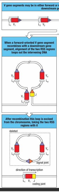
V gene segments are joined by recombination - forward
joining segments have the same transcriptional orientation - most common
the intervening DNA is loooped out and lost fromt he chromosome
there is formation of an excision signal(signal joint) and a coding joint

V geme segments are joined by recombination - revers
When segments are initially oriented in opposite transcriptional directions, alignment of the RSSs requires a coiled configuration. Thus, joining the ends of the two heptamer sequences now results in the inversion and integration of the intervening DNA into a new position on the chromosome.

why is there a 12/23 base pair rule?
may result from the asymmetric binding of RSSs to the RAG 1 and RAG 3 dimmer
there are two identical rag-1 and 2 identical rag-2
favours the 12bop - because shorter - proximity
where are rag genes expressed?
only in developin B and T cells
RAG knockout mice fail to develop B or T lymphocytes, and RAG1 or RAG2 mutations are a cause of SCID (patients lack B and T lymphocytes).

Enzymatic steps in the Rag-dependent V(D)J rearrangement
Binding of the rag-1 and rag-2 proteins as well as a protein called high mobility group.
these bind to one of the rss’s that flank the coding sequences come together
Cleavage step - the endocnuleases of RAG makes single-strand cuts in teh DNA backbone preciseling between each coding segment and its RSS.
At each cutting point this creates a 3′-OH group, which then reacts with a phosphodiester bond on the opposite DNA strand to generate a hairpin leaving a blunt double- strand break at the end of the RSS.
1 hair
1 blunt end - This is the singal joint end : Ku70:Ku80 binds the DNA ends - and joined together bu a ligase protein - to make a loop and to be the signal joiont which will get deleted later but stray blunt - the - precise


Coding joints - what happens - imprescise coding joint
KU70 and KU80 proteins need to add some nucleotides to what was cut previoously.
done by Artemis/DNA-PK endonuclease activity opens the hairpin , modifies the ends of the hairpin by adding nucleotides .
modified by terminal deoxynucleotidyl transferase (TdT; pink). TDT adds random extra nucleotides (N nucleotides; red)
V sequence end
A-T-C-A-G-T-A-C-T-G-C T-A-G-T
nicked hairpin
The two coding ends (e.g., V and J) are ligated by the DNA ligase IV/XRCC4 complex
P and N nucleotides in the VJ junction
N nucleotides are added by the enzyme terminal deoxyribonuclotuidyl transferase (Tdt)
Tdt only expressedn in developing b cells
No P Or N nucleotides int he signal junction
these help ensure the coding joint is properly ligated
and improves diversity of the coding joint
P nucleotides
palindromic nucleotides - mathces the sequence
due to the asymmetric nucking of a hairpin end
random residues
involved in diversity of the coding joint
N nucleotides
non templated nucleotides

D-J heavy chain rearrangement
same thing happens for heavy chain v-d and light chain v—j rearrangement
DNA hairpin at coding ends generated by RAG (heptamers are ligated to form the signal joint
Artemis:DNA -PK complex opens DNA hairpins generating palindromic P-nucleotides
N-nucleotide additions by Tdt
Pairing of strands
unpaired get removed by an exonuclease
DNA ligase addition of nucleotides for the gaps and then ligated together form coding joint

what contributes to the third hypervariable region
the variable addition and subtraction of nucleotides at the juncrions between gene segments
but leads to recombinational inaccuracies
Increased variability of CDR3
2/3 of rearrangements are non-productive (out of frame) this leads to in-frame stop codons.... TAA, TAG or TGA in J or C region
A trade-off between greater diversity and loss of efficiencysome can also be produyctive
because of recombination even if the v, d and j genes are the same they can produce different antigen binding sites because of this

IgM and IgD
derived from the same pre-mRNA TRANSCRIPT AND are both expressed oin the surface of mature b cells and act as receptors
b cell does not need to rearrange its genes a second time to make IgD
only one rearrangement for both - and express both of them
it is dependent ont he processing

Transmembrane and secreted forms of immunoglobulins are derived from the same heavy-chain sequence by alternative RNA processing
two poly AAA sites
either the secretion or the tranmembrane process is spliced out
different forms of rna processing takes place results in different immunoglobin forms