Learning Objectives
 Call Kai
 Learn
 Practice Test
 Spaced Repetition
 Match
 Flashcards
 Knowt Play
1/52

There's no tags or description
Looks like no tags are added yet.
53 Terms
Construct a complementary DNA strand from a given single-stranded sequence, accurately labeling 3’-5’ ends
example:
Given 5’-AAA TTT GGG CCC-3”, what is its complement aligned beneath it? Complement rewritten 5’→3’?
Complement aligned beneath it: 3’-TTT AAA CCC GGG-5’
Complement rewritten 5’ → 3’: 5’-GGG CCC AAA TTT-3’
Summarize and diagram the Avery/MacLeod/McCarty experiment, articulating its design and significance
Summary: Avery, MacLeod, and McCarty investigated which molecule carries the information that transforms harmless R bacteria into disease-causing S bacteria.
S bacteria have a protective capsule and cause disease.
R bacteria lack that capsule and do not cause disease in this experiment.
Material extracted from heat-killed S bacteria could cause living R bacteria to develop a capsule. This inherited change is called transformation.
The researchers identified DNA as the transforming material.
Design: They prepared an extract from heat-killed S bacteria and divided it into separate samples. They treated each sample with an enzyme that destroyed a particular molecule, then added the treated extract to living R bacteria.
Significance: Destroying DNA prevented transformation, while destroying protein or RNA did not. This showed that DNA carried the inherited instructions needed for capsule production.
The experiment went beyond Griffith’s finding: Griffith showed that an inherited trait could transfer between bacteria; Avery, MacLeod, and McCarty identified DNA as the substance responsible. It provided strong evidence that DNA is genetic material.
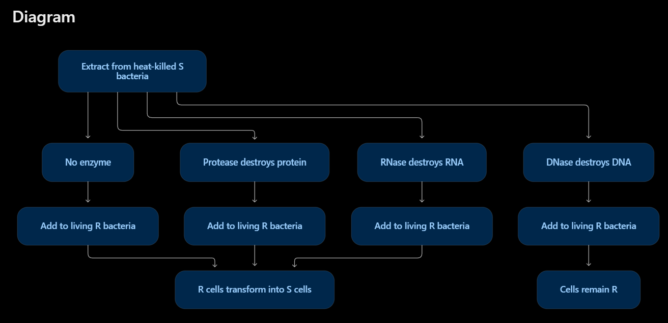
Infer and justify the expected outcomes of the Aver/McLeod/McCarty experiment if genetic information were composed of protein or RNA instead of DNA
If genetic information were composed of protein or RNA instead of DNA, the expected outcomes of the Avery/MacLeod/McCarty experiment would have shown that treating the heat-killed S bacteria extract with enzymes that destroyed proteins or RNA would still allow transformation of R bacteria into S bacteria, resulting in capsule production. This would indicate that the inherited characteristics were carried by proteins or RNA, rather than DNA.
Summarize and diagram the Hershey-Chase experiment, detailing its methods and conclusions
Hershey and Chase tested whether DNA or protein carries genetic information using bacteriophages
They labeled phage DNA with radioactive phosphorus (³²P) and protein with radioactive sulfur (³⁵S).
After infection, a blender removed attached phage coats, and a centrifuge separated bacteria into a pellet from the surrounding liquid.
Most labeled DNA was in the bacterial pellet, while most labeled protein remained in the liquid.
DNA entered the bacteria and directed production of new phages, supporting DNA as the genetic material.

Diagram the Central Dogma of molecular biology, naming all relevant molecules and processes involved
DNA —transcription→ RNA —translation→ Protein
DNA —replication→ DNA
replication: DNA is copied into DNA
transcription: RNA polymerase uses DNA as a template to make RNA
translation: a ribosome reads mRNA, while tRNAs deliver amino acids to build a protein
Summarize the different levels of chromatin compaction and analyze how each level influences gene expression
CORE NUCLEOSOME - DNA double helix 2nm: DNA before additional packaging, relatively exposed
CORE NUCLEOSOME - Nucleosomes “beads on a string” 11 nm: DNA wraps around histone cores; wrapped regions become less accessible
HISTONE H1 REQUIRED - Chromatin fiber of packed nucleosomes 30 nm: nucleosomes pack together, generally reduces access
Looped and further condensed chromatin 300 nm: higher-order structure, access to transcription reduced
Condensed section of chromosome 700 nm: highly compacted, gene expression is greatly inhibited due to lack of accessibility.
Mitotic chromosome 1400 nm: maximum condensation; transcription reduced
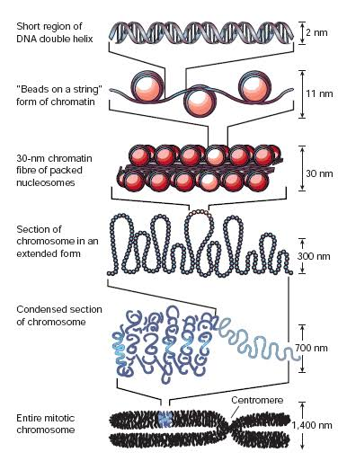
Diagram the arrangement of DNA on histone proteins
histones are associated with DNA to form nucleosomes (structural unit of chromatin, consisting of a segment of DNA wound around a core of 8 histone proteins)
H1 binds near the DNA entry/exit region and helps stabilize packing; it is separate from the core octamer.

Hypothesize how histone modifications that alter histone charge affect the affinity of histones for DNA and provide a rationale for your prediction
Histones are positive, and DNA is negative.
If histones were less positive, there would be a weaker attraction to DNA and greater accessibility and transcription. If histones were more positive, there would be a stronger attraction to DNA, and reduced accessibility and transcription,
Explain the foundational experiments that demonstrated DNA replication is semi-conservative
Meselson and Stahl tested this by growing bacteria in heavy nitrogen (¹⁵N), which became incorporated into their DNA. They then transferred the bacteria to light nitrogen (¹⁴N) and used density-gradient centrifugation to separate DNA by density.
Before replication: there’s one heavy DNA band containing 2 strands of 15N
After one generation: there’s one intermediate band where each molecule contains one heavy strand and one light strand
After two generations: one intermediate band and one light band; half the molecules are heavy-light and half are light-light
Conservative replication: The original two heavy strands would stay together, and the two new light strands would pair together. After one generation, you would see a heavy band and a light band. Instead, scientists saw one intermediate band, so this model was ruled out.
Dispersive replication: Every strand would contain mixed pieces of old heavy DNA and new light DNA. You would keep seeing one band that gets lighter each generation. Instead, after two generations, scientists saw two bands—intermediate and light, so this model was ruled out.
illustrate a piece of DNA, labeling the 5’ and 3’ ends of each strand

illustrate DNA open for replication, with RNA primers attached, and accurately label the 5’ and 3’ ends of the RNA primers
illustrate the synthesis of the new leading strand during replication
Helicase separates the parental strands. Primase makes short RNA primers because DNA polymerase cannot start a strand on its own.
The key is the primer’s free 3′-OH: DNA polymerase adds DNA there. Both leading and lagging strands need primers.
Leading strand:
Its template runs 3′ → 5′ toward the fork.
DNA polymerase synthesizes the new strand 5′ → 3′, toward the moving fork.
Synthesis is continuous after the initial primer.

depict the placement of the next RNA primers, labeling their 5’ and 3’ ends

Illustrate DNA synthesis on the lagging strand (Okazaki fragments). Describe the process of RNA primer removal and explain the role of ligase in sealing the resulting gap
As the fork opens, primase repeatedly places new primers on the lagging-strand template, near the fork.
For a rightward-moving fork:
Each new lagging-strand primer has its 5′ end nearest the fork.
Its 3′ end points away from the fork, toward the left.
DNA polymerase extends from that 3′ end to make another fragment.
Why repeated primers? DNA polymerase only synthesizes 5′ → 3′, so this strand must be built in separate sections as more template becomes exposed.

identify and describe the functions of the key enzymes involved in DNA replication
Enzyme or protein | Function |
|---|---|
Initiator proteins | Recognize replication origins and help assemble the replication machinery |
Helicase | Separates the two DNA strands |
Single-strand binding proteins | Keep separated strands from reannealing |
Topoisomerase | Relieves twisting and tension ahead of the fork by temporarily breaking and resealing DNA |
Primase | Makes RNA primers that provide a free 3′-OH |
DNA polymerase III | Main bacterial replication enzyme; synthesizes DNA 5′ → 3′ and proofreads 3′ → 5′ |
Sliding clamp | Holds DNA polymerase on the DNA so it can synthesize efficiently |
DNA polymerase I | Removes bacterial RNA primers and replaces them with DNA |
DNA ligase | Seals nicks between adjacent DNA sections |
Proofreading: Polymerase removes an incorrectly added nucleotide from the growing strand’s 3′ end, then resumes synthesis.
Describe the function of telomerase in DNA replication
Telomerase helps maintain the ends of linear chromosomes.
The problem: after the final RNA primer on the lagging strand is removed, there is no upstream 3′-OH from which DNA polymerase can fill the terminal gap. Without a solution, chromosome ends shorten over successive replications.
Telomerase:
Binds the chromosome’s 3′ end.
Uses its own RNA as a template to add repetitive telomere DNA.
Creates extra template that allows primase and DNA polymerase to synthesize more of the complementary strand.
Remember: Telomerase is a reverse transcriptase—it makes DNA using an RNA template. It extends the existing 3′ end rather than directly filling the final gap.
Illustrate 2 mechanisms of DNA repair
Nonhomologous end joining (NHEJ) | Homologous recombination (HR) | |
|---|---|---|
Basic process | Process the broken ends, then join them | Use an intact homologous DNA molecule to guide repair |
Template required? | No | Yes—usually the sister chromatid |
Accuracy | Can lose or add nucleotides at the repair site | Usually accurate because missing information is copied |
Timing | Can occur throughout much of the cell cycle | Most available after DNA replication |

Understand the basic mechanisms of transcription in eukaryotes and prokaryotes
Transcription copies a gene’s DNA sequence into RNA. It has three stages:
Initiation: RNA polymerase binds at a promoter and opens the DNA.
Elongation: It reads the template strand 3′ → 5′ and synthesizes complementary RNA 5′ → 3′.
Termination: Transcription ends, and the RNA and polymerase are released.
RNA polymerase does not require a primer. Only a small region of DNA opens at a time, and the DNA strands rejoin behind it.
Compare and contrast the mechanisms and regulatory features of prokaryotic versus eukaryotic transcription
Feature | DNA polymerase | RNA polymerase |
|---|---|---|
Building blocks | Deoxyribonucleotides: dATP, dTTP, dCTP, dGTP | Ribonucleotides: ATP, UTP, CTP, GTP |
Sugar | Deoxyribose | Ribose |
Product | DNA | RNA |
Main role | DNA replication and repair | Transcription |
Primer required? | Yes | No |
Synthesis direction | 5′ → 3′ | 5′ → 3′ |
Both use a DNA template, add nucleotides to a 3′-OH, and use complementary base pairing.
Explain how RNA polymerase identifies the key DNA strand for transcription and analyze why only certain DNA regions are transcribed into RNA
The orientation of the promoter determines which strand serves as the template and the direction of transcription.
For a particular gene, only one DNA strand acts as the template.
Different genes can use opposite strands.
Promoter recognition and regulatory proteins determine where transcription begins.
Terminators and other termination signals determine where transcription ends.
In eukaryotes, chromatin accessibility also affects whether transcription machinery can reach a gene.
construct the resulting RNA sequence from a given promoter site and DNA template
Identify the transcription direction from the promoter, read the template 3′ → 5′, and write complementary RNA 5′ → 3′.
Strand | Example |
|---|---|
Coding DNA | 5′–AGGTCCACG–3′ |
Template DNA | 3′–TCCAGGTGC–5′ |
RNA | 5′–AGGUCCACG–3′ |
RNA matches the coding strand except U replaces T. It is complementary and antiparallel to the template strand.
Identify and describe the different types of RNA, discussing their functions and significance in cellular processes
RNA type | Main function |
|---|---|
mRNA | Carries the information used to make a protein |
rRNA | Forms the ribosome’s structural and catalytic core |
tRNA | Matches mRNA codons with the correct amino acids |
snRNA | Helps the spliceosome remove introns from pre-mRNA |
snoRNA | Guides processing and modification of rRNA |
miRNA and siRNA | Regulate gene expression by reducing translation or promoting RNA degradation |
lncRNA | Performs various regulatory functions, including regulation of chromatin and transcription |
Not all genes encode proteins: some produce functional RNA as their final product.
Define and interpret a promoter, outlining its sequence characteristics and role in transcription initiation
A promoter is a DNA sequence where transcription machinery assembles to initiate transcription.
It is generally located upstream of the transcription start site, labeled +1.
Its asymmetry determines transcription direction.
Its sequence influences how efficiently a gene is transcribed.
Bacterial promoters often contain −10 and −35 elements.
Some eukaryotic promoters contain a TATA box, about 25 bases upstream of the start site.
A promoter is different from a start codon: the promoter helps start transcription; the start codon starts translation.
compare and contrast the mechanisms and regulatory features of prokaryotic versus eukaryotic transcription
Feature | Prokaryotes, especially bacteria | Eukaryotes |
|---|---|---|
Location | Cytoplasm/nucleoid region | Nucleus |
RNA polymerases | One main type | Three main nuclear types |
Promoter recognition | Sigma factor helps RNA polymerase recognize the promoter | General transcription factors recruit polymerase |
DNA packaging | No nucleosomes in bacteria | Chromatin must be accessible |
mRNA processing | Usually limited | Capping, splicing, and polyadenylation |
Relationship to translation | Translation can begin during transcription | Transcription and translation are separated |
Typical mRNA | Can encode several proteins | Usually encodes one protein |
The three main eukaryotic RNA polymerases are:
Pol I: makes most rRNA.
Pol II: makes pre-mRNA and some noncoding RNAs.
Pol III: makes tRNA, 5S rRNA, and some other small RNAs.
For Pol II initiation, TFIID, containing TATA-binding protein, helps assemble the initiation complex. TFIIH helps open the DNA and phosphorylates Pol II’s tail, allowing it to enter elongation.
illustrate and label the structural elements of prokaryotic and eukaryotic genes, including regulatory and coding regions
These elements appear in the following order when read in the direction of transcription:
Bacterial protein-coding gene | Eukaryotic protein-coding gene |
|---|---|
Regulatory sites and promoter | Regulatory sites, including enhancers, and promoter |
Transcription start site, +1 | Transcription start site, +1 |
5′ untranslated region, including a ribosome-binding site | 5′ untranslated region |
Start codon | Start codon |
Protein-coding region | Protein-coding region distributed across exons, with intervening introns |
Stop codon | Stop codon |
3′ untranslated region and termination signals | 3′ untranslated region, poly(A) signal, and downstream termination region |

Describe and explain the steps involved in eukaryotic RNA processing, such as capping, splicing, and polyadenylation
Processing occurs in the nucleus, and much of it begins while transcription is still underway.
Process | What happens | Why it matters |
|---|---|---|
5′ capping | A modified guanine, 7-methylguanosine, attaches to the 5′ end | Protects RNA and supports export and translation initiation |
Splicing | Introns are removed and exons joined | Produces the mature RNA sequence |
Polyadenylation | The RNA is cleaved downstream of the poly(A) signal, and poly(A) polymerase adds a tail to the new 3′ end | Supports RNA stability, export, and efficient translation |
The poly(A) tail itself is added after cleavage; RNA polymerase does not directly transcribe the tail. Polymerase continues downstream before transcription terminates.
distinguish and explain the structural and functional differences between introns and exons in eukaryotic genes
Introns: Sequences removed from pre-mRNA during splicing.
Exons: Sequences retained in mature RNA.
Exons are not necessarily entirely protein-coding: they can include untranslated regions.
Alternative splicing joins different combinations of exons, allowing one gene to produce different mRNAs and potentially different proteins.
Sketch and annotate the splicing reaction, demonstrating the process and molecular components involved
The starting pre-mRNA contains:
Exon 1 — intron containing a branch-point A — Exon 2
The spliceosome, made of snRNAs and proteins organized into snRNPs, carries out two reactions:
The 2′-OH of the branch-point adenine attacks the 5′ splice site, cutting that junction and forming an intron lariat, or loop.
The free 3′-OH of exon 1 attacks the 3′ splice site, joining exon 1 to exon 2 and releasing the intron lariat.
The products are:
Joined exons + released intron lariat
Most spliceosomal introns begin with GU and end with AG in the RNA. The spliceosome’s RNA contributes directly to catalysis.

Identify the first (start) and last (stop) codons for any protein sequence
Start codon: AUG, which establishes the reading frame and codes for methionine.
Stop codons: UAA, UAG, UGA, which recruit release factors and do not encode amino acids.
Explain and differentiate the mechanism by which eukaryotic and prokaryotic ribosomes recognize the start codon during translation initiation
Eukaryotes | Bacteria |
|---|---|
The small subunit is recruited near the 5′ cap | The small subunit recognizes a Shine–Dalgarno sequence |
It scans 5′ → 3′ for an AUG in a suitable Kozak sequence context | Base pairing between the Shine–Dalgarno sequence and ribosomal RNA positions the start codon |
Usually initiates one main protein per mRNA | Can initiate separately at several coding regions on one mRNA |
describe and sequence the major steps involved in ribosome assembly on an mRNA
For eukaryotic initiation:
The small ribosomal subunit, initiation factors, and initiator tRNA carrying methionine assemble.
The complex is recruited to the mRNA’s 5′ end.
It scans until the initiator tRNA pairs with the start codon.
Initiation factors leave, and the large subunit joins.
The initiator tRNA sits in the P site, leaving the A site available for the next charged tRNA.
The initiator tRNA enters the P site directly.
Explain and illustrate the functions of the E, P, and A sites on a ribosomes
Site | Function |
|---|---|
A: aminoacyl site | Receives the incoming tRNA carrying the next amino acid |
P: peptidyl site | Holds the tRNA carrying the growing protein chain before peptide-bond formation |
E: exit site | Holds the empty tRNA briefly before it leaves |
During elongation:
A charged tRNA pairs with the codon in the A site.
Ribosomal RNA catalyzes a peptide bond, transferring the growing chain from the P-site tRNA to the A-site tRNA.
The ribosome moves one codon along the mRNA.
The chain-bearing tRNA moves A → P, and the empty tRNA moves P → E and exits.
The ribosome is a ribozyme: its RNA catalyzes peptide-bond formation.
sketch and label a polysome
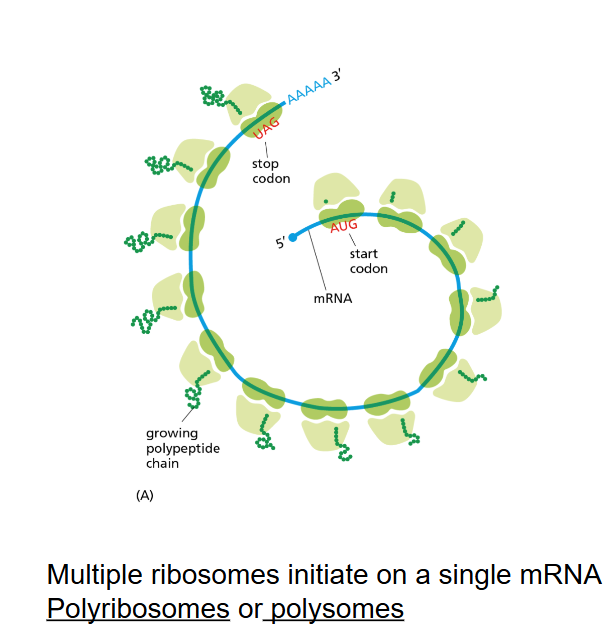
describe and analyze distinct regulatory steps starting from DNA that modulate protein expression
Regulatory step | How it changes protein expression |
|---|---|
Chromatin accessibility | Controls whether transcription machinery can reach the gene |
Transcription | Changes how much RNA is produced |
RNA processing | Changes which mature RNA forms, including through alternative splicing |
RNA export and localization | Controls whether and where the RNA becomes available for translation |
mRNA stability | Changes how long an mRNA remains available |
Translation | Changes how often ribosomes produce protein from the mRNA |
Protein processing | Folding, modification, and subunit assembly determine protein function |
Protein degradation | Changes how long the protein remains in the cell |
Explain and summarize the role of ubiquitin in tagging proteins for degradation and analyze the function of proteasomes in maintaining cellular protein quality
Ubiquitin is a small protein that can be covalently attached to a target protein. An appropriate polyubiquitin chain marks a protein for degradation by the proteasome.
The proteasome:
Recognizes the tagged protein.
Removes and recycles ubiquitin.
Unfolds the protein and feeds it into its internal chamber.
Breaks it into peptides, which other enzymes can further break down into amino acids.
This removes damaged or misfolded proteins and controls the lifespan of proteins that should act only briefly.
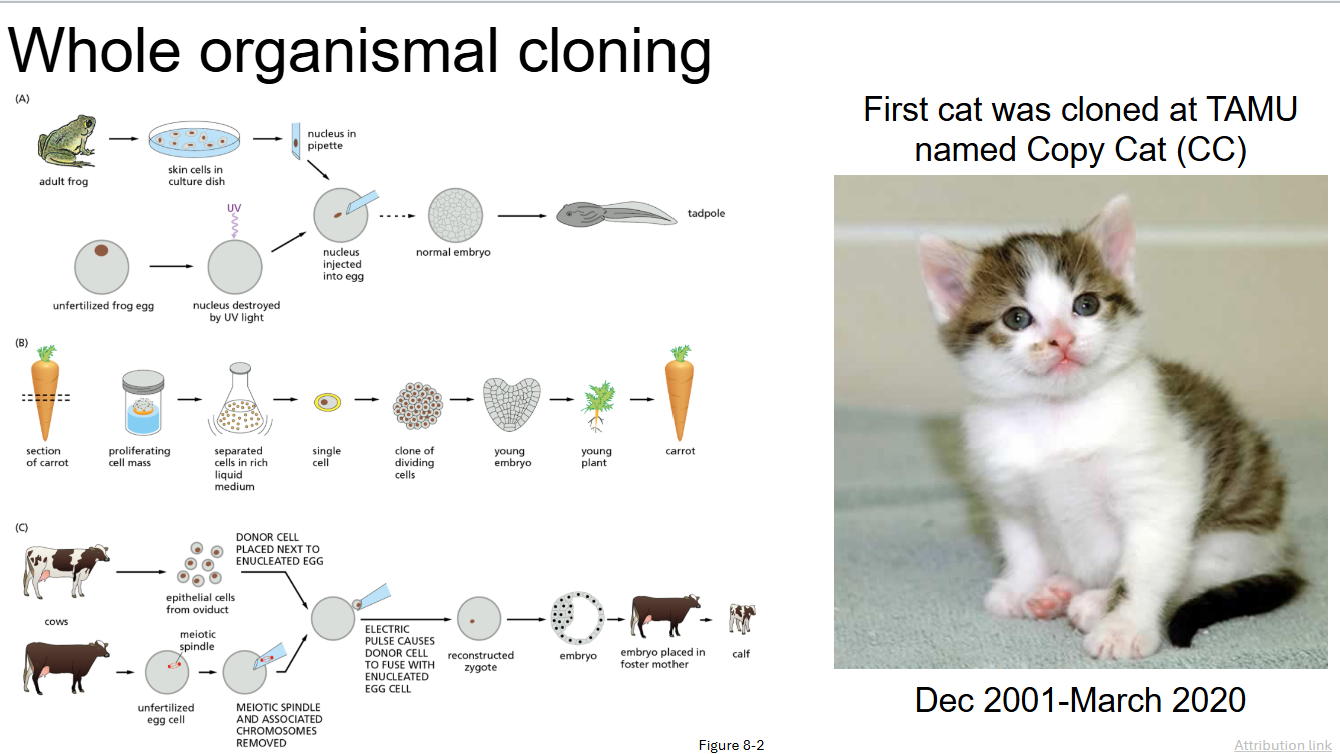
illustrate and interpret the basic steps involved in cloning a plant or animal, demonstrating understanding of the procedures and their significance
Animal cloning: somatic cell nuclear transfer
Take a somatic cell—a body cell—from the animal being cloned.
Remove the nucleus from an unfertilized egg.
Transfer the somatic cell’s nucleus into that egg.
Stimulate the egg to divide and form an embryo.
Implant the embryo into a surrogate mother.
The resulting animal has essentially the same nuclear DNA as the nucleus donor. Its mitochondrial DNA comes mainly from the egg donor.
Plant cloning
Isolate cells from a plant, such as a carrot.
Grow them in a nutrient medium with appropriate plant hormones.
Allow the cells to divide and develop shoots and roots.
Transfer the resulting plant to soil.
Significance: Differentiated cells generally retain the genetic information needed to produce an entire organism. Different cell types arise mainly because they express different genes, rather than possessing entirely different genes. Cloning requires resetting the cell’s gene expression program.
identify and classify at least one type of DNA-binding motif found in proteins, such as the helix-turn-helix or zinc finger
A DNA-binding motif is a recurring protein structure that helps a protein bind DNA. Transcription regulators use these structures to recognize regulatory sequences.
Motif | Structure and function |
|---|---|
Helix-turn-helix | Two α-helices connected by a short turn. A recognition helix fits into the DNA’s major groove and recognizes particular sequences. |
Zinc finger | A zinc ion stabilizes a folded structure that contacts DNA. Several fingers can recognize a longer sequence together. |
Proteins recognize the chemical features exposed by bases in the major groove, usually without separating the DNA strands.
Many DNA-binding proteins act as dimers, which can increase binding strength and sequence specificity.

explain and summarize the basic mechanism of the bacterial tryptophan and lactose operons, detailing regulatory elements and gene expression outcomes
An operon is a group of genes controlled together by a shared promoter, usually transcribed into one mRNA.
Key parts include:
Promoter: Where RNA polymerase binds to initiate transcription.
Operator: A regulatory DNA site where a repressor binds.
Structural genes: Encode the proteins needed for a shared function.
Tryptophan, or trp, operon
The trp operon encodes enzymes that make tryptophan.
Condition | What happens | Outcome |
|---|---|---|
Low tryptophan | Repressor is inactive and does not bind the operator | Operon ON; enzymes make tryptophan |
High tryptophan | Tryptophan binds and activates the repressor, allowing it to bind the operator | Operon OFF |
Tryptophan acts as a corepressor. This is negative feedback: once enough tryptophan exists, the cell stops making more.
Lactose, or lac, operon
The lac operon encodes proteins that help bacteria take up and break down lactose. It responds to both lactose and glucose.
Negative regulation by the lac repressor:
No lactose: The repressor binds the operator and blocks transcription.
Lactose present: Its derivative, allolactose, binds the repressor and reduces its ability to bind the operator.
Positive regulation by CAP:
Low glucose: cAMP increases and binds CAP. The CAP–cAMP complex binds DNA and helps recruit RNA polymerase.
High glucose: cAMP is low, so CAP provides little activation.
Lactose | Glucose | lac operon expression |
|---|---|---|
Absent | High or low | Essentially OFF |
Present | High | Low |
Present | Low | High |
Remember: The trp operon turns off when its product is abundant. The lac operon is expressed most strongly when lactose is available and glucose is scarce.

Compare and contrast bacterial and eukaryotic transcription regulation, analyzing key similarities and differences in control mechanisms
Both use sequence-specific DNA-binding proteins that activate or repress transcription. Environmental signals can change the activity of these regulators.
Feature | Bacteria | Eukaryotes |
|---|---|---|
Gene organization | Related genes may form an operon | Genes usually have individual promoters |
Regulatory sites | Often close to or overlapping the promoter | Can be close to or far from the promoter |
Activation | Activators often help RNA polymerase bind | Activators recruit transcription machinery, coactivators, and chromatin-modifying proteins |
Repression | Repressors often block polymerase binding or activity | Repressors can interfere with activators or recruit proteins that make chromatin less accessible |
DNA packaging | No nucleosomes in bacteria | Nucleosomes strongly affect access to DNA |
Initiation machinery | RNA polymerase uses sigma for promoter recognition | RNA polymerase II requires general transcription factors |
In eukaryotes, DNA looping brings distant enhancers and their bound activators near a promoter. Histone modification and chromatin remodeling can then make the promoter more accessible.
Define and evaluate the concept of combinatorial control in gene expression, assessing its impact on cellular regulation
Combinatorial control means a gene’s expression depends on the combined actions of several regulatory proteins.
For example, a gene might be strongly expressed only when:
Activator A is present.
Activator B is present.
Repressor C is absent.
Different genes respond to different combinations. Therefore, a relatively small collection of regulators can generate many distinct gene expression patterns.
This allows:
Cell specialization: Neurons and liver cells express different combinations of genes.
Coordinated responses: One regulator can influence multiple genes that share its binding sequence.
Development: Changing combinations of transcription factors guide cells toward different identities.
Gene expression can also vary in degree: combinations of regulators determine how much RNA is made, not simply whether a gene is on or off.
Distinguish and analyze various types of regulatory RNAs (e.g., miRNA, siRNA, incRNA) and explain their functions in gene expression control
Regulatory RNAs control gene expression without being translated into proteins.
RNA | Main characteristics | Effect on gene expression |
|---|---|---|
miRNA: microRNA | Usually produced from an endogenous RNA precursor that folds into a hairpin | Guides a protein complex to target mRNAs, reducing translation and promoting mRNA degradation |
siRNA: small interfering RNA | Usually produced from longer double-stranded RNA | Guides a protein complex to a highly complementary RNA, commonly causing its cleavage |
lncRNA: long noncoding RNA | Longer than about 200 nucleotides; has diverse structures and functions | Can recruit regulatory proteins, organize complexes, or influence chromatin and transcription |
miRNA and siRNA mechanism:
An RNA precursor is processed into a short RNA duplex; Dicer participates in this processing.
One strand becomes the guide RNA in RISC, the RNA-induced silencing complex.
The guide pairs with a target RNA.
RISC reduces expression by promoting RNA degradation or inhibiting translation.
Key distinction: miRNAs often pair imperfectly with several different mRNAs, whereas siRNAs commonly pair extensively with a particular target.
lncRNA example: Xist
Xist helps silence one X chromosome by coating it and recruiting proteins that promote repressive chromatin formation.
Distinguish and explain the differences between somatic cells and germ line cells, and analyze the consequences of mutations in each cell type
Somatic cells are body cells that do not directly contribute DNA to offspring, whereas germline cells are cells that give rise to eggs or sperm.
Somatic mutations | Germline mutations |
|---|---|
Occur in body cells | Occur in reproductive cell lineages |
Affect the individual | Can affect offspring |
Not normally inherited | Can be inherited |
May cause cancer or altered cell function | Can introduce heritable genetic variation |
Key concept: A mutation in a germline cell can be passed to offspring, whereas a mutation in a somatic cell generally affects only the individual.
The diagram on slide 11 illustrates that a germline mutation can appear in offspring throughout their body, while a somatic mutation affects only the descendants of the mutated body cell.

identify, describe, and classify types of mutations, and compare their potential effects on cellular function and heredity
A. Coding sequence mutations
These occur in protein-coding regions of DNA and can affect the resulting protein.
Mutation | Description | Potential effect |
|---|---|---|
Silent (synonymous) | Changes a codon without changing its amino acid | Often no change in protein sequence |
Missense | Changes one amino acid to another | May alter protein function |
Nonsense | Changes an amino acid codon into a stop codon | Produces a shortened protein |
Frameshift | Insertion or deletion shifts the reading frame | Alters downstream amino acids, often severely |
Important: Frameshift mutations occur when nucleotides are inserted or deleted in numbers that are not multiples of three.
B. Regulatory mutations
These occur in DNA sequences that control gene expression.
Can change when a gene is expressed.
Can change where a gene is expressed.
Can change how much of a gene product is produced.
Unlike coding mutations, regulatory mutations may change the amount or location of protein production without changing the protein's amino acid sequence.
C. Larger-scale genetic changes
Gene duplication: Produces an additional copy of a gene that can evolve a different function.
Exon shuffling: Combines exons from different genes to produce new protein-domain combinations.
Transposition: Moves mobile genetic elements to new DNA locations.
Horizontal gene transfer: Transfers genetic material between organisms without parent-to-offspring inheritance.
Exam takeaway: Be able to recognize the mutation type from a DNA diagram and explain whether it affects protein structure, gene expression, or genome organization.
interpret and evaluate neutral mutations, and assess their impact on genome evolution
A neutral mutation is a DNA change that has little or no effect on an organism's reproductive success.
Neutral mutations can occur in coding or noncoding DNA.
Examples include:
A silent mutation that does not change the amino acid.
A mutation in a noncoding region that does not significantly affect gene function.
A missense mutation that does not meaningfully affect protein function.
Why are neutral mutations important?
They contribute to genetic variation.
They can accumulate over generations through genetic drift.
They provide DNA differences that scientists can compare to study evolutionary relationships.
Important distinction:
Beneficial mutation: increases reproductive success in a particular environment.
Harmful mutation: decreases reproductive success.
Neutral mutation: has little or no effect on reproductive success.
The lecture's lactose tolerance example (slide 24) demonstrates a beneficial regulatory variant: continued lactase expression in adulthood became advantageous in certain populations with histories of cattle domestication.
Explain and summarize the origin of gene families, and illustrate the mechanisms that contribute to gene family diversification
A gene family is a group of related genes that originated from a common ancestral gene and have similar DNA sequences.
Mechanisms contributing to diversification
1. Unequal crossing-over
Homologous chromosomes misalign during recombination.
One chromosome receives an extra copy of a gene.
The duplicated gene can accumulate mutations independently.
2. Gene duplication and divergence
A gene is duplicated.
One copy can retain its original function.
The other copy can accumulate mutations and potentially develop a new function.
3. Whole-genome duplication
An organism gains additional copies of its entire genome.
Common in plants.
Examples from the lecture include potatoes (4N), wheat (6N), and some strawberries (8N).
4. Exon shuffling
Exons from existing genes are recombined.
This produces new combinations of protein domains and potentially new protein functions.
Example: Globin gene family
The globin gene family evolved through gene duplication and divergence.
An ancestral globin gene duplicated.
Different globin genes evolved, including α-globin and β-like globin genes.
Different globin proteins have specialized functions in oxygen transport.
Fetal γ-globin has a higher oxygen affinity, allowing oxygen transfer from mother to fetus.
Exam takeaway: Gene duplication provides extra genetic material that can evolve new functions without necessarily eliminating the original gene's function.

Draw and model the process of a transposon copying an exon from one gene to another, and explain how this event can affect genome evolution
Transposons are mobile genetic elements, sometimes called jumping genes, that can move to new locations in the genome.
Many DNA transposons encode transposase, an enzyme that recognizes transposon sequences and facilitates their movement.
Consequences for genome evolution
Creates new combinations of exons.
Can produce proteins with new functions.
Increases genetic diversity.
May disrupt existing genes when inserted into coding or regulatory regions.
Types of transposition
Type | Mechanism |
|---|---|
Cut-and-paste | DNA transposon moves from one location to another |
Replicative | Transposon is copied, with a copy inserted elsewhere |
Retrotransposition | DNA → RNA → DNA copy, which inserts at a new location |
Exam takeaway: Transposons can move genetic material between genes, producing new gene structures and contributing to genome evolution.

Construct and illustrate the mechanism of plasmid transfer between two bacteria, and describe its implications for genetic diversity
Horizontal gene transfer is the transfer of genetic material between organisms rather than from parent to offspring.
One mechanism is bacterial conjugation, which transfers plasmid DNA between bacteria.

Describe how sequence analysis tools can be used to locate and identify an open reading frame in a genomic sequence
An open reading frame (ORF) is a stretch of DNA that could encode a protein because it contains a continuous reading frame without an in-frame stop codon.
How to identify an ORF
Examine the DNA sequence in all six possible reading frames (three on each strand).
Look for a potential start codon, usually ATG in DNA.
Identify the sequence of codons that follows.
Find an in-frame stop codon (TAA, TAG, or TGA in DNA).
Evaluate whether the ORF could encode a functional protein.
Predict and compare evolutionary relationships by analyzing and interpreting DNA sequences across different organisms
Scientists compare DNA sequences to determine how closely organisms are related through common ancestry.
General rule: Organisms with more similar homologous DNA sequences are usually more closely related.
How to determine evolutionary relationships
Obtain homologous DNA sequences from different organisms.
Align the sequences.
Identify nucleotide similarities and differences.
Compare the number and pattern of mutations.
Construct a phylogenetic tree representing their likely evolutionary relationships.
define and describe key laboratory methodologies, including restriction enzymes, PCR, cDNA, and CRISPR
A. Restriction enzymes
Slides 5–6
Restriction enzymes are proteins that recognize specific DNA sequences and cut DNA at or near those sites.
Often recognize short, specific DNA sequences.
Can produce sticky ends (single-stranded overhangs) or blunt ends (no overhangs).
Allow scientists to cut DNA into fragments for cloning and analysis.
Key concept: Restriction enzymes act as molecular scissors that cut DNA at specific sequences.
B. Polymerase chain reaction (PCR)
Slides 13–15
PCR is a laboratory technique used to amplify a specific DNA sequence, producing millions of copies from a small amount of starting DNA.
PCR requires a DNA template, primers, nucleotides, and a thermostable DNA polymerase.
It involves three repeating steps:
Denaturation: Heat separates the two DNA strands.
Annealing: Cooling allows primers to bind to complementary DNA sequences.
Extension: DNA polymerase synthesizes new DNA strands.
Key concept: PCR amplifies a specific DNA region so it can be detected or studied.
C. Complementary DNA (cDNA)
Slide 16
cDNA is DNA synthesized from an RNA template using the enzyme reverse transcriptase.
Typically made from mature mRNA.
Represents genes that were being expressed in the sampled cells.
Generally lacks introns because mature mRNA has already undergone splicing.
Can be used to study gene expression or clone protein-coding sequences.
Key concept: cDNA represents RNA-derived sequences, whereas genomic DNA contains the organism's original DNA sequences, including introns and regulatory regions.
D. CRISPR-Cas genome editing
Slide 27
CRISPR-Cas is a genome-editing system that allows scientists to target and modify specific DNA sequences.
A guide RNA directs a Cas protein to a complementary target sequence.
The Cas protein can cut the DNA.
Cellular DNA repair processes can produce mutations or enable a targeted genetic modification.
Key concept: CRISPR allows targeted changes to DNA and is used to investigate gene function.
Summarize the process and purpose of techniques like gel electrophoresis and Sanger sequencing.
A. Gel electrophoresis
Slides 7–8
Gel electrophoresis separates DNA fragments based on their size.
How it works:
DNA samples are placed into wells in a gel.
An electric current is applied.
Negatively charged DNA migrates toward the positive electrode.
Smaller DNA fragments move through the gel faster and travel farther than larger fragments.
DNA bands are visualized and compared with a DNA ladder to estimate fragment sizes.
Purpose: To separate, visualize, and estimate the sizes of DNA fragments.
B. Sanger sequencing
Slides 17–18
Sanger sequencing is a method used to determine the nucleotide sequence of DNA.
It uses normal DNA nucleotides (dNTPs) and special chain-terminating nucleotides (ddNTPs).
How it works:
DNA polymerase synthesizes complementary DNA strands.
Occasionally, a fluorescently labeled ddNTP is incorporated.
Because ddNTPs lack a 3′-OH group, DNA synthesis stops when one is incorporated.
DNA fragments of different lengths are separated.
The fluorescent labels are detected to determine the DNA sequence.
Purpose: To identify the order of nucleotides (A, T, G, C) in a DNA fragment.
Exam takeaway: Gel electrophoresis separates DNA by size, while Sanger sequencing determines the nucleotide order of DNA.
Illustrate how polymerase chain reaction (PCR) is used in laboratory and clinical settings
PCR is used to make many copies of a specific DNA region, allowing scientists to analyze DNA even when the original sample is very small.
Step | What happens | Typical temperature |
|---|---|---|
1. Denaturation | Double-stranded DNA separates into single strands | ~95°C |
2. Annealing | Primers bind to complementary DNA sequences | ~50–65°C |
3. Extension | DNA polymerase synthesizes new DNA strands | ~72°C |
Applications
Laboratory applications:
Amplifying DNA for sequencing.
Detecting specific genes or genetic variants.
Preparing DNA fragments for cloning.
Clinical applications:
Detecting genetic mutations associated with disease.
Identifying pathogens in patient samples.
Supporting diagnosis of inherited genetic conditions.
Exam takeaway: PCR repeatedly separates DNA, binds primers, and synthesizes new strands to amplify a specific DNA sequence.

Compare the differences between genomic DNA and cDNA and discuss their applications
Genomic DNA vs. cDNA
Feature | Genomic DNA | cDNA |
|---|---|---|
Source | DNA extracted from cells | RNA converted into DNA |
Introns | Present in genes that contain introns | Generally absent when made from mature mRNA |
Exons | Present | Present in processed transcript |
Regulatory regions | Present | Generally absent |
Represents | Genome, including coding and noncoding regions | RNA sequences expressed in sampled cells |
Enzyme used to produce it | Not required for extraction | Reverse transcriptase |

Describe the workflow needed to clone a gene using plasmids and discuss how transgenic animal models can be used to study human disease
Gene cloning workflow
Isolate the gene of interest: Obtain the DNA sequence that scientists want to clone.
Cut the DNA and plasmid: Use restriction enzymes to produce compatible DNA ends.
Join the DNA fragments: DNA ligase joins the gene of interest to the plasmid, creating recombinant DNA.
Introduce the plasmid into bacteria: The recombinant plasmid enters bacterial cells through transformation.
Select transformed bacteria: Use a selectable marker, such as antibiotic resistance, to identify bacteria carrying the plasmid.
Allow bacteria to reproduce: As bacteria divide, they replicate the plasmid, producing many copies of the inserted gene.
Key concept: Plasmids act as DNA carriers, while DNA ligase joins DNA fragments to create recombinant plasmids.
1. Transgenic animals (slide 25)
Scientists introduce a gene into an animal to study how its expression affects the organism.
2. Knockout mice (slide 26)
A knockout mouse has a specific gene inactivated.
Scientists observe what happens when the gene no longer functions.
This helps determine the gene's normal role.
The resulting phenotype may provide insight into human disease.