Bio Bases based on study guide
 Call Kai
 Learn
Learn Practice Test
 Spaced Repetition
 Match
Match Flashcards
 Knowt Play
1/71

There's no tags or description
Looks like no tags are added yet.
Name | Mastery | Learn | Test | Matching | Spaced | Call with Kai | Chat |
|---|
No analytics yet
Send a link to your students to track their progress
72 Terms
Reasons a behavior occurs.
Unconscious sensory processing which triggers the nervous system
Hormonal triggers
Outside stimulus
Processes that occurred anywhere from seconds and minutes to days and months ago
Frontal lobe development including learning hx, base rate of trauma and PTSD
Interoceptive info
Intention
Define interoceptive information.
Perception and interpretation of the body’s internal signals such as hunger, pain, emotional states.
Prisoner’s Dilemma v Wall Street Game v Communist Game.
Identify and describe Luria’s Functional Units
Attention: Regulation of cortical tone (mental alertness/wakefulness) involves the brain stem, reticular activating system. Regulates how much arousal/activation for a particular stimuli. Operates according to Law of Strength.
2. Responsible for receiving/analyzing info -> operates via simultaneous and successive processing. Involves parietal, occipital, and temporal lobes. Operates law of sequences, which creates a sequential/series order.
3.Promotes/regulates intentions and plans. Executes the plans and verifies outcome compared to plan -> frontal lobe/prefrontal area.
Law of Strength
A strong stimulus causes a strong response, weak stimulus causes a weak response
Law of Simultaneous Processing
Information is pieced together all at one time into a unified and holistic view (big picture)
Law of Successive Processes
Information is processed in an ordered and sequential manner, usually chain-like where one progression is related to the next
Name two types of cells in the CNS
Glia + neurons
Four morphological components of the neuron.
Soma, dendrite, axon/axon hillock, end bouton
Describe cell body, dendrites, axons, myelin sheath, end bouton.
Cell body: also known as the soma, contains cellular structures such as the nucleus, golgi apparatus, ribosomes, and the mitochondrion.
Dendrites: branch-like structures protruding from the soma. They receive electrical/chemical signals from other nerve cells and carry them to the cell body (soma).
Axons: originates at axon hillock, conducts electrical impulses away from neurons
Myelin sheath: envelops axon and propagates speed of action potential via nodes of ranvier. Keeps electricity inside axon.
End bouton: site at end of axon where signals are passed from one neuron to another. Translates electrical messages into chemical.
Draw a neuron with all of its structures.

How is the resting potential of a cell maintained?
The resting potential of a cell is maintained through the selective permeability of the cell to K+ via diffusion, with 3 Na+ being let out for every 2 K+. Basically, through a combination of ion transportation and passive ion diffusion.
How is achievement of the critical threshold determined?
The threshold is determined around -55mv, when the balance of sodium coming into the cell overpowers that of the outward potassium current.
How is the speed of an action potential determined?
The speed of the action potential is propagated by the electrical reaction of the action potential within the Nodes of Ranvier and the environment outside the cell as they are opposite and cause the AP to “hop” down the axon.
What is the role of the presynaptic membrane?
Site of where chemical signaling is initiated/sends the neurotransmitters, recycling and reuptake and neurotransmitters, has autoreceptors that act as a way to inhibit the release of more NTs
What is the role of the postsynaptic membrane?
Receives neurotransmitters on their receptors, activates and releases ionotropic or metabotropic to those receptors, create either excitatory synaptic potential for firing of an action potential or inhibitory synaptic potential to make firing less likely.
Explain the process of neurotransmitter release, crossing the synaptic cleft, and connecting to the postsynaptic membrane.
1. AP reaches the presynaptic axon terminal causing depolarization in the presynaptic membrane and an influx of Ca+
2. Vesicles filled with neurotransmitters are triggered by the Ca+ to merge with the presynaptic membrane
3. Vesicles release neurotransmitters into the presynaptic cleft
4. They diffuse across the cleft and into the receiving cell and bind to specific receptors on the postsynaptic membrane using a lock/key method
5. Leftover neurotransmitters are taken via reuptake
Neurotransmitter specificity of sending neuron.
All neurons are equipped with all neurotrans
Principles of dynamic polarization and connection specificity.
Dynamic polarization: states that electrical signals within a nerve cell flow in only one direction: from the postsynaptic sites of the neuron, usually the dendrites and cell body, to the trigger region at the axon
Connection specificity: states that nerve cells do not connect randomly with one another in the formation of networks but make specific connections—at particular contact points—with certain postsynaptic target cells and not with others.
Explain the characteristics of unipolar, bipolar, anaxonal, and multipolar neurons.
Unipolar: single primary process, highly sensory, gives rise to many branches, highly efficient
Bipolar: 2 distinct processes, oval soma, dendritic structure receives signals from other neurons and axon carries it towards the CNS. Largely sensory, short, and efficient
Multipolar: multiple dendrites plus a single extended axon. Receives information from many sources, is the most abundant of the neurons, very efficient, varies in size and length.
Anaxonal: has no axon, with multiple processes, signals from the cell body but doesn’t transmit long distance information; Modulates to integrate information in short distances.
Describe the three nerve cell classifications.
Sensory: carry information from the body's peripheral sensors into the nervous system for the purpose of both perception and motor coordination (afferent)
Motor: carry commands from the brain or spinal cord to muscles and glands (efferent information)
Interneuron: relay and local. Relay neurons have are long and convey signals over considerable distances while local neurons form connections with nearby neurons in local circuits
Describe the two types of interneurons.
relay and local. Relay neurons have are long and convey signals over considerable distances while local neurons form connections with nearby neurons in local circuits
Describe the different types of glial cell, along with their roles.
Astrocyte: provides scaffolding
Microglia:
Roles of the nucleus and mitochondria in cell body.
The nucleus holds the DNA of the cell
Mitochondria produces the ATP needed to power the cell
Dendritic spines.
Tiny, branch-like structures which extend out from the dendrites and increase surface area and exposure for receiving incoming electrical or chemical signals
Axon hillock.
Conducts AP, enveloped by myelin sheath, is the accountant of the cell (makes sure excitatory and inhibitory processes are accounted for)
Nodes of Ranvier.
Structures in the axon which are not covered by the myelin sheath, propagate an action potential through opposite electrical charge
Electrochemical properties of the neuron at resting potential.
-65/70mv
Graded potentials.
depolarizations or hyperpolarizations that occur in dendrites, soma, or near the axon terminals, occur when ion channels open or close causing ions to enter or leave the neuron, their size or amplitude is directly proportional to the strength of the triggering event.
Identify the six stages of the action potential.
Na+ enters the cell as cell channels open
K+ leaves the cell via electrostatic diffusion
Na+ channels become refractory as no more electrical charge is needed for an action potential
K+ continues exiting the cell, polarizing it
K+ channels close, Na+ channels reset
Any remaining K+ lingering outside the cell is cleaned up by microglia
Describe the process of synaptic transmission.
a. Neurotransmitter is synthesized and then loaded into synaptic vesicles
b. The vesicles fuse to a presynaptic terminal
c. The NT spills into the synaptic cleft and binds to postsynaptic receptors, but only to those they are keyed for
d. A biochemical or electrical response is elicited in postsynaptic cell
e. The neurotransmitter is removed via enzymes or reuptake by the presynaptic neuron or glial cell
Ionotropic v metabotropic receptor characteristics.
Ionotropic receptors mediate fast and efficient transmission. They can generate postsynaptic potentials, are part of the receptor directly controlling the connection to the postsynaptic membrane and provide a quick change of ionic charge. Part of the same ionic channel that a neurotransmitter will lock/key on.
Metabotropic receptors require extra proteins to open and connect to an ionic channel, energy is brought in alongside the proteins to open the channel, and are less efficient and slower than ionotropic due to the extra steps involved in synaptic transmission
Processes (2) involved in NT re-uptake.
Ionic charge releases the connection between a neurotransmitter and the postsynaptic membrane. Then enzymes begin to break them down to return to the vesicle or be treated as waste product. NT are returned to presynaptic membrane via transport proteins
Polarization v hyperpolarization v depolarization.
Polarization: resting state of -70mv
Hyperpolarization: Comes back from 30mv and overshoots past -70mv
Depolarization: stimulus brings it to -50mv due to Na+ flowing in, moves it to 30mv
Neuronal integration.
process by which multiple synaptic potentials combine within one postsynaptic neuron, in some cases triggering one or more action potentials
Autoreceptors.
regulate activity of sending neuron, how much of a neurotransmitter is sent out. Excitatory or inhibitory depending on need
Heteroreceptors.
receive NT received by other neurons. sensitive to neurotransmitters and hormones that are not released by the cell on which it sits
Fissure v sulcus v gyrus
Fissure: division, divides brain structures
Sulcus: shallow grooves along the cortex
Gyrus: bumps along the cortex
Be able to describe the various planes/directional indicators for human neuroanatomy.

Longitudinal fissure
Divides the brain in 2
Four lobes of the cerebral cortex
Frontal, temporal, parietal, occipital
Be able to diagram the primary, secondary, and tertiary cortex in each lobe, and explain their general functions.
Primary: takes raw data of sensation or motor function
Secondary: puts together raw data w/other info of likeminded neurons (synthesized)/(secondary association)
Tertiary: largely parietal lobe + integrates with other data available big picture is put together in almost reverse order

Location and function of the medulla.
HR, BP, sneezing, some consciousness, coughing, vomiting, base of reticular activating system: arousal, sleep
well-protected but can be pushed/shut-down w/brain swelling

Location and function of the pons.
fine motor tuning, role in pain mediation, analgesic, respiration, arousal of facial expressions, motor messages from cortex to cerebellum

What is Locked in syndrome?
Disease primarily affecting the pons in which you are conscious and aware of yourself (cognitively all there) but have no ability to move or control your limbs or functions
Pathway of the RAS and functions.
Starts in medulla in upper brainstem, is about two inches long. Extends into the pons and reaches into midbrain. From there it splits into two paths: one into the thalamus and the other directly in the cerebral cortex. (ascending signals go to general sensory tracts and descending motor projections go to spinal cord)

Location of the midbrain.
upper part of brainstem

Functions of the tegmentum.
responsible for integration + pain, movement, arousal, relays inhibitory signals to the thalamus and basal nuclei preventing unwanted body movement
Structures within the tectum and their functions.
Superior colliculi: visual reflect center, helps visually track moving objects and control reflexes in response to visual stimulus
inferior colliculi: auditory reflex centers, control reflexive response to sound
Location and function of the cerebellum.
coordinates movement, fine tunes/smoothes out movements + movement timing

Symptoms associated with damage to the cerebellum.
Intention tremor, difficulty with balance, ataxia, gait issues
Identify the structures in the Limbic System.
Hippocampus
Thalamus
Amygdala
Limbic cortex
Hypothalamus
Role of Amygdala.
assigns emotional significance to stimuli
Role of Nucleus Accumbens.
in middle of central sulcus and communicates with cortex. Plays a role in reinforcing properties of stimuli as a dopaminergic system and plays role in motor learning
Role of the hippocampus.
Within the inferior temporal lobe, involved in formation of new memories (verbal + nonverbal), susceptible to stress, plays role in episodic memory, spatial navigation, temporal coding and allows for multi-sensory memory system.
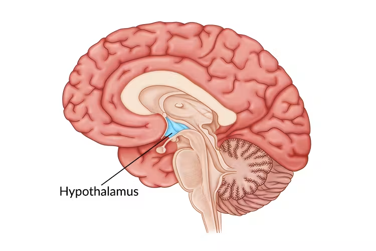
Location and function of the hypothalamus.
inferior to thalamus (look for egg), integrates info from NT and hormones indicating a change in bodily state. Wants to establish homeostasis based on inputs. Controls pituitary gland, produces male/female growth hormones, regulates sleep, antidieuretic hormone

Location and function of the thalamus.
one half in each hemisphere and divided by third ventricle, sensory relay station for all senses except olfaction. Participates in sleep cycle activation and the RAS
Location and structures of the basal ganglia.
Caudate nucleus and putamen (together called striatum), globus pallidus, connected to substantia negra

Structures comprising the striatum.
Caudate nucleus and putamen
Functions of the caudate nucleus and putamen.
Caudate nucleus: plays role in associative learning, involved in motor control (selects and initiates voluntary movements
putamen: initiates, coordinates, fine tunes movement, also plays role in procedural learning habits
Role of substantia nigra.
A cell group in the midbrain that uses dopamine as a neurotransmitter and innervates the striatum, so it plays a function in motor control
Basal ganglia disorders.
resting tremors, bradykinesia, parkinson’s, Huntington’s, micrografia, tardive dyskinesia, choreoathetoid, dystonia
Role of cranial nerves.
They have different motor and sensory functions including aiding in smell, vision, facial sensations, swallowing and speech, and hearing and balance
Vagus nerve.
longest cranial nerve that goes down to heart and stomach from the brainstem, aids with swallowing, speech, and cardiac innervation, both a sensory and motor nerve
Functions of the ventricles.
CSF production, storage, circulation, nutrition delivery to brain, buoyancy/buffer for brain, waste management
Location and role of choroid plexus.
In the 3rd and 4th ventricles, basal ganglia area.
Role: produces CSF, maintains the blood-brain barrier

Flow of cerebrospinal fluid.
lateral ventricle → inter-ventricular foramen → third ventricle → cerebral aqueduct → fourth ventricle → Luschka and Magendie foramens → subarachnoid space → subarachnoid granules → blood stream via sinuses
Layers of the meninges (DAP).
dura mater, arachnoid mater, and pia mater
Functions of the meninges.
serve as a shock absorber to not damage cortex, provides nutritional support (O2, blood, glucose), structural (holds brain in place)
Describe the blood supply distribution of anterior, middle, and posterior cerebral arteries. In broad terms, understand the deficits you might find with damage to these areas.
Anterior cerebral artery supplies to
Describe the major arteries which bring blood to the brain.
69.
Know the major conclusions for the three in-class hormone presentations.
Estrogen: supports regulation back to emotional baseline, dendritic growth
Cortisol: long-term exposure to cortisol is highly correlated with alterations in brain structure and cognitive impairment