CARE: SKIN INTEGRITY (PRESSURE INJURIES AND CONTACT DERMATITIS)
 Call Kai
 Learn
Learn Practice Test
 Spaced Repetition
 Match
Match Flashcards
 Knowt Play
1/91

There's no tags or description
Looks like no tags are added yet.
Name | Mastery | Learn | Test | Matching | Spaced | Call with Kai | Chat |
|---|
No analytics yet
Send a link to your students to track their progress
92 Terms
pressure injury
Skin and/or underlying tissue damage caused by prolonged pressure or pressure combined with shear.
commonsites for pressure injuries
Sacrum
Heels
Trochanters
Ischial tuberosities
Elbows
Occiput
what may pressure wounds present as
intact skin, an open wound, deep discoloration, pain, or device-shaped injury.
when are pressure injuries preventable
when risk is identified early and prevention is consistent
pressure
Tissue compressed between bone and surface
EX: sacral injury from bedrest
nursing prevention for pressure
Reposition, offload, support surfaces
shear
Skin stays in place while deeper tissue slides
EX: sliding down in bed with HOB elevated
nursing prevention for shear
Lift, don’t drag; limit HOB when appropriate
friction
skin rubs against surface
elbows/heels rubbing on linens
nursing preventions for friction
Protective dressings, smooth linens, careful transfers
moisture
Skin becomes overhydrated and fragile
EX: incontinence- associated dermatitis
nursing prevention for moisture
Cleanse, dry, barrier cream, toileting plan
who is vulnerable for pressure injuries
Immobility: unable to shift weight
Poor perfusion : PVD, shock, diabetes
Altered sensation: spinal cord injury, neuropathy
Moisture: incontinence, diaphoresis
Poor nutrition: low protein/calories, dehydration
Age/frailty: thin skin, reduced reserve
Medical devices: oxygen tubing, masks, casts
Medications: sedatives, vasopressors, steroids
what does the braden scale do
Identifies risk level and guides prevention bundles for pressure injuroes
for the braden scale the lower the score
the higher the risk
braden scale categories
Sensory perception
Moisture
Activity
Mobility
Nutrition
Friction/shear
what does the braden score not replace
Focused skin assessment
Clinical judgment
Device checks
Patient-specific interventions
when is a risk score useful
only if it triggers appropriate prevention
wound assessment components
Location: Anatomic site; relationship to bony prominence or device
Size: Length × width × depth in cm
Wound bed: Granulation, epithelialization, slough, eschar
Drainage: Amount, color, consistency, odor
Edges/margins: Attached, rolled/epibole, undermining, maceration
Periwound skin: Erythema, warmth, induration, moisture, pain
Tunneling/undermining: Clock method and depth
wound assessment what to look for
Persistent color change
Purple, blue, gray, or ashen tone
Localized shiny or taut skin
wound assessment what should you feel
Temperature difference
Boggy or firm tissue
Swelling or induration
what should you ask the patient during a wound assessment
Pain
Burning
Tingling
Change in sensation
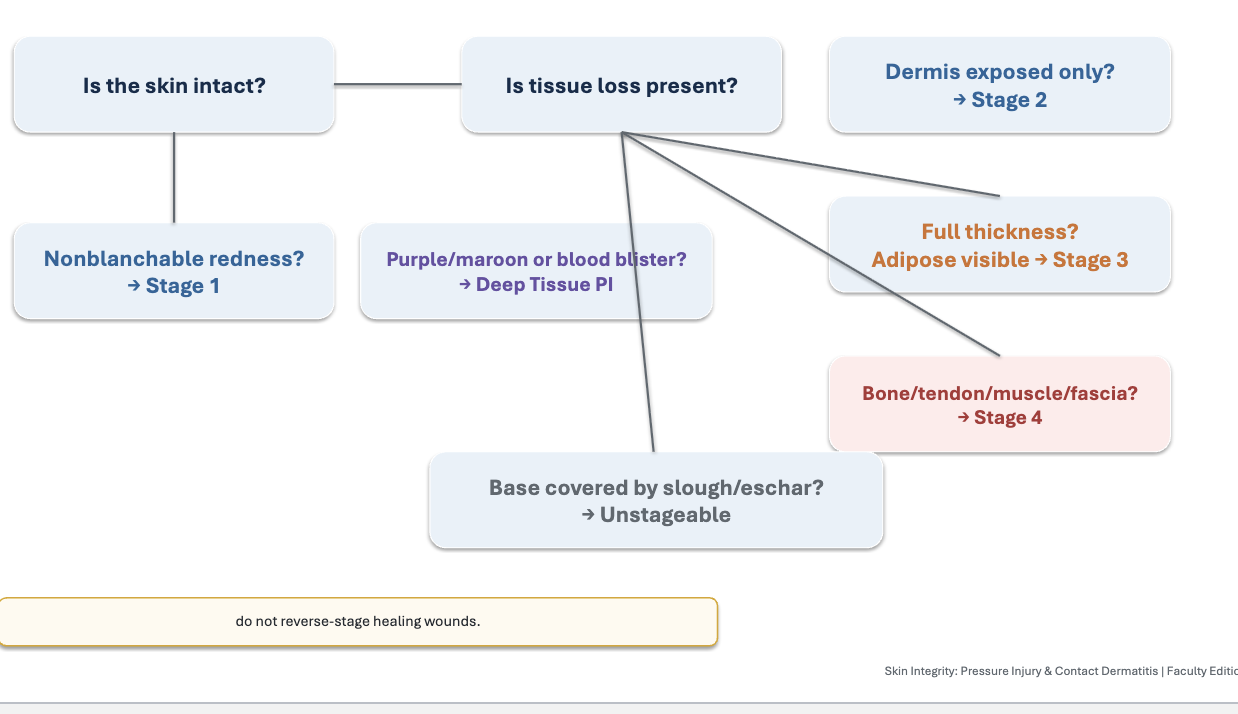
Stage 1 pressure ulcer key finding
Nonblanchable erythema of intact skin
stage 1 pressure ulcer what you see
Skin intact; may show temperature, sensation, or firmness changes
Stage 2 pressure ulcer key finding
Partial-thickness skin loss with exposed dermis
Stage 2 pressure ulcer what you see
Pink/red moist wound bed or serum-filled blister; no adipose tissue
Stage 3 pressure ulcer key finding
full thickness skin loss
Stage 3 pressure ulcer what you see
Adipose may be visible; slough/eschar may appear; no exposed bone/tendon/muscle
Stage 4 pressure ulcer key finding
Full-thickness skin and tissue loss
Stage 4 pressure ulcer what you see
Exposed or palpable fascia, muscle, tendon, ligament, cartilage, or bone
unstageable pressure ulcer key finding
depth is obscured
unstageable pressure ulcer what you see
Slough or eschar prevents visualization of wound base
deep tissue pressure injury key finding
Persistent deep red/maroon/purple discoloration
deep tissue pressure injury what you see
Intact or non-intact skin; damage begins in deeper tissue
pressure injury staging decision tree
Medical Device-Related Pressure injury
Caused by diagnostic or therapeutic devices.
Shape often matches device.
Stage using usual staging system
mucosal membrane pressure injury
Occurs on mucous membranes from device use.
Examples: ET tube, NG tube, urinary catheter.
Cannot be staged
STOP Stable Heel Eschar
Dry, adherent, intact eschar on ischemic heel/limb is usually left intact unless ordered otherwise.
device related injuries are
preventable
inspect under and around devices
Stage 1 Pressure Injury: Non- Blanchable Erythema of Intact Skin (NPIAP)
• Intact skin with a localized area of non-blanchable erythema.
• May be preceded by blanchable erythema or by changes in sensation, temperature, or firmness
• Does not include purple or maroon discoloration
• May appear differently in darkly pigmented skin.
Stage 2 Pressure Injury: Partial-Thickness Skin Loss with Exposed Dermis (NPIAP)
• Partial thickness loss of skin with exposed dermis
• Wound bed is pink or red and moist
• May occur as an intact or ruptured serum filled blister
• Adipose or deeper tissues are not visible
Stage 3 Pressure Injury: Full-Thickness Skin Loss (NPIAP)
• Full thickness loss of skin
• Granulation tissue and epibole often present
• Slough and or eschar may visible
• May be undermining or tunneling
• Depth varies by anatomic location and degree of adiposity
The injury can extend down to, but not through underlying fascia; there is no exposed muscle or bone.
Stage 4 Pressure Injury: Full-Thickness Skin and Tissue Loss (NPIAP)
• Exposed or directly palpable fascia, muscle, tendon, ligament, cartilage, or bone in the wound.
• Slough and or eschar may be visible
• Epibole, undermining, and or tunneling may occur
• Depth varies by anatomic location
Pressure Injury Prevention Bundle
Risk assess: Braden + clinical judgment
Inspect skin: head-to-toe + under devices
Reposition: turn schedule; offload heels
Manage moisture: cleanse, dry, barrier product
Reduce shear: lift, don't drag; HOB mindful
Support surfaces: mattress/chair cushions
Nutrition: protein, calories, hydration
Mobilize: ambulate/ROM as able
These preventions work best as a bundle not individual interventions
What not to do to a pressure injury
✕ Do not massage bony prominences: May damage fragile tissue.
✕ Do not drag patients across linens: Increases friction and shear.
✕ Do not use donut cushions: Can concentrate pressure.
✕ Do not leave heels resting on bed: Offload heels completely.
✕ Do not ignore device pressure: Assess under tubing, masks, braces, collars.
✕ Do not remove stable heel eschar: Unless ordered/clinically indicated
why is protein and calories needed for for pressure injuries
Needed for tissue repair, immune function, and collagen synthesis.
what is hydration needed for pressure injuries
Supports perfusion and skin turgor
what to monitor during hydration
Monitor intake, output, and contraindications
what micronutrients are needed for pressure injuries
Vitamin C, zinc, copper, vitamin A may be considered when deficient or ordered.
when is wound healing impaired
when perfusion, nutrition, or infection control is inadequate
wound care principles: think TIME
TIME
TIME stand for
Tissue
infection/inflammation
mositure balance
edge
tissue clincial question
Is nonviable tissue present?
tissue nursing action
Debridement when appropriate; protect stable heel eschar
infection/inflammation clinical question
Are there local or systemic infection cues?
infection/inflammation nursing action
Culture/antibiotics if ordered; report fever, odor, purulent drainage
moisture balance clinical question
Is the wound too dry or too wet?
moisture balance nursing action
Select dressing to maintain moist healing and protect periwound
Edge clincial question
are edges advancing
edge nursing action
Monitor size/depth, epibole, undermining; consult WOC nurse
dry wound bed dressing goal
Add/maintain moisture as ordered
heavy exudate wound dressing goal
Absorb drainage and protect periwound skin
Fragile surrounding skin dressing
Use gentle adhesive or non-adhesive options
tunneling/dead space wound goal
Fill dead space without overpacking
infection concern dressing
Assess/report; antimicrobial dressing may be ordered
high risk bony area dressing
Protective foam may reduce friction/shear
When to consult WOC nursing
Unstageable or deep tissue pressure injury
Stage 3 or Stage 4 pressure injury
Rapidly worsening wound
Complex dressing needs
Heavy exudate, odor, tunneling, or undermining
Concern for infection or delayed healing
Medical device-related injury
Need for patient/family education on wound care
team members for WOC nursing
WOC nurse, provider, dietitian, PT/OT, case management, patient/family
WOC nursing prioirty
Escalate early. Waiting often increases wound severity and cost
Complications of Pressure Injury
Local infection: increased pain, erythema, drainage, odor
Cellulitis: spreading warmth/redness/swelling
Osteomyelitis: bone involvement, delayed healing
Sepsis: fever, tachycardia, hypotension, altered LOC
Pain/QOL: sleep disruption, mobility limits, distress
Cost/LOS: longer hospitalization and more intensive care
systemic infection cues from pressure injuires require
prompt escalation
Irritant Contact Dermatitis: mechanism
Direct skin damage from irritant
Irritant Contact Dermatitis: common triggers
Soaps, detergents, chemicals, frequent hand hygiene
Irritant Contact Dermatitis: timing
Can occur quickly after exposure
Irritant Contact Dermatitis: symptoms
Burning, pain, redness, dryness/fissures
Irritant Contact Dermatitis: mangament
Remove irritant, protect barrier, moisturize
Allergic Contact Dermatitis: mechanism
Delayed immune hypersensitivity reaction
Allergic Contact Dermatitis: commn triggers
Latex, nickel, poison ivy, adhesives, fragrances
Allergic Contact Dermatitis: timing
Often delayed 24–72 hr after exposure
Allergic Contact Dermatitis: symtoms
Intense itching, erythema, vesicles, oozing/crusting
Allergic Contact Dermatitis: management
Avoid allergen, topical steroids if ordered, antihistamines for itching
what are you doing when doing an assessment of contact dermitisis
find the exposure pattern
contact dermatitis history
New soaps, detergents, gloves, plants, metals, adhesives, workplace chemicals, skin products.
contact dermatitis distribution
Where is the rash?
Does it match a device, jewelry, glove, plant exposure, or topical product
contact dermatitis complications
Scratching can lead to secondary infection.
Assess for drainage, warmth, fever,
spreading erythema.
Contact Dermatitis Management
remove
relieve
treat
protect
Contact Dermatitis Management: remove
Stop exposure to irritant/allergen
Contact Dermatitis Management: relieve
Cool wet compresses, reduce itching
Contact Dermatitis Management: treat
Topical corticosteroid or antihistamine if ordered
Contact Dermatitis Management: protect
Barrier creams, gloves, trigger avoidance
contact dermatitis patient teaching
Avoid scratching. Keep nails short. Report infection signs.
Contact Dermatitis: safety→ allergy
Latex allergy requires clear documentation and latex-free supplies.