Neuropsychology
 Call Kai
 Learn
Learn Practice Test
 Spaced Repetition
 Match
Match Flashcards
 Knowt Play
1/212

There's no tags or description
Looks like no tags are added yet.
Name | Mastery | Learn | Test | Matching | Spaced | Call with Kai | Chat |
|---|
No analytics yet
Send a link to your students to track their progress
213 Terms
Brain & Behaviour throughout time
Aristotle: nonmaterial mind guides behaviour
Descartes: mind (pineal gland) decides movement of the body
Darwin: behaviour originates in brain activity
Natural selection: environment plays a role
Today: mind originates from brain function
Behaviour depends on both
Donald Hebb
Considered the ‘father of neuropsychology’
Hebbian theory = neural pathways develop based on experiences; as pathways are used more, they become faster and stronger
Early neuropsychology was closely linked to brain injury and dementia research and diagnosis
Relationship between loss of brain function and change in thoughts / behaviors easier to observe
Modern neuropsychology includes a variety of disorders
The Hebbian assumption of change
Environment, culture, customs, family history, lifestyle, and more shape who we are
In neuropsychology, we focus a lot on thoughts, behaviours, and their relationship to the brain
However, much relevant information is found outside psychology and the brain that influence both
For example, cloned mice do not have identical behaviours
Plasticity, flexibility, and adaptability are fundamental properties of the brain
Neuropsychology is founded upon the assumption of change
There is no rehabilitation unless the brain can change
Therapies would not work unless psychologies can change
Hebbian plasticity is the foundation of neuropsychology

Neurological examination
Patient’s history
State of awareness
Alert, drowsy, stupor, confused
Speech abnormalities, facial asymmetries, body posture
Emotions (agitated, anxious, depressed, apathetic, restless)
Physical examination
Blood pressure, brain imaging, reflexes, pain, muscle movement, smell, etc.
Disorders
Strokes, injuries, and lesions may show asymmetry, loss of function
Parkinson’s may show loss of smell and motor changes
Dementia may show memory loss, disorientation, or agitation
Biopsychosocial model of neuropsychological assessments
Neuropsychological assessment
Combines many tests depending on patients’ symptoms
May include IQ, cognitive, and psychometric tests
Biopsychosocial model
Social support networks (friends, family) influence outcomes
Patients’ sense of wellbeing influence outcomes
Sometimes a mismatch between the patients’ needs and their social network (e.g., patient wanted to stay home, family thinking they should work) or environment (e.g., needing a quiet place to sleep but living somewhere noisy) can add stress that may impair healing
How did the biopsychosocial model change neuropsychology?
Premise: A person is not made of isolated organs, but functions as a whole
You need good biological, social, and psychological circumstances to have good mental health
First conceptualized in 1950 by Roy Grinker - applied "bio" to psychology
Proposed as a medical perspective by George L. Engel in 1977 - applied "social" to medicine
Example from Dementia
Dementia
Umbrella term for impaired memory, cognition, and decision-making
Common causes: Alzheimer’s pathology, Huntington’s disease, multiple sclerosis (MS)
Symptoms include poor mood and perception
May include depression, apathy, and hallucinations
Neuropsychiatric Inventory (NPI)
Used to characterize dementia in the clinic
Assesses frequency and severity of symptoms
Assesses changes in behavior
Is it getting worse?
Biopsychosocial perspective in dementia
Usually applied as part of treatment plan
External triggers are assessed through history interview
For example, one study found that 80% of dementia symptoms had external triggers
Social support and environmental well-being considered in treatment plan
Biopsychosocial perspective helped lead to other social-clinical models
Part of our social well-being is our cultural fit
Beliefs from our community shape our experience of medicine
Not all symptoms are symptoms
Certain beliefs are cultural
Sometimes hallucinations are even considered culturally appropriate
Cultural Formulation Interview (CFI)
Used to determine whether something is pathological
Example from Dementia
Might assess whether apparent ‘hallucinations’ or ‘delusions’ are considered odd by family members
Do the experiences described have a cultural place?
Might assess relationship to healthcare system
Do they believe that Western biomedicine is a valid approach?
Do they feel safe?
Asking these questions can reduce psychosocial distress for some patients
Fissures and Poles

Limbic system

Ventral and Dorsal Streams

Directional terms

Anatomical planes

Lesion studies
Broca – lesions in a particular region (left front side of the brain), that resulted in not being able to produce words, despite knowing the object / what to communicate (cognition intact)
Broca’s aphasia
What the lesions told us
Clinical cases (like Broca’s aphasia) led to discovery that certain types of damage was consistent with certain types of symptoms
Localization: Which area
Lateralization: In one hemisphere
Distribution of function: Compensation
Hierarchical organization: Sophistication of functions very depending on whether a ‘higher’ or ‘lower’ brain area is damaged → Brain processes start with lower levels and are processed through increasingly higher levels
Localisation example
Lateralisation example

Distribution of functions

Hierarchical organisation
Processing begins with lower (relevant) brain regions then moves to higher brain regions (hindbrain → midbrain → forebrain)
Loss of function in higher brain regions = dissolution (brain can compensate with lower regions, simplified behaviors)
Lower brain areas can sometimes compensate but output issimpler

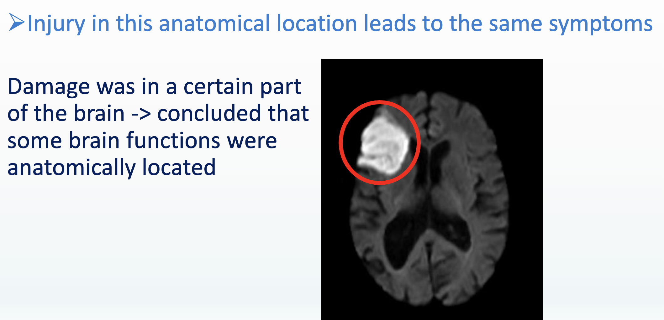
Brain lesion
Brain lesion = area of brain damage
Can result from stroke, loss of blood flow, tumor, injury, etc.
This is what it looks like in an MRI scan
(left) White = damage from reduced blood flow caused by a stroke
(right) Black = tumor tissue

Genetics – Recessive

Genetics – Dominant

Genetics – X / Y chromosomes

Biopsychosocial model and stress

Stress and genes

Interaction genes – environment (modification)

Stress and Epigenetics
Epigenetics: changes to the expression of genome.
Stress has larger biological consequence than originally thought
Structural and temporal (functional) changes
Stress can influence epigenetic changes and mental health
Within lifespan
Across generations
Epigenetics
Changes to the expression of genome
Example: specialisation after cel migration – to nerve or muscle cells (same DNA, other function).
Puberty: body undergoes extensive changes, without DNA changing: hormones regulate gene expression
Applications to neuropsychology
Behaviour is caused by genetics, epigenetics, and experience-based learning
Influences on cardiovascular health can also be derived from genetics, epigenetics, or experience (e.g., diet)
Cardiovascular health directly affects brain function
What is a neuropsychologist?
A clinician and/or scientist who...
Uses neurology, neuroscience, and psychology
To understand how behaviours correlate with brain function
To assess ‘normal’ and ‘impaired’ cognitive, social, physical, and emotional functioning
Neuropsychology is usually clinical in nature
Neuropsychologists are not medical doctors
Clinical tasks are usually diagnostic
Referrals to specialists may be given for treatments
Research tasks may include investigating causes of a disorder, its brain/behavioral/cognitive processes, its diagnostic approaches, the efficacy of treatments (how well they work), etc.
Many neuropsychologists are also scientists
Use neuroimaging to study relationship between brain and psychology
Functional neuroanatomy is crucial
Working knowledge of relevant biological and psychological theories
May study healthy and patient populations
The Hebbian Assumption of Change in research and practice
Learning and skill development across the lifespan
Healing after a brain injury (physical)
Treating a disorder (psychological)
Heritable traits passed down across generations
Patients in neuropsychology
Dementia
Broca’s aphasia
Wernicke’s aphasia
Brain lesions / Stroke
Why start with neurons?
Neuropsychologists rely primarily on information about larger brain structures and functions
Global functions appear trait-based (i.e., fixed, unchanging)
Neuropsychology relies on change, which is not always big enough to be observed on this level of organization
Understanding nerve cells is crucial
Principles of change are observed through understanding cellular processes
Many disorders have cellular causes
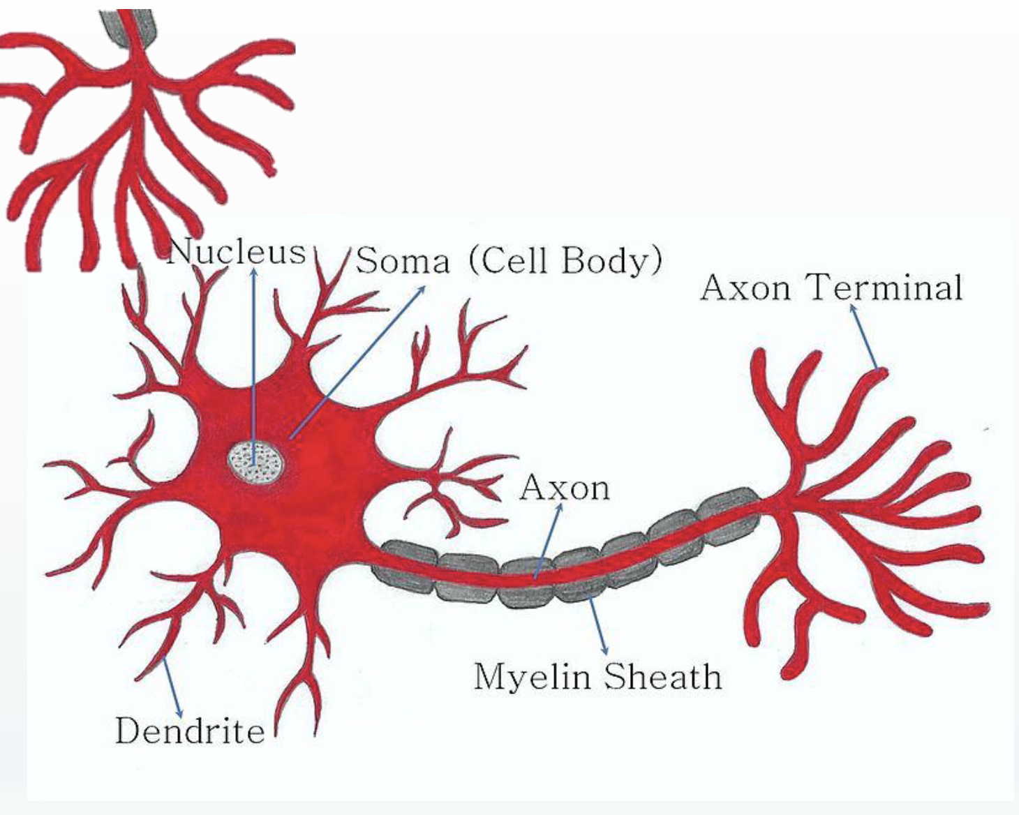
Neuron
Electrical and chemical signals pass from the axon terminal of one neuron to the dendrites of the next neuron
Soma → gray matter
Myelin sheath → white matter
Both electrical and chemical responses can be measured by brain imaging devices
Both gray and white matter activations can be measured by brain imaging devices
There is a correlation between electrical signals and blood flow
Types of neurons

Grey Matter and White Matter

Pyramidal cells

Other types of human multipolar cells

How do neurons function?
Signal is received from neighboring cell
Terminal bouton release neurotransmitters across the synapse to nearby dendrites
Receiving neuron produces 4 signals (i.e., chemical translations) / stages
Input signal = receives messages from other neurons - dendrites
Trigger signal = integrates the received message - soma
Conducting signal = sends information down axon to terminal boutons
This signal goes awry in MS
Output signal = chemical neurotransmitters (NTs) are released – terminal boutons
This step occurs when neurons fire
Nerve cells: global function neurons

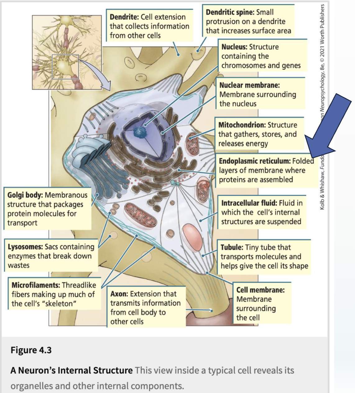
Neuron as a mini-factory
Nucleus contains genetic information
Endoplasmic reticulum uses genetic information as though it is a set of instructions for assembling proteins
Chemical signs - proteins

Back propagation
Hebbian plasticity = brains learn from life experiences
Neurons back propagate
In artificial neural networks (ANNs), it results in adaptive changes
Networks “learn how to learn” through back propagation
Theorized to act similarly in neuron

Discovery of the neural transmission
Discovered by J.Z. Young (1932)
Alan Hodgkin and Andrew Huxley (1939) began studying squid neurons
The modern history of ion channels began in 1952 - seminal papers on the theory of the action potential in the squid giant axon
When do neurons fire?

“All or None” activation
The axon generates an action potential only if the resting potential crosses a threshold (e.g. from -70mV to -60mV)
The magnitude of the action potential is always the same! (like a light switch) → ALL or NONE
Channels, gates, and pumps
Through the use of pumps and channels, an action potential travels along the axonto the synapse, and is there transmitted to the next neuron

Myelin
Efficiency of signal transport depends on “insulation” of the axon
Myelin shaft – thicker is more effective (jumps vs. little steps)
Damage to myelin common in Multiple Sclerosis (MS)
More effective = more used
Every experience has a specific synaptic pattern (see A → B)
Synaptic growth and myelination:
first perception and motor neurons
later associative areas
last areas for cognition and learning
Less used pathways are pruned away

NTs are released and received
A neuron transfers its action potential to other neurons through the synapse

Synapse

Mechanisms of neuroplasticity
Neuroplasticity vs neural flexibility
Neural flexibility = adaptive functional changes (temporary)
Neuroplasticity = structural (physical) reorganization after learning or injury
Mechanisms of plasticity
homologous area adaptation = opposite hemisphere takes over a sensory/cognitive process
cross-modal reassignment = loss of one sensory / cognitive process heightens the remaining senses processes
map expansion = some areas involved strengthen to compensate for loss
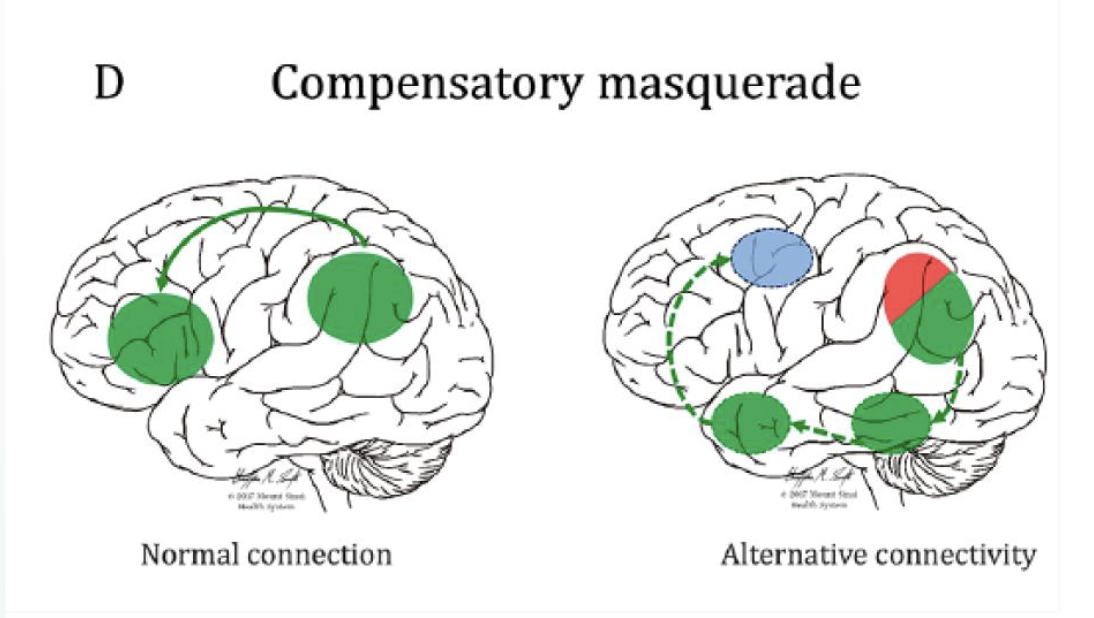
compensatory masquerade = brain uses alternative pathways / strategies for processing (alternative routes)
Homologous area adaptation
opposite hemisphere takes over a sensory/cognitive process
Cross-modal reassignment
loss of one sensory / cognitive process heightens the remaining senses / processes
Map expansion
areas that were involved grow stronger
Compensatory masquerade
brain uses alternative pathways / strategies for processing (taking a different route to get there)
old areas that were always part of the network may help enough that tasks can still be performed, but the performance isn’t as good
What might prompt brain changes?
Environment / exposure / experience sampling
Learning (changes to neurons / pathways)
Therapy (changes to neurons / pathways)
Biological changes
Disease progression / accidents
Medical treatments / surgeries
Medications
Psychosocial factors
Stress (methylation changes / other epigenetic changes)
Emotions (physiological changes / epigenetic changes)
Epigenetic changes
The epigenome mains, modifies, and regulates genes
Determines which genes are expressed (turns genes “on” and “off”)
“On” = protein is produced; “Off” = no protein is produced
DNA makes RNA makes proteins
Genes don’t change, but phenotypes do
Phenotype = observable traits that result from a gene x environment (GxE) interaction
The same genotype interacting with the environment can producemany different phenotypes
3 main mechanisms of change
DNA methylation
Histone methylation
mRNA modification
DNA methylation
Methyl groups are added to the DNA molecule
Methyl group = 1 carbon and 3 hydrogen atoms
Adding a methyl group blocks promoters from expressing the gene
Can change the activity of a DNA segment without changing the sequence
Typically acts to repress gene transcription

Histone methylation
Histone = a type of protein that wraps (“spools”) itself around DNA
Changes in histone methyl groups can induce or repress gene expression
Left image “spooled” DNA (not expressed), right image “unspooled” DNA (expressed)
Methylation increases density (more “spooling”, less expression)

mRNA modification
mRNA = messenger RNA = dynamic regulators of gene expression
Primarily refers to 13 different chemical alterations
Affect translation or stimulate other regulatory processes like mRNA degradation or localisation
Small changes can large differences in outcome
MULTIPLE SCLEROSIS (MS)
MS is caused by damaged myelin → Cumulative damage produces brain lesions
Immune cells attack the myelin sheath
Myelin acts as lubricant
Exposed axon is scarred (sclerosis = scars)
Information transmitted by that neuron is disrupted
MS is a heterogeneous disorder = patients have different symptoms & experiences depending on lesion size and location, and how their individual brain is functionally organised
Common symptoms of MS
Affects brain and spinal cord
Common early symptoms:
Vision problems
Numbness/tingling sensations
Motor symptoms: clumsiness, weakness of limbs
Balance and coordination
Fatigue
Common later symptoms:
Mobility problems: muscle spasms, stiffness, weakness, or paralysis
Bladder or bowel problems
Cognitive problems: difficulties thinking, learning, and/or planning (language preserved)
Pain
Speech and swallowing difficulties
Sexual problems
Emotional problems: depression and anxiety
Most people with MS only have a few of these symptoms
Average lifespan is 7 years shorter than general population
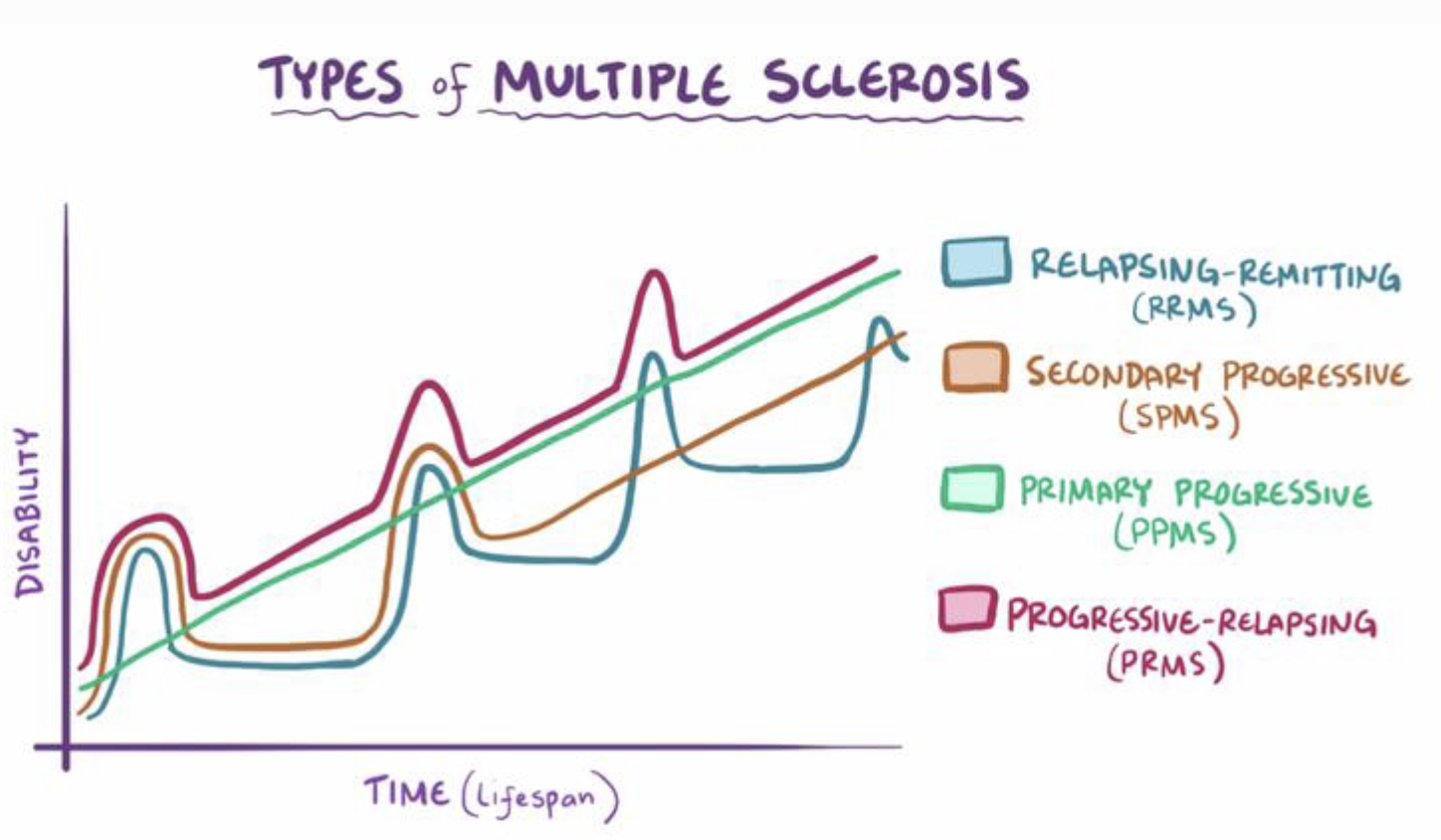
Types of MS
Primary progressive (PPMS-green)
Symptoms progressively get worse after onset
No periods of remission
Relapsing remitting (RRMS-blue)
Relapse = active symptoms / myelin damage
Remission = no current symptoms / myelin damage
Symptoms disappear and reappear across time
Progressive relapsing (PRMS-red)
Similar to RRMS but symptoms become increasingly disabling with each relapse
Very rare, severely disabling
Secondary progressive (SPMS-orange)
Occurs after patient previously had RRMS
Remissions stop, symptoms get steadily worse
Overview neurotransmitters

Cholinergic system
ACh facilitates neuronal communication → Memory formation and learning, attention and arousal
Projected to hippocampus and limbic system
Alzheimer’s disease: substantial loss of cholinergic neurons in the basal forebrain → cognitive decline
Parkinson’s disease: Higher levels of ACh are suggested to cause dyskinesia, uncontrolled and involuntary movement.

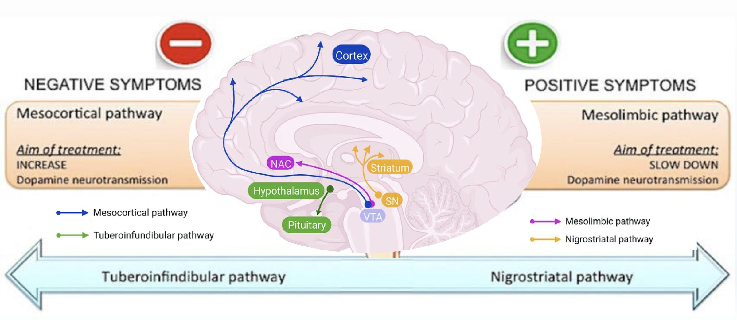
Dopaminergic system

Dopamine and schizophrenia symptoms
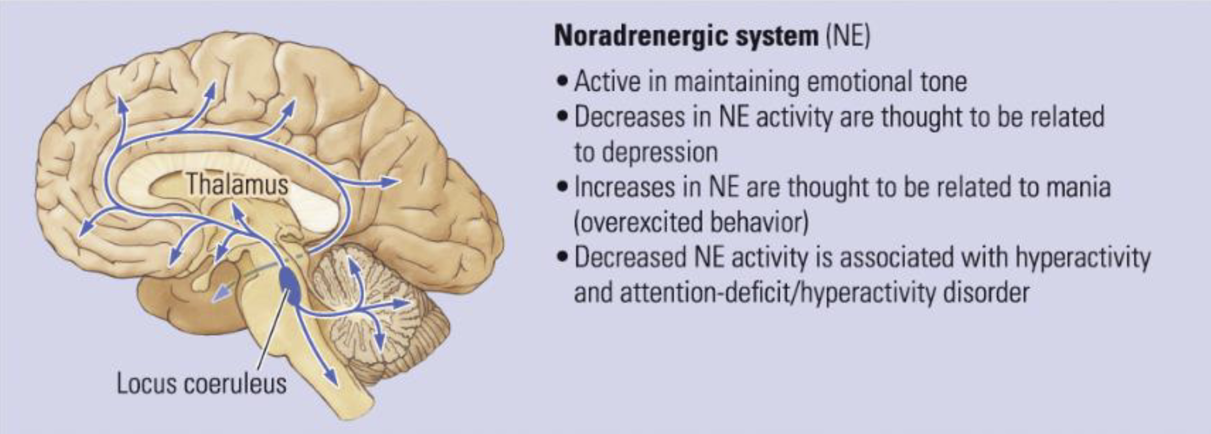
Noradrenergic system
Epinephrine, also known as adrenaline, is both a hormone and neurotransmitter
Influences mood, motivation, and arousal
Low – depression ; High – mania
Associated with ADHD
Norepinephrine vs. Epinephrine

Serotonergic system

Serotonin and psychopathology
Serotonin (5-HT, 5-hydroxytryptamine) modulates a broad spectrum of functions, including mood, cognition, anxiety, learning, memory, reward processing, and sleep.
depression, anxiety, tremors, schizophrenia, OCD

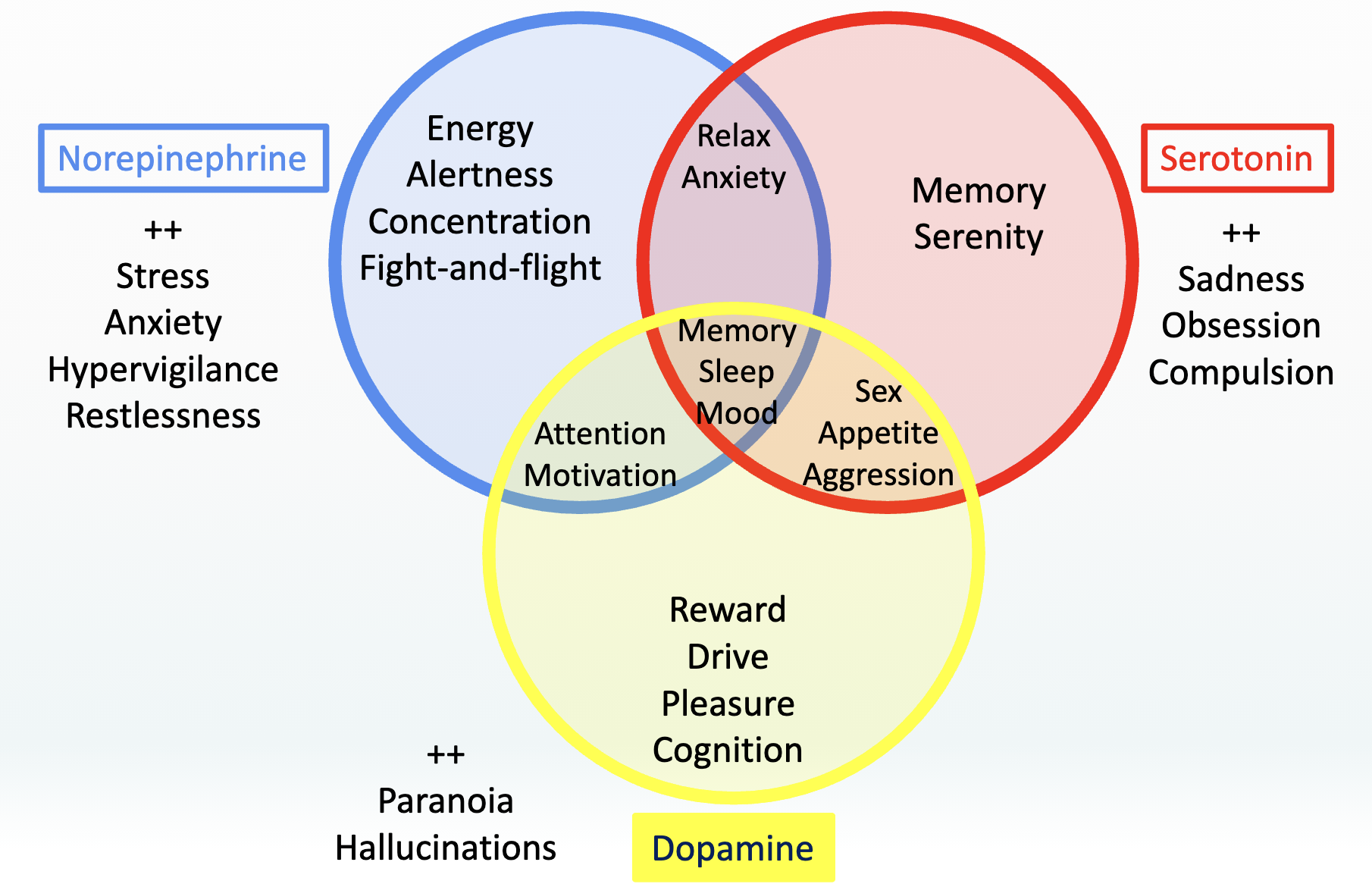
Combination of neurotransmitters
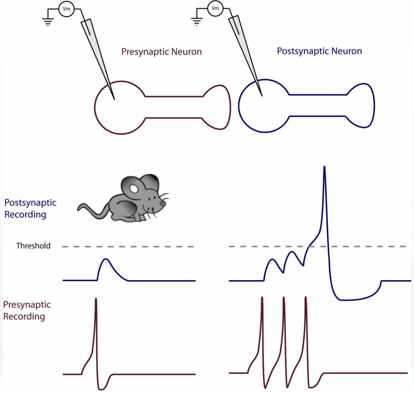
Single-cell recording
Intracellular vs. extracellular recordings
Microelectrode is placed next to a single neuron (almost touching) or inside the cell
Electrical charge is recorded
Direct measure of brain activity
Invasive, cannot be used to study humans
Electroencephalogram (EEG)
One or more electrodes placed on scalp (and around eye)
Electrical charges of neurons near scalp are recorded (extracellular)
Direct measure of brain activity
Good temporal resolution
We know when activation occurs
Poor spatial resolution
Scalp is a bad conductor of electricity
Hard to know where the activation occurs
Non-invasive
Can also be used to study children, portable
Event Related Potential (ERP)

Magnetoencephalography (MEG)

Magnetic resonance imaging (MRI)

Structural and functional MRI

Diffusion tensor imaging (DTI)

Spatial and temporal resolution

(Hind brain) - Spinal cord

(Hind brain) - Brainstem
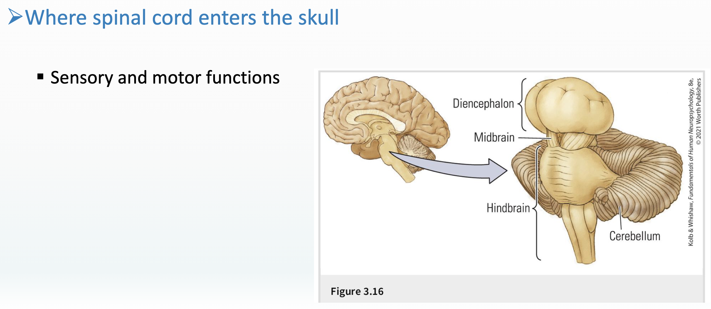
(Hindbrain) - Cerebellum
Holds more than half of the neurons in your whole body.
Grey and white matter
Subcortical nuclei
Lobes, fissures etc.
Balance, coordination, movement, and motor skills
Integrates them with mental processes

(Hindbrain) - other structures
Vital functions
Arousal
Consciousness
Bridge to brain

Medulla - Crossing neural circuits
Visualizations of decussation
90% of motor and somatosensory fibers cross over
Left brain controls right body, Right brain controls left body
Remember: many Broca’s infarct have right side body impairments...

Midbrain
Sensory input from eyes and ears (basic)
Movement control
Reflexes, bypass the brain,
direct uncontrollable action

Midbrain - Diencephalon
Thalamus – central hub connecting sensory systems to cortex
Hypothalamus – motivated behavior
Connection pituitary gland
Hormones / chemical messages
Epithalamus
Pineal gland – body rhythm
Habenula – hunger & thirst

Forebrain – Telencephalon – “the brain”
Basal ganglia
Reward and associative learning
Connect sensory to motor systems
Regulate (smooth) movement
Limbic system
Amygdala – emotions
Hippocampus – personal memory and spatial navigation
Cingulate cortex – social interactions, decision making and executive functions
Cortex
Motor cortex
Visual cortex
Thinking, perceiving, communicate
Consciously interact with world
Complex high functions
Connecting brain regions

Function of brain regions - hierarchy

The brain’s blood flow
Blood supply is important for healthy tissue and cellular functioning
Blood carries oxygen and glucose
Some neuroimaging devices measure changes in blood flow to infer which areas of the brain are more active
The assumption is that greater neural activity requires greater blood flow
Asymmetrical blood flow can affect function
Anterior cerebral circulation - provides blood flow to the eyes and the anterior brain. Blood source include these (among others):
Middle cerebral artery (MCA) ↓
Anterior cerebral artery (ACA)
Posterior cerebral circulation - provides blood flow to the the occipital lobes, cerebellum, brainstem and posterior brain. Blood source include these (among others):
Vertebral arteries (smaller arteries from the shoulders, lateral chest, and arms)
Posterior inferior cerebellar artery (PICA) & Anterior inferior cerebellar artery (AICA)
Superior cerebellar artery (SCA) & Posterior cerebral artery (PCA)
Basilar artery (blood flow to the midbrain and cerebellum)
Pontine branches (includes circle of Willis)
Circle of Willis
Joins several arteries at the inferior side of the brain
Internal carotid arteries branch into smaller arteries
Supplies oxygenated blood flow to over 80% of the cerebrum
Main aqueduct for the brain’s hemodynamic response (what’s measured by fMRI and fNIRS)

Main arteries
Anterior cerebral artery (ACA): Supplies blood to the medial parts, including the superior frontal and anterior parietal lobes.
Middle cerebral artery (MCA): Supplies blood to lateral parts, the temporal and lateral-parietal lobes.
Posterior cerebral artery (PCA): Supplies blood to posterior parts, including the occipital lobe, thalamus and midbrain.
Carotid arteries are major blood vessels in the neck that deliver blood to the brain (internal carotid artery - ICA), face, and neck

Ventricles
Interconnected cavities that produce and circulate cerebrospinal fluid (CSF)
Lateral: C-shaped in each hemisphere
Third: between the two halves of the thalamus
Fourth: between the brainstem and cerebellum

What is laterality?
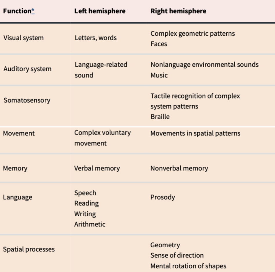
Lateralization of brain function = the brain’s two hemispheres have separate functions
Cerebral asymmetry
Neuronal asymmetry
Genetic asymmetry
Not all brain functions are lateralized
Brain functions that are going to lateralize usually have by age 6-7
Asymmetrical functions maybe lateralized = usually appearing on one side (e.g., language is usually processed in the left hemisphere)
What is lateralized?
Cerebral Asymmetry
Anatomical differences (of most people...)
Left hemisphere is larger, heavier, and has more gray matter (cell bodies)
Right hemisphere has more cortical surface area*
Left hemisphere has a thicker cortex+
Language and music are oppositely lateralized, as are corresponding brain regions (Broca’s has more sulci* on the left side)
Asymmetry in the temporal lobe corresponds with asymmetry in the thalamus
Distribution of neurotransmitters is asymmetrical
Cerebral torque = the right side of the brain is slightly forward compared to the left side
*More sulci = bigger surface area → theorized to mean more intelligence/more advanced
+Cortex → theorized to reflect social skills and processes
Neuronal & Genetic Asymmetry
These asymmetries are difficult to study, and only general principles are known...
Neuronal Asymmetry = Neural branches and connections differ across hemispheres
Genetic Asymmetry = Gene expression occurs differently in each hemisphere
Both are difficult to study because of the sheer numbers of neurons in the brain (and ethics?)
Behavioural asymmetries in the intact brain
1. Visual asymmetry
Information presented to only one visual field is processed most efficiently by the hemisphere specialized to receive it
words in left hemisphere (LH)
faces in right hemisphere (RH)
2. Auditory asymmetry
When different stimuli are presented simultaneously to path wars, as preferreateral access, and the ipsilateral pathway is slower
Right ear advantage for words (LH)
Left ear advantage for music (RH)
Example:
"Ga" goes directly to the language processing area in the LH.
"Ba" travels first to the RH, then crosses over to the LH. Path takes longer.
→ Right ear dominance for words
Music other way around
3. Somatosensory asymmetry
Performance = Shape and pattern recognition (cf. Braille)
The left hand of a right-handed participant is superior at nearly all tasks of this type
Identification = See what you feel
right-hand advantage for letters (LH)
left-hand advantage for other shapes (RH)
NB. LH dominance for words / language
4. Motor asymmetry
Everyone has motor asymmetries
Easier to study in patient groups
☆ Behaviour does NOT correspond fully with biological/anatomical measures
☆ Laterality might be a question of which information is processed first (timing)
☆ Personal preference for strategies may influence outcomes
Right vs left ear advantage

Aphasia
A language disorder caused by damage in a language area of the brain
Aphasia leaves a person unable to communicate effectively with others
Affects language expression or comprehension via speech or writing
Most often caused by a stroke
Most recovery (if any) occurs within a few months of onset
Broca’s aphasia
Broca’s aphasia
Difficulty forming words or sentences
Loss of grammatical structure (“telegraphical”)
Intact understanding of language
Noticed in the clinic
Brain lesion observed in frontal lobe
Patients often have right-side weakness or paralysis due to lesion’s impact on motor function
Hierarchy:
Speech is hard to produce but usually not impossible (advanced)
Other language functions, like understanding speech as well as simple motor speech routines
Production of familiar songs (or automated vocal behaviors) often remain intact (basal)