Skin, Hair & Nail Assessment (Exam 2)
 Call Kai
 Learn
 Practice Test
 Spaced Repetition
 Match
 Flashcards
 Knowt Play
1/63

There's no tags or description
Looks like no tags are added yet.
64 Terms
What are the five main functions of the integumentary system?
Protection, temperature regulation, sensation, absorption and excretion, and vitamin D production.
What is the primary protective function of the skin?
It acts as a barrier to protect the body from injury and outside substances.
How does the skin help regulate body temperature?
Through sweating and changes in blood flow near the skin's surface.
How does the skin provide sensation?
It contains sensory receptors that detect touch, pressure, pain, and temperature.
What does the skin absorb and excrete?
It can absorb certain substances and excrete sweat and other substances.
What vitamin does the skin help produce?
Vitamin D.
What are the two main techniques used during a skin assessment?
Inspection and palpation.
What should the nurse inspect during a skin assessment?
Skin color, lesions, bruises, wounds, and rashes.
What should the nurse palpate during a skin assessment?
Temperature, moisture, texture, turgor, and edema.
What is skin turgor?
The skin's elasticity and ability to return to its normal position after being pinched.
What should the nurse assess when examining a wound?
Location, size, tissue, edges, drainage, odor, surrounding skin, and symptoms.
Why is it important to assess the entire patient instead of only looking at a wound?
A wound can be related to underlying problems such as poor circulation, diabetes, infection, or pressure.
What causes jaundice?
Liver dysfunction and buildup of bilirubin.
What is hyperpigmentation?
Darkening of the skin.
What are possible causes of hyperpigmentation?
Diabetes and Addison's disease.
What is hypopigmentation?
Lightening or loss of skin pigmentation.
What are possible causes of hypopigmentation?
Vitiligo and tinea versicolor.
A patient has pale skin and pale nail beds. What should the nurse suspect?
Pallor, which may be associated with anemia or shock.
A patient has red, warm skin around a wound. What should the nurse suspect?
Erythema, which may indicate inflammation or infection.
A patient has bluish discoloration of the skin. What should the nurse suspect?
Cyanosis, which may indicate poor oxygenation.
A patient has yellow discoloration of the skin. What should the nurse suspect?
Jaundice, potentially related to liver dysfunction or bilirubin buildup.
A patient develops darkened skin pigmentation. What is this called?
Hyperpigmentation.
A patient develops lighter patches of skin. What is this called?
Hypopigmentation.
What is diaphoresis?
Excessive sweating.
What are possible causes of diaphoresis?
Fever, physical exertion, anxiety, and myocardial infarction (MI).
What can dry or flaky skin indicate?
Dehydration, aging, hypothyroidism, or decreased oil production.
What are possible causes of cool skin?
Cold exposure and poor circulation.
What are possible causes of hot skin?
Infection, inflammation, burns, and fever.
A patient is sweating heavily and appears pale. What should the nurse consider?
Diaphoresis, which can occur with fever, anxiety, exertion, or myocardial infarction.
A patient has cool skin and weak pulses. What should the nurse assess?
Circulation and peripheral perfusion.
A patient has hot, red skin surrounding a wound. What should the nurse suspect?
Possible inflammation or infection.

What is a scar?
Fibrous tissue that replaces injured skin.
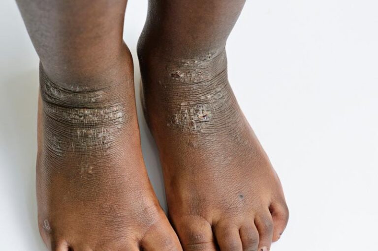
What is lichenification?
Thickened skin with accentuated skin markings.
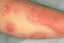
What is erosion?
Superficial loss of the epidermis.

What is excoriation?
A superficial abrasion, often caused by scratching.

What is a fissure?
A linear crack that extends into the skin.

What is a crust?
Dried serum, blood, or exudate on the skin.

What is a scale?
Flakes of accumulated stratum corneum.

What is a wheal?
A temporary, raised area of dermal edema.

What is a vesicle?
A small, fluid-filled skin lesion less than 1 cm.
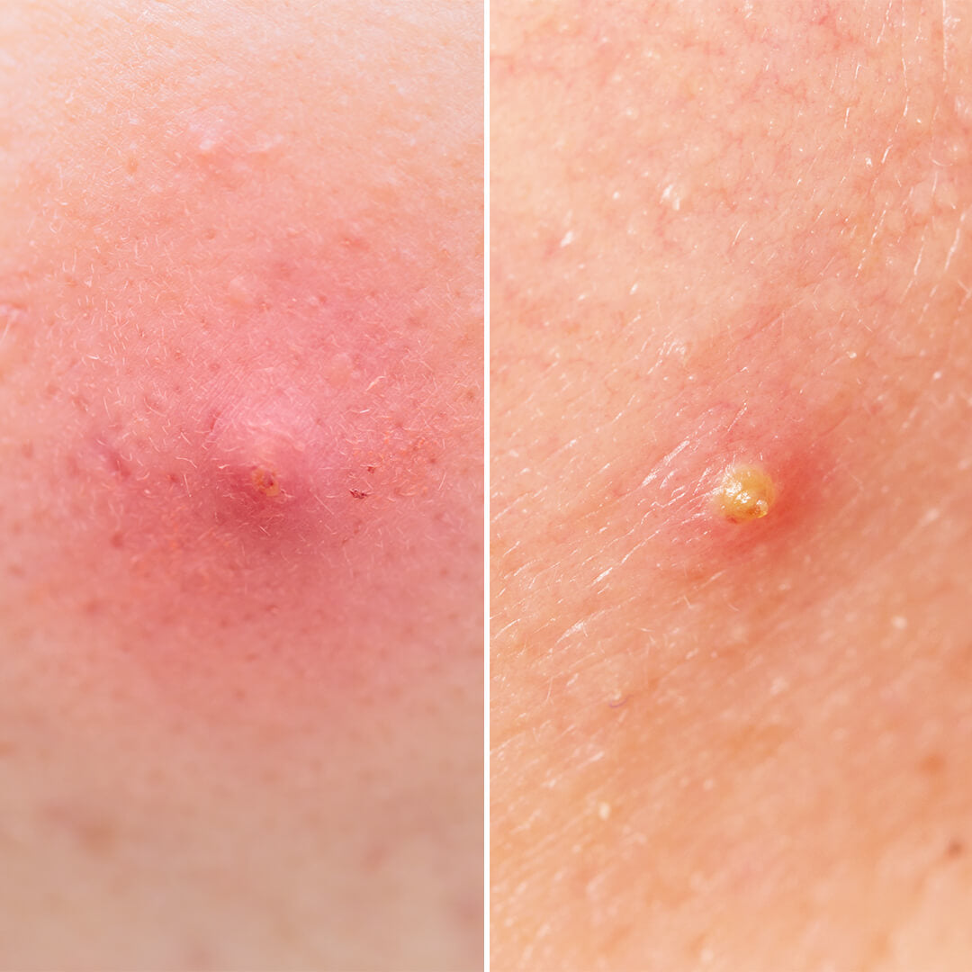
What is a pustule?
An elevated skin lesion containing purulent (pus-like) material.
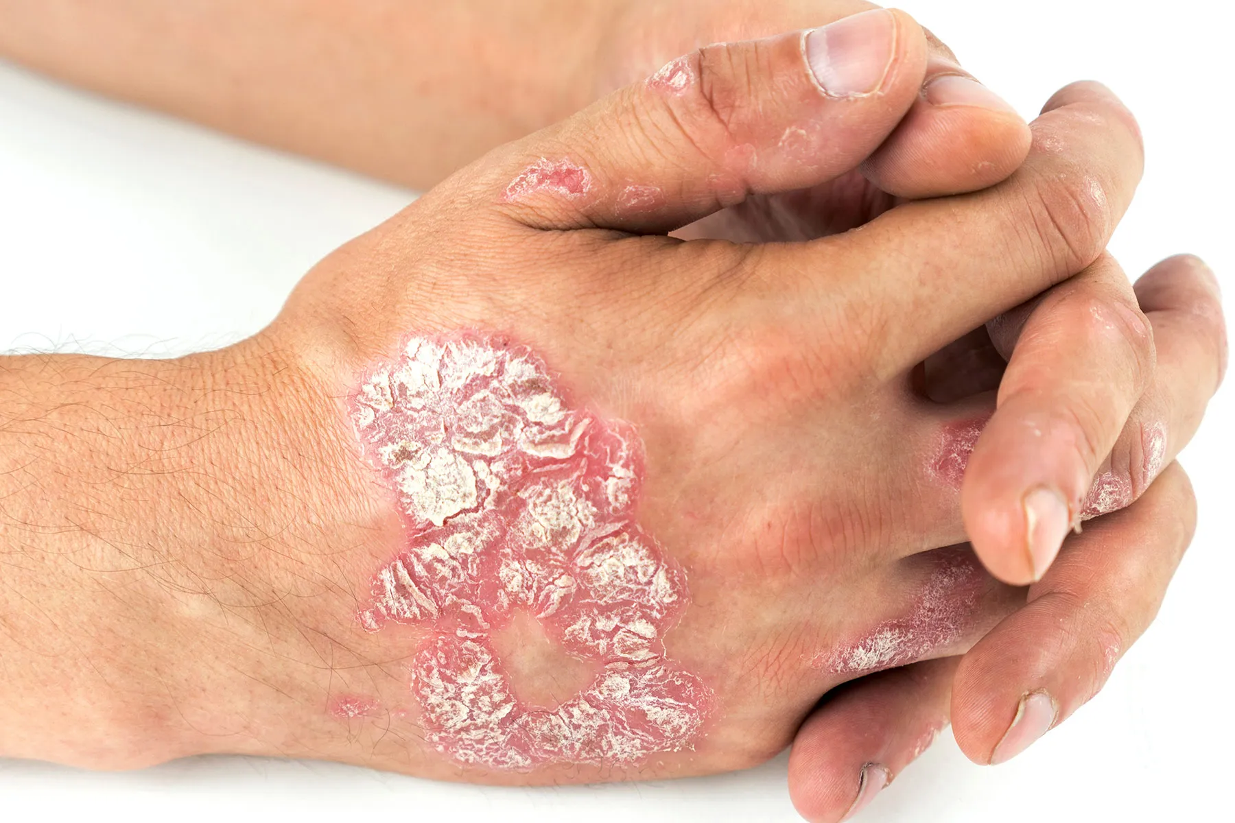
What is a plaque?
A raised, broad, flat-topped skin lesion.
What is a papule?
A small, raised, solid skin lesion.
What is a patch?
A flat area of color change greater than 1 cm.
What is a nodule?
A solid, palpable lesion that extends deeper into the skin.
What is a macule?
A flat, nonpalpable color change less than 1 cm.
What is a bulla?
A fluid-filled skin lesion greater than 1 cm.
What is the difference between a vesicle and a bulla?
A vesicle is less than 1 cm, while a bulla is greater than 1 cm. Both contain fluid.
What is the difference between a macule and a papule?
A macule is flat and nonpalpable, while a papule is raised and solid.
What is the difference between a patch and a macule?
A patch is a flat color change greater than 1 cm, while a macule is less than 1 cm.
What is the difference between a pustule and a vesicle?
A pustule contains pus, while a vesicle contains clear fluid.
What is the difference between erosion and excoriation?
Erosion is superficial loss of the epidermis, while excoriation is a superficial abrasion often caused by scratching.
What is the difference between a crust and a scale?
A crust is dried serum, blood, or exudate, while a scale is flakes of accumulated stratum corneum.
What should the nurse document when assessing a skin lesion?
Location, size, color, borders, distribution, and symptoms.
What are the four common types of ulcers discussed in the lecture?
Venous, arterial, pressure, and diabetic foot ulcers.
What is a venous ulcer?
A wound commonly located on the lower leg that is associated with venous circulation problems.
Where are venous ulcers commonly located?
The lower leg, often around the ankle area.
What are common findings of venous ulcers?
Edema, brown hemosiderin staining, and often increased wound drainage (exudate).
What is hemosiderin staining?
Brown discoloration of the skin associated with venous disease.
What should the nurse assess before applying compression to a patient with a venous ulcer?
Arterial perfusion and circulation, including pulses and appropriate vascular assessment.
Why must arterial perfusion be assessed before compression?
Compression can worsen tissue injury if the patient has inadequate arterial blood flow.
What is an arterial ulcer?
A wound caused by inadequate arterial blood supply to the tissue.
Where are arterial ulcers commonly located?
Distal areas of the foot and toes.
What are common findings of arterial ulcers?
Cool feet, diminished pulses, and pain at rest or with elevation.