Ch. 6 - Bone Growth
 Call Kai
 Learn
 Practice Test
 Spaced Repetition
 Match
 Flashcards
 Knowt Play
1/65

There's no tags or description
Looks like no tags are added yet.
66 Terms
skeletal cartilages
provide support and cushioning
appositional growth
adds to overall thickness
only CT and cartilages
interstitial growth
adds to overall length
only CT and cartilages
long bone
longer than it is wide
humerus
irregular bone
doesn’t look like any other bones
vertebra
flat bone
thin and light
mostly in skull, sternum, and parts of the pelvic bone
short bone
equal in length and width; often where there’s lots of force
talus
bone functions
support
protection: surrounds organs / tissues
movement: skeletal muscles must cross joints to move
mineral storage and balance: calcium phosphate, balancing pH
blood cell production: in marrow
fat storage: stored in yellow marrow of long bones
yellow bone marrow
fat
red bone marrow
blood producing
periosteum
membrane of bone that is very tightly attached
pulls on bone and makes processes
appositional growth
dense irregular fibrous CT
perforating (sharpey’s) fibers
hold the periosteum to the surface of bone
diploë
spongy bone that is found between layers of flat bones
contains red bone marrow
helps distribute stress evenly and absorb shock because it rearranges faster
osteogenic cell
stem cells that are destined to become bone
O2% determines cell type
lays down matrix; cells get trapped in matrix and harden
osteoblast
matrix-synthesizing cell responsible for bone growth
osteocyte
mature bone cell that maintains the bone matrix
connected to neighbors which helps with vascularization and repairs (4-6 weeks)
osteoclast
bone - resorbing cell
multi-nucleated
ruffled border that is an extension of cell membrane (looks for things to dissolve w/ acid)
circumferential lamellae
the layers connect and go all the way around the bone
interstitial lamellae
fills in the gaps between circumferential lamellae
true osteons
only found in compact bone
key feature of compact bone
osteons
compact bone structure
tube within tube structure
lacuna
small, hollow cavity embedded within the mineralized extracellular matrix of bone that houses a single mature bone cell
osteon
the tiny, cylinder-shaped building block of compact bone
trabecualae
little beam / scaffolding of spongy bone
red lines
body weight stress
purple lines
stress from muscles pulling
hydroxyapatite crystals
main inorganic component of bone matrix
bone matrix is 35% organic and 65% inorganic
parietal bone
brain pushing on fetal parietal bone stimulates growth
fetal skeleton
most is hyaline cartilage that ossifies
major ossification centers at 12 weeks
intramembranous ossification
the direct conversion of embryonic connective tissue (mesenchyme) into bone without first forming a cartilage intermediate
occurs mainly in flat bones of the skull and clavicle
intramembranous ossification
ossification centers appear in the fibrous connective tissue membrane
bone matrix (osteoid) is secreted within the fibrous membrane and calcifies
woven bone( network of trabeculae) and periosteum form
lamellar bone replaces woven bone, just deep to the periosteum; red marrow appears
endochondral ossification steps
bone collar forms around hyaline cartilage model
cartilage in the center of the diaphysis calcifies and then develops cavities
the periosteal bud invades the internal cavities and spongy bone begins to form
the diaphysis elongates and a medullary cavity forms as ossification continues; secondary ossification centers appear in the epiphyses
the epiphyses ossify; when completed, hyaline cartilage remains only in the epiphyseal plate and articular cartilages
bone grows
increases thickness by appositional growth
adds more concentric lamellae
bone growth zones
proliferation zone
hypertrophic zone
calcification zone
ossification zone
proliferation zone
cartilage cells undergo mitosis
hypertrophic zone
older cartilage cells enlarge
calcification zone
matrix becomes calcified, cartilage cells die, and matrix begin deteriorating
ossification zone
new bone formation is occurring
parathyroid glands
release parathyroid hormone (PTH)
stimulates osteoclasts
blood calcium homeostasis
9-11mg / 100ml
calcitriol
vitamin D
increases blood levels of Ca++
encourages calcium absorption
produced by melanocytes
PTH
increases blood levels of Ca++
important as we age
calcitonin
decreases blood levels of Ca++ when levels are high
stimulates osteoblasts
more active when younger
hypercalcemia
higher than normal levels of calcium in the blood
occurs when too much calcium enters the extracellular fluid or when the kidneys fail to excrete it normally
excess PTH signals osteoclasts to break down bone tissue faster than it can rebuild → weakens bones over time
hypocalcemia
lower than normal level of calcium in the blood serum
occurs due to disruptions in the hormone or mineral systems that regulate blood calcium (PTH and vitamin D)
the body pulls stored calcium from the skeleton to maintain normal levels in the blood → weakens bones over time
growth hormone (GH)
children: signals cartilage and bone cells to multiply and lengthen the epiphyseal plates, determining final height
adults: metabolism regulation, body structure maintenance, triggers release of insulin like growth factor
GH hyposecretion
dwarfism
GH hypersecretion
gigantism - before puberty
acromegaly - adulthood
thyroid hormone (TH)
responsible for maintaining metabolic rate
sex hormones
responsible for eunuchs (growth spurts)
testosterone
estrogen
progesterone
insulin like growth factors
produced primarily by the liver in response to growth hormone (GH), it drives bone, cartilage, and organ growth
rickets
a childhood bone disorder that causes the softening and weakening of growing bones, usually due to a prolonged deficiency in vitamin D, calcium, or phosphorus
results in bowed legs
scurvy
a disease caused by a severe deficiency of vitamin C after prolonged inadequate dietary intake of fresh fruits and vegetables
often causes teeth to fall out because collagen is broken down
wolff’s law
bone remodels in response to mechanical stress
mechanical stress: physical force and load placed on bones during movement, gravity, and muscle contraction
bone repair
a hematoma forms
fibrocartilaginous callus forms
bony callus forms
bone remodeling occurs
hematoma
a collection of pooled, clotted blood that forms outside of blood vessels due to trauma, impact, or a bone fracture
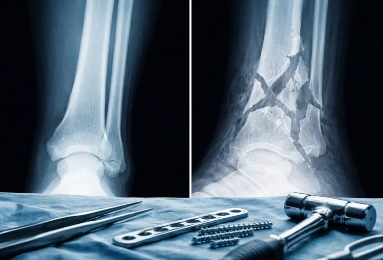
comminuted fracture
bone fragments into three or more pieces
common in the elderly and those with osteoporosis
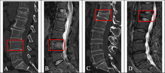
compression fracture
bone is crushed
common in porous bones (osteoporotic bones) subjected to extreme trauma like a fall
spiral fracture
ragged break occurs when excessive twisting forces are applied to a bone
common in sports

epiphyseal fracture
epiphysis separates from the diaphysis alone the epiphyseal plate
occurs where cartilage cells are dying and calcification of the matrix is occurring
common in children

depressed fracture
broken bone portion is pressed inward
common in the skull

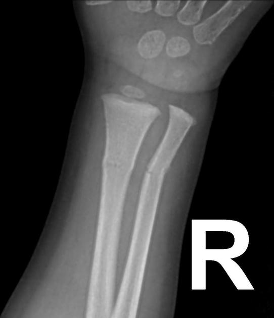
greenstick fracture
bone breaks incompletely, only one side of the shaft breaks and the other side bends
common in children whose bones have relatively more organic matrix and are more flexible
osteogenesis imperfecta
also known as brittle bone disease, is a rare genetic disorder present from birth that causes bone to break easily —often from little or no apparent trauma
most cases are caused by mutations in the COL1A1 or COL1A2 genes, which disrupt the production or structure of type I collagen—the main protein that provides strength and flexibility to bones and connective tissues.
osteomyelitis
a painful infection and inflammation of the bone or bone marrow most commonly caused by bacteria such as Staphylococcus aureus
osteoporosis
a systemic bone disease where bones become weak, thin, and brittle due to a decrease in bone mineral density and the deterioration of bone tissue, greatly increasing the risk of fractures
caused by:
inadequate nutrition
lack of calcium
too much PTH