The Central Nervous System (Brain)
 Call Kai
 Learn
Learn Practice Test
 Spaced Repetition
 Match
Match Flashcards
 Knowt Play
1/54

There's no tags or description
Looks like no tags are added yet.
Name | Mastery | Learn | Test | Matching | Spaced | Call with Kai | Chat |
|---|
No analytics yet
Send a link to your students to track their progress
55 Terms
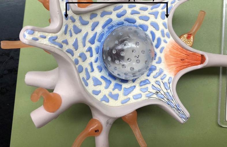
Cell body (soma)
The cell’s metabolic and genetic control center

Axon
Transmits electrical impulses away from the neuron’s cell body to other neurons, muscles, or glands
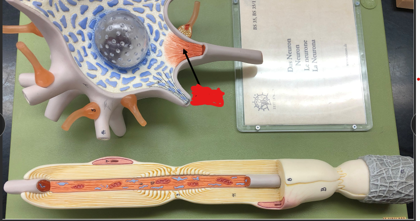
Axon Hillock
Neurons' decision-making center by adding up incoming electrical signals, and if they are strong enough, triggering an action potential to travel down the axon

Dendrites
The branch-like structures of a neuron that receive electrical and chemical signals from other cells and carry them to the cell body

Neurilemma
The thin outermost layer of cytoplasm containing the nucleus of a Schwann cell that wraps around peripheral nerve fibers to protect them and help them regenerate after an injury (not in CNS)

Myelin
Protective fatty layer that wraps around axons to insulate nerve fibers and speed up the travel of electrical signals through the nervous system

Nodes of Ranvier
small, uninsulated gaps in the myelin sheath along an axon that contain high concentration of ion channels to re-energize and speed up electrical signal as they hop down the nerve
Synaptic Knobs
bulb-like swelling at the very end of an axon that store and release neurotransmitters to pass chemical messages across the gap to the next cell
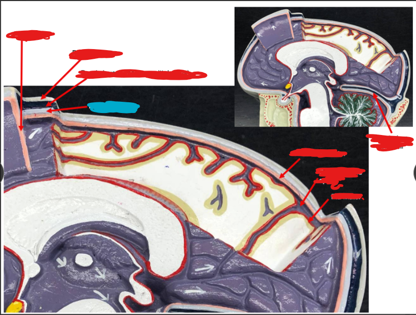
Meninges (Brain)
Protect, support, and anchor the brain and spinal cord within the CNS

Dura Mater
Provides a tough, protective, and stabilizing outer covering for the CNS

Periosteal layer of the Dura Mater
The outer sublayer of the tough membrane that lines the inner surface of the skull

The Meningeal Layer of the Dura Mater
the inner sublayer of the dura mater that folds inwards to form protective partitions inside the brain protective

Dural Sinuses (Superior Sagittal Sinus)
Large blood-filled channels located between the two layers of the dura mater inside the skull

Arachnoid Mater
Sits right between the tough dura mater and the inner tightly fitted pia mater; surrounds and protects the spinal cord
Subarachnoid Space
The cerebral spinal fluid-filled interval located between the middle protective layer of the brain (the arachnoid mater) and the delicate innermost layer (the pia mater)
Cerebral Spinal Fluid
Clear colorless liquid that cushions and protects your brain and spinal cord fills the subarachnoid space

Pia Mater
The delicate innermost layer of the meniges, follows every fold, groove, and curvve
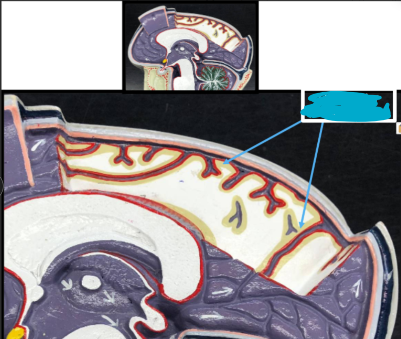
Gray Matter
unmyelinated neuron cell bodies, dendrites, and synapses that process information, memories, and thoughts in the central nervous system
White Matter
Bundles of myelinated axons (nerve fibers) that act as communication highways, connecting different gray matter regions

Telencephalon
The largest and most developed region of the brain commonly known as the cerebrum

Sulci/Sulcus (singular)
Shallow grooves on the surface of the brain that increase the cortex’s surface area to pain in more neurons

Central Sulcus
prominent deep groove that runs down the side of the brain separating the frontal lobe from the parietal lobe and dividing the primary motor cortext from the primary somatosensory cortex cortex

Gyrus/Gyri (singular)
Raised ridges or folds on the surface of the brain that work alongside sulci to maximize the surface area of the cerebral cortex

Precentral Gyrus
prominent ridge located at the back of the frontal lobe directly in front of the central sulcus. Houses the primary motor cortec resposible for controlling the volunatry movments of yoru body

Postcentral Gyrus
A prominent ridge located at the front of the parietal lobe directly behind the central sulcus that houses the primary somatosensory cortex, responsible for perceiving and processing sensory expereinces
Fissures
The deepest and largest grooves on the surface of the brain that physically divide it into large sections, such as splitting the left and the right hemispheres or separating major lobes

Longitudinal Cerebral Fissure
the deep major groove that runs down the exact middle of the brain from front to back, separating the left and right cerebral hemispheres

Frontal lobe
The largest of the brain's four major lobes is located directly behind your forehead. It is responsible for higher-level cognitive functions of the forehead.

Parietal Lobe
Located near the top back of the brain, primarily responsible for processing sensory information

Temporal Lobe
Located on the bottom sides of the brain, behind the ears. Responsible for processing auditory information, understanding spoken language, and encoding memory
Occipital Lobe
Located at the very back of the brain and serves as the primary visual processing center

Cerebral Cortex
the thin outer layer of gray matter covering the brain’s hemispheres and acts as the main processing center for high-level functions

Primary Motor Cortex
the strip of brain tissue located in the precentral gyrus of the frontal lobe that control voluntary movement in the body

Premotor Cortex / Somatic Motor Association area Area
the region in the frontal lobe located directly in front of the primary motor cortex that is responsible for planning, preparing, and organizing voluntary movements based on external sensory cues like vision and sound

Prefrontal Association Area
The front part os the frontal lobe acts as the brain’s executive control center, regulating functions such as planning, decision-making, working memory impulse control, and social behavior

Motor speech/ Broca’s Area
region in the left frontal lobe that acts as the brain’s speech planning center. It assembles words, sequences, and programs motor movements necessary to speak sequences fluently

Primary Sensory Cortex
Strip of brain tissue located in the postcentral gyrus of the parietal lobe that receives and processes sensory inputs from the body, including touch, temperature, pressure, and pain

Somatic Sensory Association Area
Located directly behind the primary somatosensory coretex. It interprets and gives meaning to raw sensory inputs

Wernicke’s area (General Interpretive Area)
Region located in the left temporal lobe (near the auditory cortex) that acts as the brain’s language comprehension center

Auditory Cortex
A region located in the temporal lobe of the brain that receives and processes sound information from the ears, allowing you to hear

Gustory Cortex
a brain structure located deep within the lateral fissure that is responsible for the cconscious perception and discrimination of taste
Olfactory Cortex
Directly processes information about smells sent from the nose

Visual Cortex
The region of the cerebral cortex responsible for processing visual information
Basal Nuclei
group of structures located deep within the brain hemispheres that smooth out coordination and control voluntary body movmements
White Matter Tracts
Bundle of myelinated nerve fibers (axons) in the central nervous system that function as communication highways, connecting different areas of grey matter to one another
Association Tracts
White matter fiber bundle (myelinated axons) that connect and transmit information between different functional regions within the same cerebral hemisphere
Commissural / Corpus Callosum
Commissural Tract: a bundle of myelinated nerve fibers (axons) that crosses the brain’s midline to connect the left and right hemispheres
Corpus Callosum is the largest commissural rac in the brain
Abducens
the sixth cranial nerve (CN VI), a purely motor nerve that controls the lateral rectus muscle to move the eye outward away from the midline.
Projection Tracts
a bundle of myelinated nerve fibers that connects the cerbral cortex with lower reions of the central nervosu system
Accessory/ IX