Post Seg Exam 3 - Know These
 Call Kai
 Learn
 Practice Test
 Spaced Repetition
 Match
 Flashcards
 Knowt Play
1/136

There's no tags or description
Looks like no tags are added yet.
137 Terms
hypertension is considered a systolic BP of ____ or diastolic or _____
>130;
>80
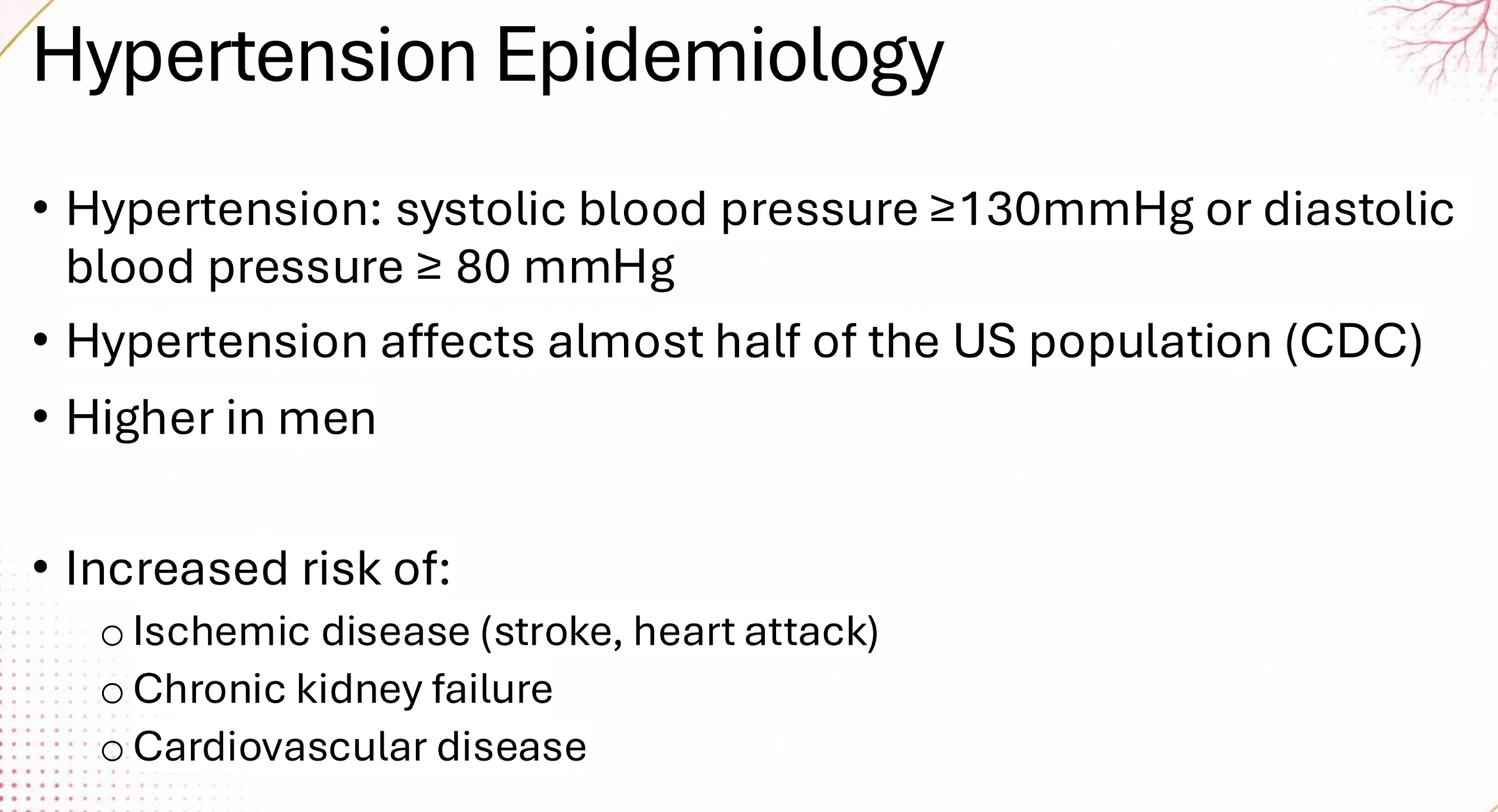
______ _____ is a focal destruction of choriocapillaris due to ischemia
Elschnig’s Spot
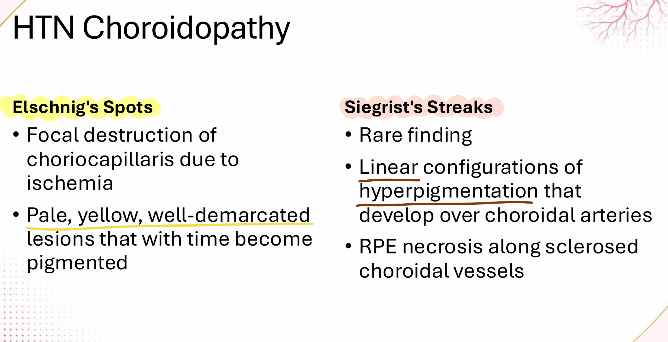
______ _____ is a linear configuration of hyperpigmentation that develops over choroidal arteries
Siegrist’s Streak

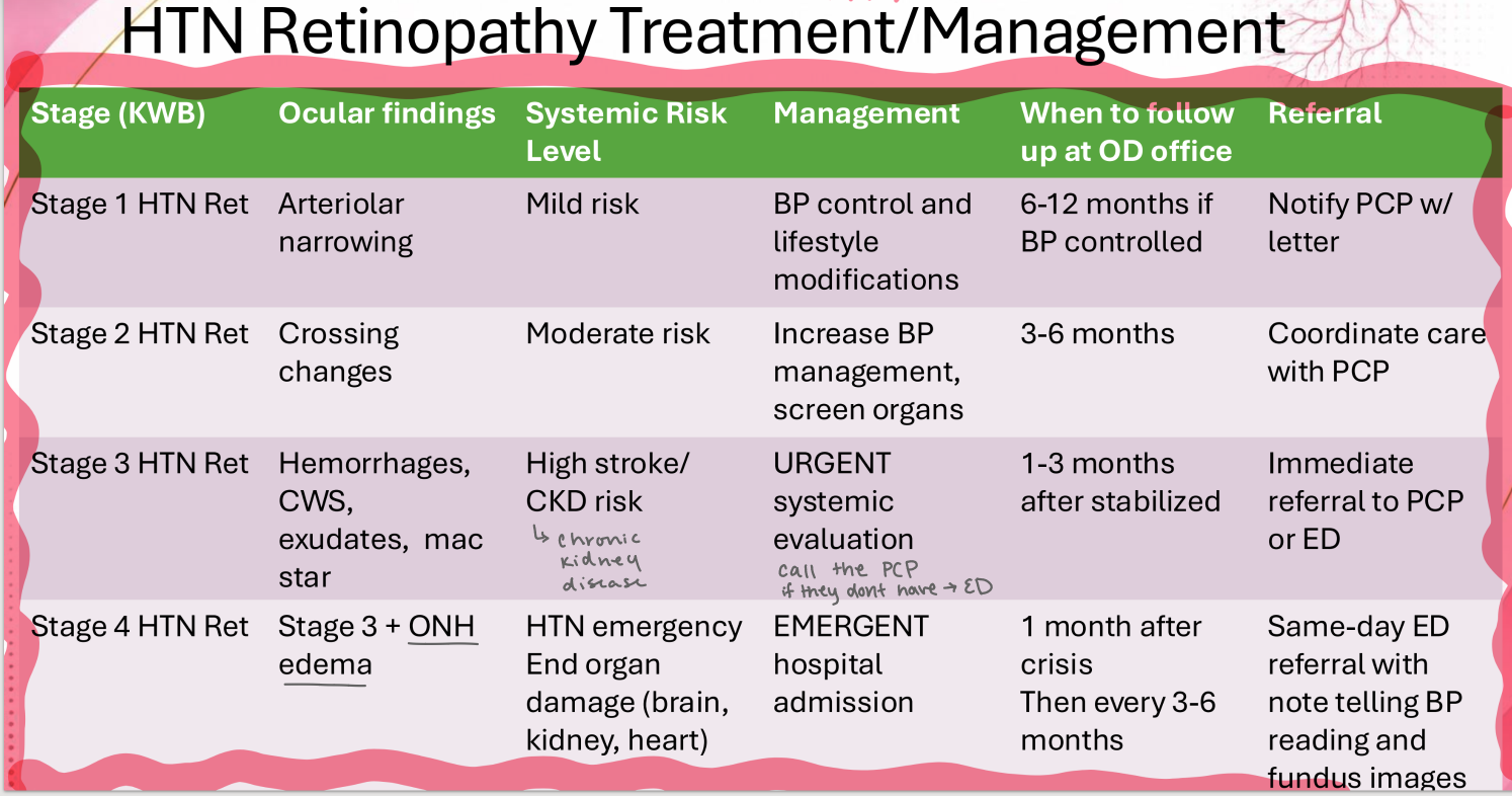
Generalized arteriolar narrowing only is Grade ___ on the Keith-Wagener Barker HTN-R scale
1

Focal narrowing and arteriovenous nicking/ nipping is Grade ___ on the Keith-Wagener Barker HTN-R scale
2

Focal narrowing and arteriovenous nicking/ nipping + exudates, hemorrhages, and CWS is Grade ___ on the Keith-Wagener Barker HTN-R scale
3

Focal narrowing and arteriovenous nicking/ nipping + exudates, hemorrhages, and CWS + optic disc swelling is Grade ___ on the Keith-Wagener Barker HTN-R scale
4

Increase in arteriolar light reflex is Grade ___ in the Scheie arteriosclerotic retinopathy grading/ chronic arteriolar sclerosis scale
1
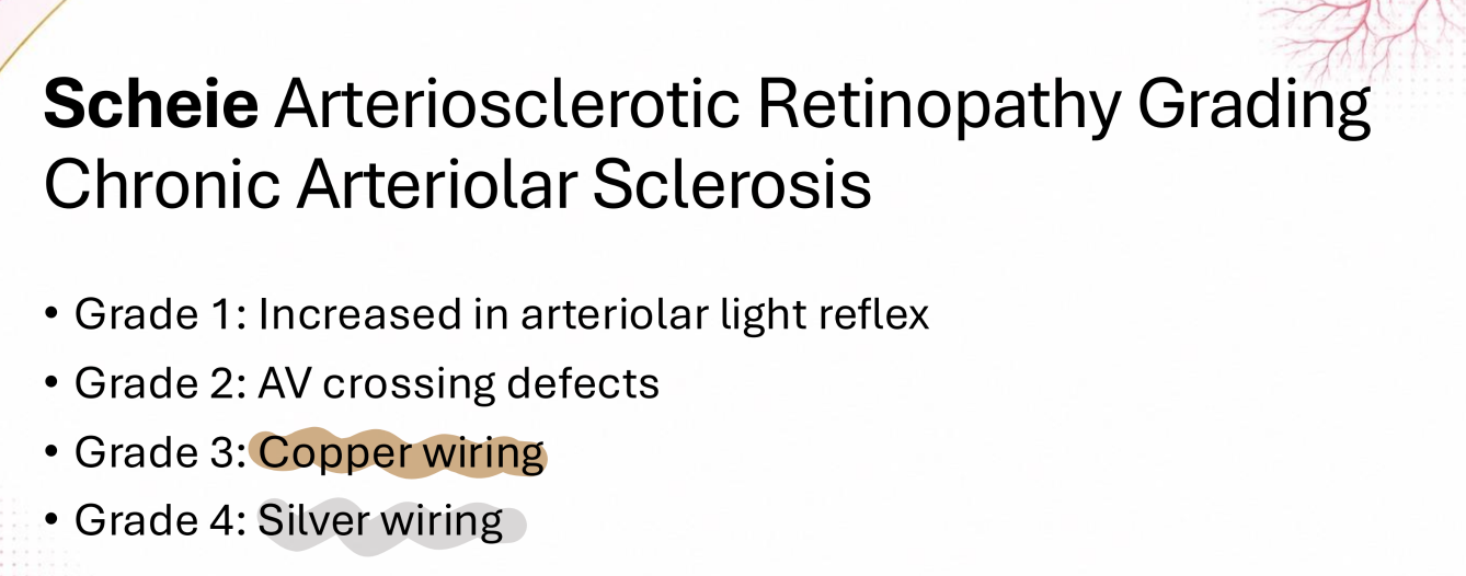
AV crossing defects is Grade ___ in the Scheie arteriosclerotic retinopathy grading/ chronic arteriolar sclerosis scale
2
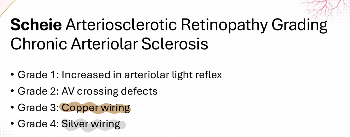
Copper wiring is Grade ___ in the Scheie arteriosclerotic retinopathy grading/ chronic arteriolar sclerosis scale
3
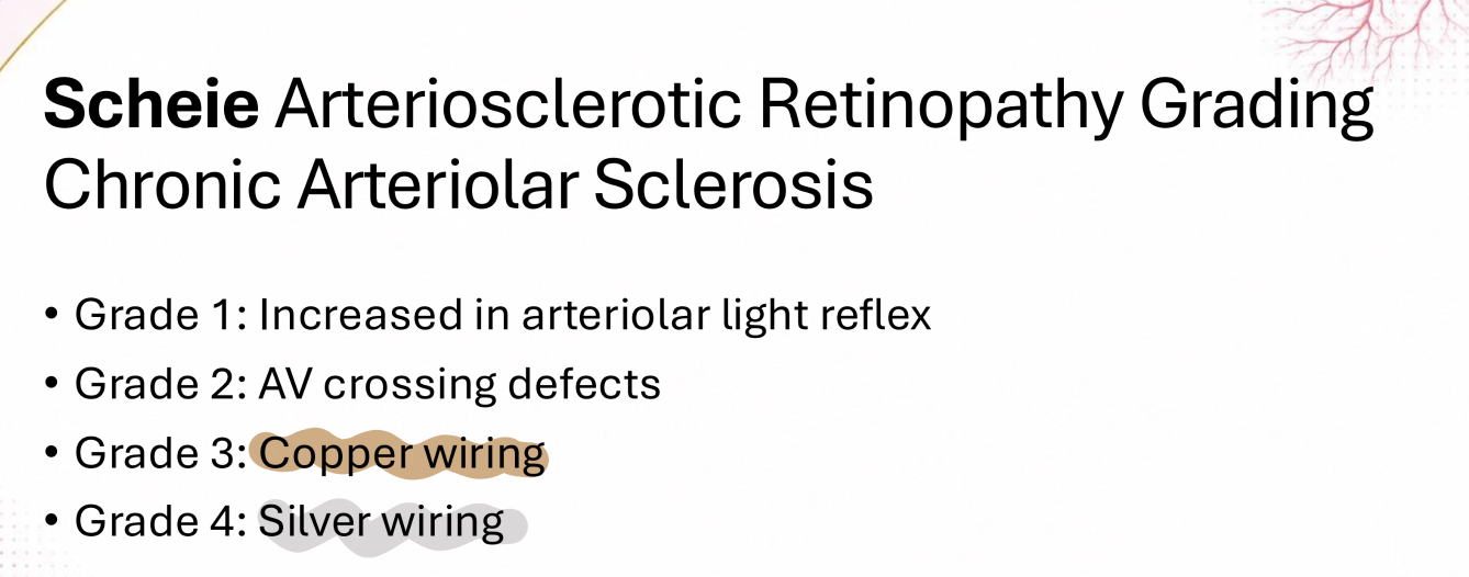
Silver wiring is Grade ___ in the Scheie arteriosclerotic retinopathy grading/ chronic arteriolar sclerosis scale
4

What systemic risk should we be worried about in a patient with stage 3 HTN ret.?
stroke; chronic kidney disease
definition of retinal arterial macro-aneurysm is an aneurysm measuring a size between ______ in diameter
100 - 300 microns

patients with symptomatic retinal artery occlusion have the highest risk of stroke when?
during the 2 weeks before and up to 1 month after the occlusion

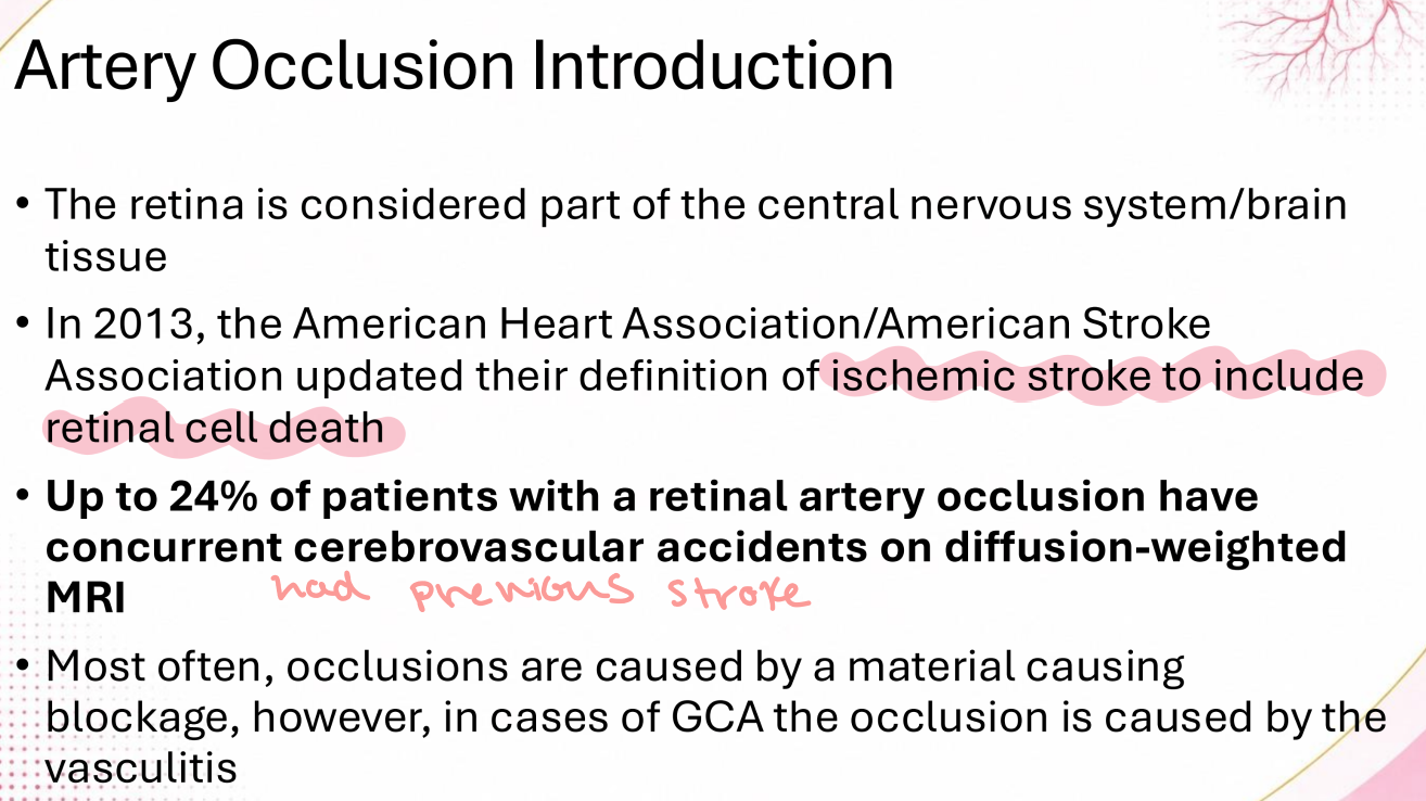
up to 24% of patients with a retinal artery occlusion have concurrent _____ ______diffusion-weighted MRI
cerebrovascular accidents
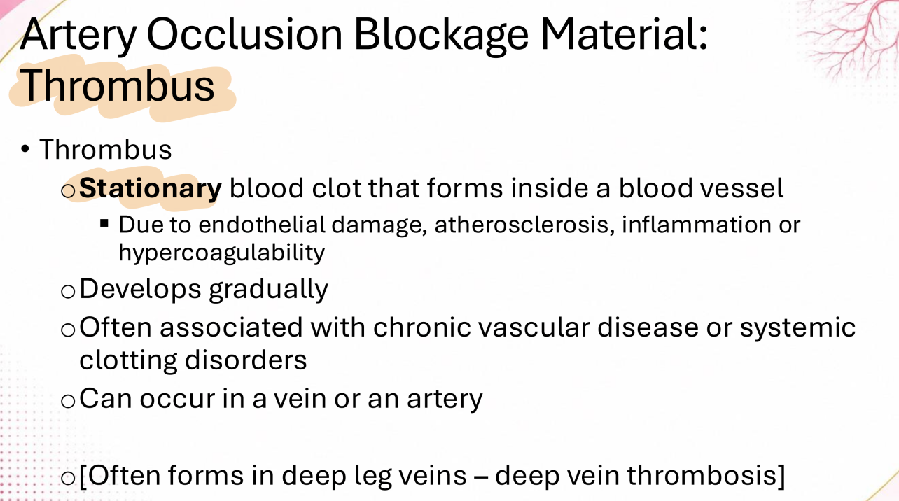
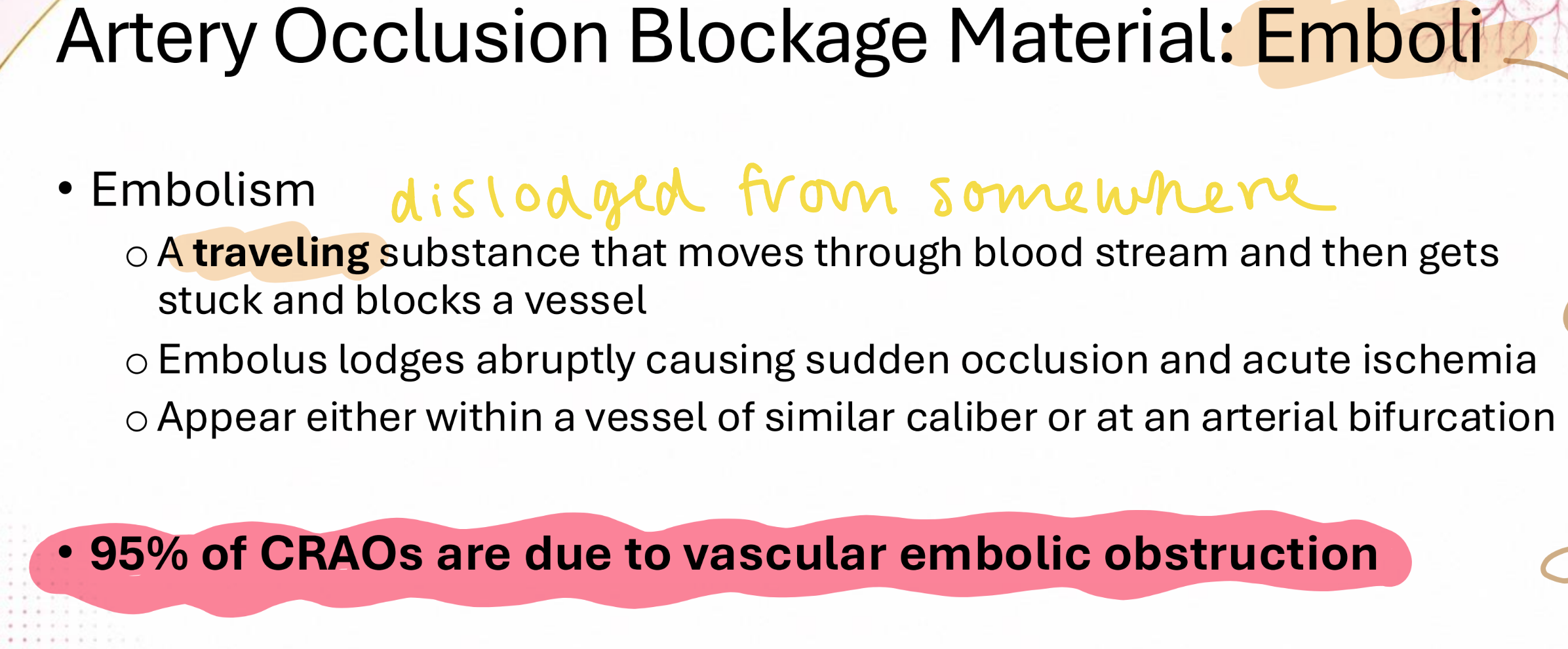
(thrombus/ embolism) is a stationary blood clot that forms inside blood vessel
thrombus
(thrombus/ embolism) is a traveling substance that moved through blood stream and then gets stuck/ blocks a vessel
embolism

95% of CRAO are due to _____ _____ _____
vascular embolic obstruction

“Hollenhorst Plaque” is what kind of emboli?
cholesterol

(embolism / thrombus) are more common
embolism
which type of emboli is less common but more severe due to its large size and impact on larger retinal artery branches?
calcific

Most common cause of a cherry red spot is:
Central Retinal Artery Occlusion (CRAO)

Central Retinal Artery Occlusion (CRAO) will typically show (normal/abnormal) appearance of veins in the acute phase
normal
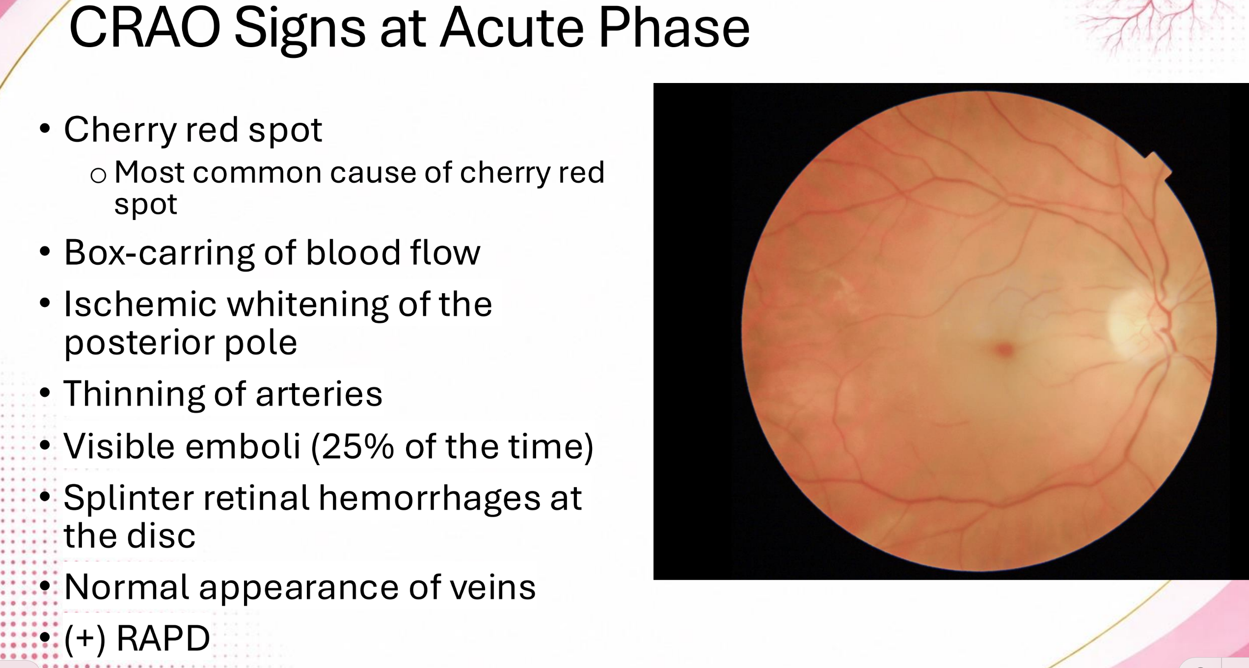
Cherry red spot is due to normally perfused (NFL / Choroid) and ischemia of (NFL / Choroid)
Choroid; NFL

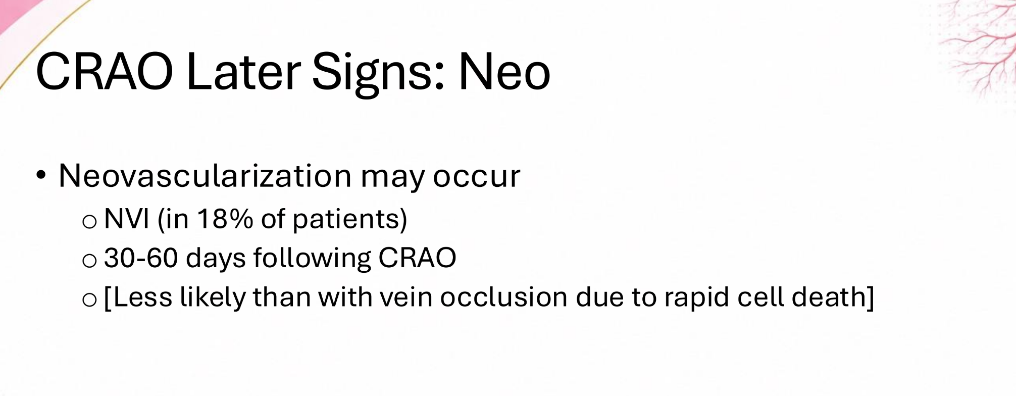
Neovascularization is more likely with (RAOs / RVOs)
RVOs
CRAOs are commonly associated with ____ artery stenosis
carotid

What ALWAYS needs to be ruled out in a case with amaurosis fugax & signs similar to CRAO
Giant Cell Arteritis (GCA)

CRAO typically occur (unilateral / bilateral)
unilateral
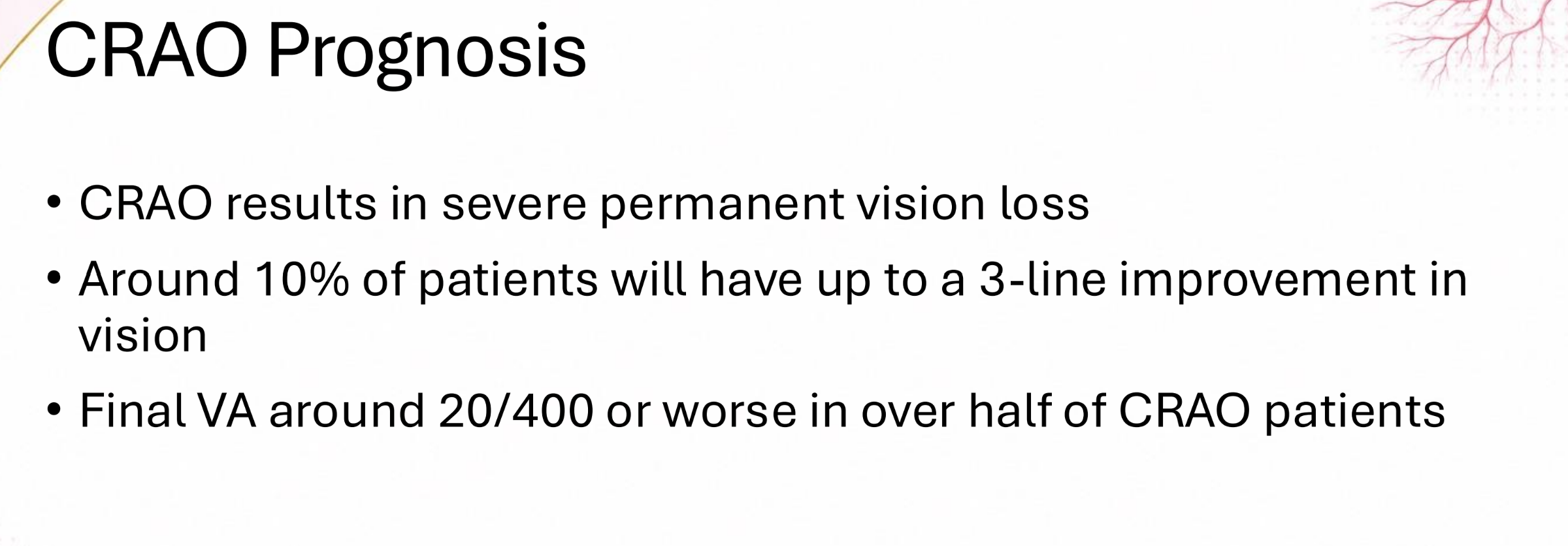
visual prognosis of Central Retinal Artery Occlusion
poor,
one-half of patients will result in VA <20/400
what is the most common cause of BRAO?
carotid emboli

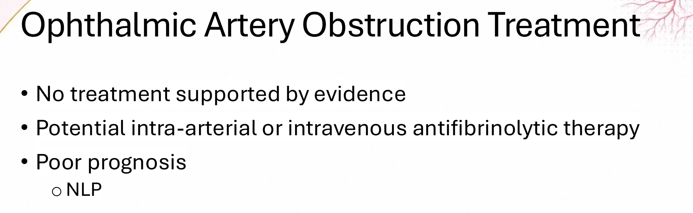
ophthalmic artery obstruction prognosis?
poor,
NLP
Cilioretinal Artery is present in what % of individuals?
30%

Cilioretinal Artery Occlusions commonly occur in combination with (CRAO / CRVO / BRAO / BRVO)s
CRVOs

top two most common retinal vascular disorders?
diabetic retinopathy,
vein occlusion

When there is compression of the vein by an atherosclerotic artery, it will cause:
Vein Occlusion

nearly 50% of vein occlusion are due to _____
hypertension

(BRVOs / CRVOs) are 6-7x more common
BRVOs

(Retinal Collaterals / Optociliary Shunt Vessels) are smaller, loopy, and arise within the retina
retinal collaterals

(Retinal Collaterals / Optociliary Shunt Vessels) are larger, tortuous, at the optic disc, and connect the retinal venous system to the choroidal circulation
optociliary shunt vessels

CRVO is due to a thrombus in the central retinal (artery / vein)
vein

why would a patient with a CRVO develop vision loss?
macular edema

Ischemic CRVO is commonly associated with what finding?
neo (disc, iris, retina)
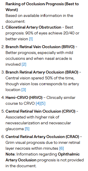
Rank the vascular occlusions from best prognosis to worst: (BRVO, CRVO, BRAO, HRVO, CRAO, Cilioretinal Artery Obstruction)
Cilioretinal Artery Obstruction,
BRVO,
BRAO,
HRVO,
CRVO,
CRAO
OIS stand for:
ocular ischemic syndrome (OIS)

patients with poor collateral circulation between the internal and external carotid systems are more susceptible to developing _____
Ocular Ischemic Syndrome (OIS)
(non-ischemic CRVO / OIS) will have dilated and tortuous retinal veins whereas (non-ischemic CRVO / OIS) will only have dilated veins
non-ischemic CRVO, OIS

hypercoagulability triad:
hypercoagulability,
vascular stasis,
vascular trauma

_______ has the highest risk for thrombotic events among inherited thrombophilias
antithrombin III deficiency

Protein C or Protein S (Excess/ Deficiency) will lead to increased clot formation/ hypercoagulability
deficiency

(Hyperhomocysteinemia / Hypohomocysteinemia) will lead to increased inflammation and thrombosis
Hyperhomocysteinemia

most common acquired thrombophilia is ____
antiphospholipid syndrome (APS)
(SS /SC) sickle cell has more severe systemic manifestations
SS (Homozygous)

(SS /SC) sickle cell has more severe ocular manifestations
SC (Heterozygous)
What are the 4 signs that have specific names in a patient with sickle cell?
“Macular Depression Sign”,
“Salmon Patches”,
“Black Sunburst”,
“Sea Fans”

the hallmark posterior segment sign of sickle cell is ____ ____ _____ aka “sea fans”
peripheral retinal neovascularization

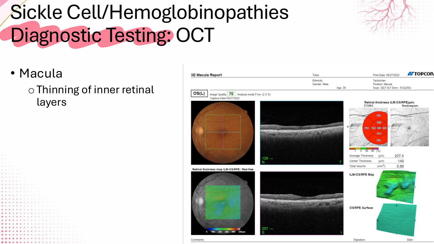
In a patient with sickle cell, you might expect to see (thinning / thickening) of the inner retinal layers in the macula
thinning
Coats’ Disease is normally (Unilateral / Bilateral)
unilateral
hallmark sign of Eales Disease:
recurrent vitreous hemes

radiation therapy damages the (endothelial cells / pericytes) while sparing the (endothelial cells / pericytes)
endothelial cells;
pericytes

retinal capillary hemangiomas have a strong association with ___ _____ ___ disease
Von Hippel Lindau (VHL)
Will retinal cavernous hemangioma show leakage on FA?
nope

T/F: Wyburn-Mason Syndrome is inherited
false


likely dx?
A-V malformation

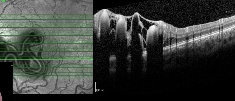

8 yo female, normal MRI
likely dx?
AV Malformation
by definition, Purtscher’s Retinopathy must be caused by _____
trauma

Optic Nerve Hypoplasia is commonly associated with which systemic condition?
Sept-Optic Dysplasia (De Morsier Syndrome)

Sept-Optic Dysplasia/ De Morsier Syndrome Triad?
Optic Nerve Hypoplasia,
Midline Brain Abnormalities (Absence of Septum Pellucidum),
Pituitary Gland Dysfunction

_______ ________ ___________ is a condition characterized by a subnormal number of optic nerve axons
Optic Nerve Hypoplasia

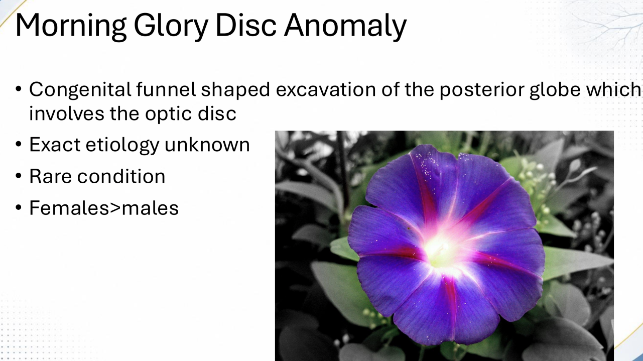
_______ ________ ___________ ___________is a condition characterized congenital funnel shaped excavation of the posterior globe, involving the optic disc
morning glory disc anomaly
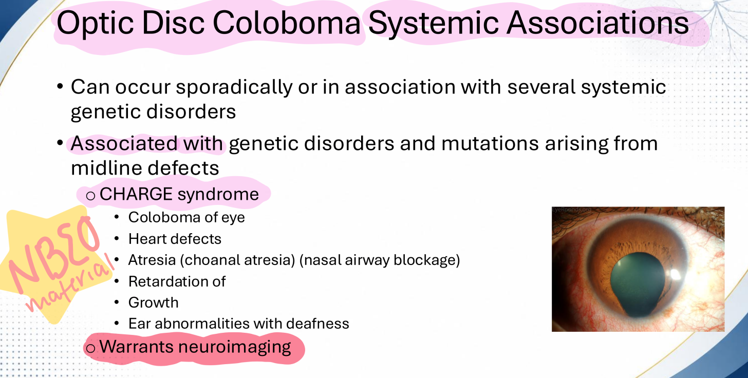
Optic Disc Coloboma is an enlarged disc within a sharply demarcated white excavation (superiorly / inferiorly), due to an incomplete closure of the ________ ______ during gestation
inferiorly,
embryonic fissure

genetic disorders and mutations associated with optic disc colobomas:
Coloboma of eye,
Heart Defects,
Atresia (choanal atresia)/ nasal airway blockage,
Retardation of Growth,
Ear abnormalities with deafness
_______ _______ is characterized as an isolated excavation in the optic nerve head
optic pit

_____ is characterized by a large optic disc (>2.5 mm)
Megalopapilla

_____ _____ is characterized by oblique insertion on optic nerve to the globe, and the retina and choroid do not border the ONH
scleral crescent

_____ _____ is characterized by oblique insertion on optic nerve to the globe, and the retina does not border the ONH
choroidal crescent

______ _____ _______ is characterized as a benign congenital finding where myelination extends past the lamina cribrosa and appears in the retina
myelinated nerve fibers

Bergmeister’s papilla is a benign glial remnant positioned over the ONH and is a remnant of the ______ ______
hyaloid artery

_____ ______ _______ is characterized as a benign neoplasm over the optic nerve
Optic Nerve Melanocytoma
Leber Hereditary Optic Neuropathy arises from an inherited point mutation in ___ _____
mitochondrial DNA

Leber Hereditary Optic Neuropathy has a (maternal / paternal) inheritance pattern
maternal
Leber Hereditary Optic Neuropathy has a strong (male / female) predilection
male

The most common hereditary optic neuropathy is . . .
autosomal dominant optic atrophy

most cases of autosomal dominant optic atrophy are caused by a mutation of _____
OPA1 gene on chromosome 3q

(LHON / ADOA) is a hereditary optic nerve condition that has mitochondrial (maternal) inheritance
LHON (Leber’s Hereditary Optic Neuropathy)
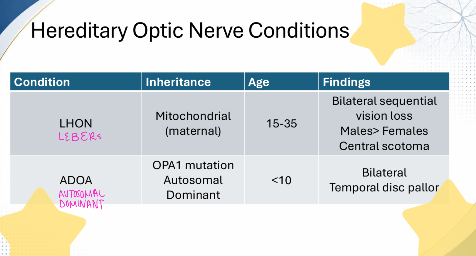
(LHON / ADOA) is a hereditary optic nerve condition that has a OPA1 mutation that is autosomal dominant inheritance
ADOA (Autosomal Dominant Optic Atrophy)

(LHON / ADOA) is a hereditary optic nerve condition that shows bilateral sequential vision loss as well as a central scotoma, and is more common in males
LDON (Lebers Hereditary Optic Neuropathy)

(LHON / ADOA) is a hereditary optic nerve condition that shows bilateral temporal disc pallor
ADOA (Autosomal Dominant Optic Atrophy)

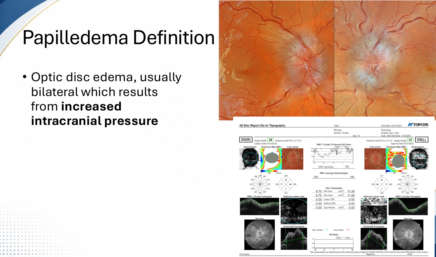
In order to be true papilledema, it needs to be caused by _____ _____ ______
increased intracranial pressure
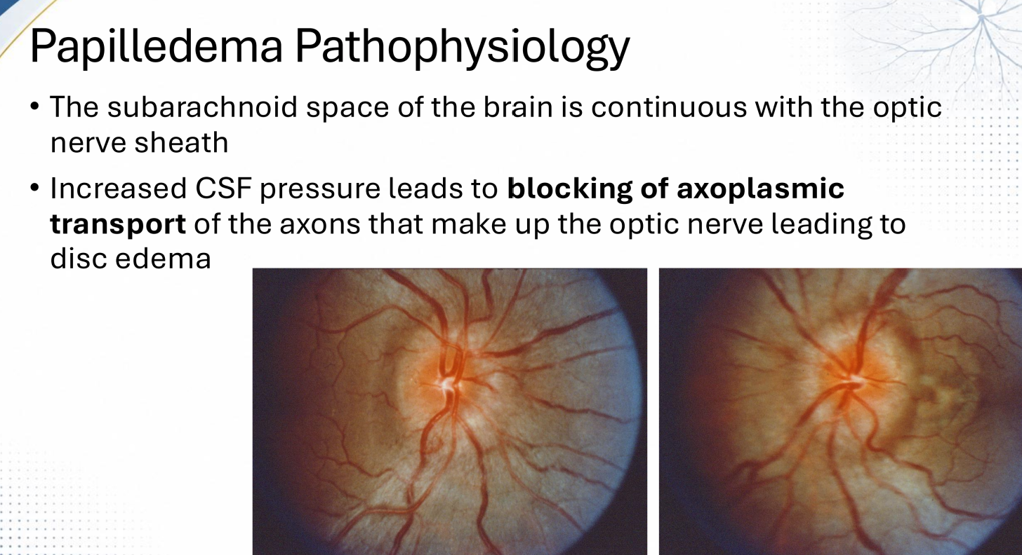
The _____ space of the brain is continuous with the optic nerve sheath
subarachnoid

Papilledema disc edema starts with (increased/decreased) cerebral spinal fluid pressure
increased

What causes the disc edema in papilledema?
increased cerebral spinal fluid pressure that leads to blocking of axoplasmic transport of ON axons
Papilledema is (always bilateral / always unilateral / either bilateral or unilateral)
always bilateral

When examining the ONHs and you see disc hyperemia, disc swelling, blurred margins, and NLF blurring OU, what is the likely stage of papilledema?
early

When examining the ONHs and you see gross elevation of the optic nerve, engorged veins, peripapillary splinter hemorrhages, less disc hyperemia, and hard exudates OU, what is the likely stage of papilledema?
full developed

When examining the ONHs and you see fewer hemorrhages, less disc hyperemia, hard exudates, what is the likely stage of papilledema?
chronic
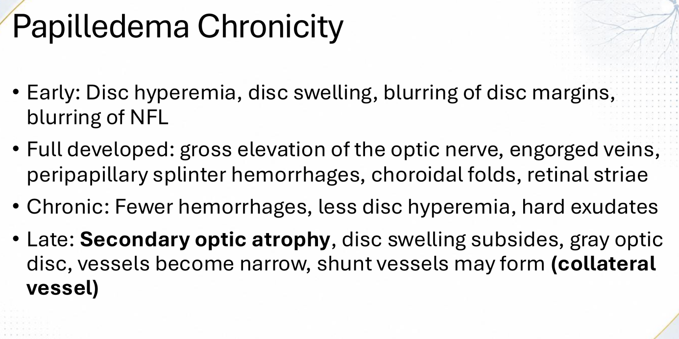
When examining the ONHs and you see secondary optic atrophy, disc swelling subsided, gray optic disc, narrowed vessels, and collateral vessels, what is the likely stage of papilledema?
late

When examining the ONHs and you see nasal margin blurring and a normal temporal margin OU
what is the papilledema grading using Frisen?
Grade 1

When examining the ONHs and you see blurring of all borders/ peripapillary halo OU with significant nasal elevation normal temporal margin OU
what is the papilledema grading using Frisen?
Grade 2

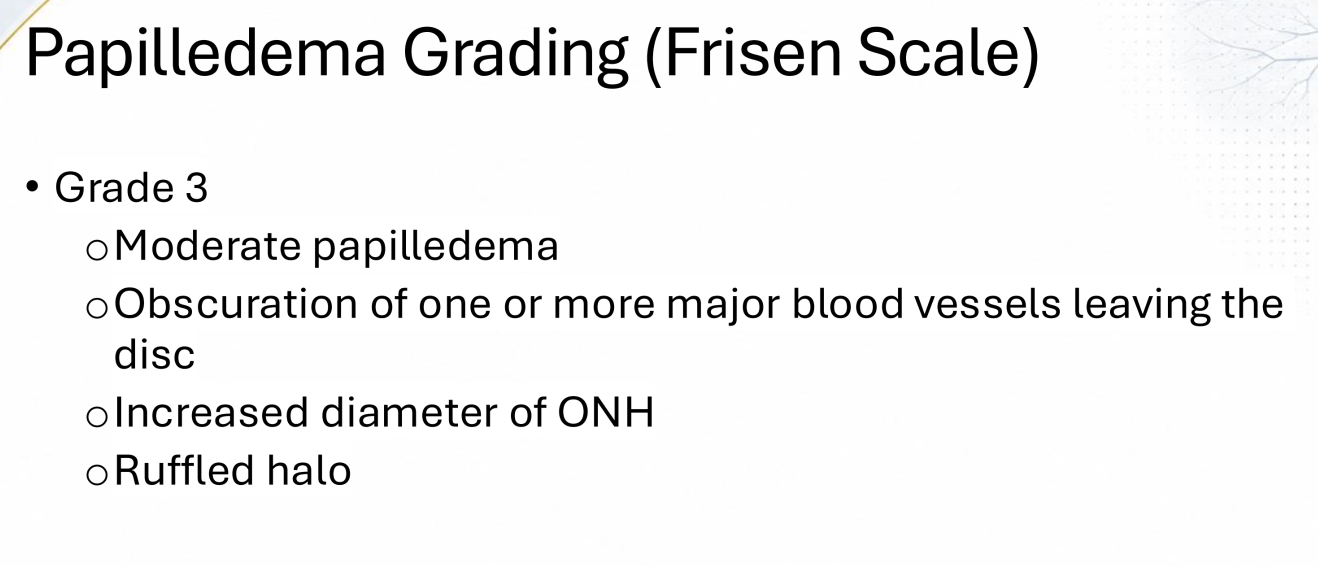
When examining the ONHs and you see obscuration of one or more major blood vessels leaving the disc OU as well as an increased diameter of the ONH OU and a ruffled halo OU
what is the papilledema grading using Frisen?
Grade 3
When examining the ONHs and you see obscuration of major vessels on the disc OU, and elevation of entire ONH OU, as well as CWS and hemes
what is the papilledema grading using Frisen?
Grade 4
