A and P Exam 3
 Call Kai
 Learn
 Practice Test
 Spaced Repetition
 Match
 Flashcards
 Knowt Play
1/80

There's no tags or description
Looks like no tags are added yet.
81 Terms
Muscle Function
Turn chemical energy into mechanical force
Maintain
Posture
Stabilize joints
Generate heat
Muscle Excitability
Ability to receive and respond to stimuli
Muscle Contractibilty
Ability to shorten
Muscle Extensibility
Ability to lengthen
Muscle Elasticity
Ability to recoil and return to original shape
Types of muscle - all come from ______
Mesoderm
Skeletal muscle
Cardiac muscle
Smooth muscle
Myoblasts
Precursors to skeletal muscle
Multi-nucleated Myotube
Phase in between myoblast and myofiber
Myofiber
Cylindrical, multinucleated mature muscle cell
Nervous system must form a synapse/motor end plate in order for myotube to become myofibril
Skeletal Muscle
Fibers: striated, tubular, multi nucleated
Voluntary
Usually found attached to skeleton
Vascularized and innervated by a plexus (network of blood vessels and nerves)
Direct (A) Attachment Type
Straight attachment from muscle to bone
Attachment via periosteum
Attachment via perichondrium during development
Indirect (B, C) Attachment Type
Attachment via tendon
Attachment via tendon to bone
Aponeurosis attachment: sheet of connective tissue usually for wider-sheet like muscles; attaches sheet like muscles needing a wide area of attachment (lumbar, abs, scalp)
Cardiac Muscle
Fibers: Striated, branched, and mono or multinucleated
Involuntary
Calcium mainly from caveolae
Only found covering walls of the heart
Smooth Muscle
Fibers: Non-striated, spindle-shaped, uninucleated, no sarcomeres, no troponin/tropomyosin, has calmodulin and myosin light chain kinase, intermediate filament cytoskeleton,
Calcium mainly from caveolae
Involuntary
Usually found covering the walls of internal organs
Organization of Muscle

Epimyseum
Outermost sheath that surrounds the entire muscle
Perimyseum
Dense irregular connective tissue sheet that surrounds fascicles
Endomysium
Smallest sheath, loose areolar connective tissue surrounding each muscle fiber
Fasicle
Bundle of muscle cells with motor units
Motor unit - A motor neuron and all of the muscle fibers that it innervates
Muscle Fiber
Individual muscle cell. Extremely large. Has sarcoplasm (cytoplasm for muscle cells). Has sarcolemma (cell membrane for muscle cells). Made of myofibrils
Myofibril
Organelle, only in striated muscle. Consists of sarcomeres, rod shaped
Sarcomere
Smallest unit of muscle, contractile unit of muscle with actin and myosin
Sarcomere photo

Second sarcomere photo

H-Zone
No actin overlap, no thin filaments, only thick filaments. Shortens during contraction

M-Line
Represents the middle of the sarcomere Structural proteins, anchors thick filament and whole sarcomere
Z-Disk
Represents the edges of the sarcomere. Tolerates a lot of mechanical stress
A-Band
Represents the Portion of Myosin
Anisotropic” Lack of light, lots of proteins + mitochondria. Thick and thin filaments
I-Band
Represents the portion of actin that has no myosin overlap
“Isotropic”. Allows light. Less proteins. Z-Disk + some actin filaments
Shortens when muscle contracts
Thin Filaments
String of globular actin proteins
Has myosin binding sites
Binding sites blocked by tropomyosin - regulatory protein, allows muscles to relax
Troponin complex - positions tropomyosin, relaxed = actin binding sites covered, contract = pull on tropomyosin to uncover myosin binding sites
Thick Filaments
Myosin
2 proteins
Binds to actin, pulls, causes contraction
Sarcoplasmic Reticulum
Where muscles stores, releases, and sequesters calcium (initiates contraction)
It is a special Ed smooth endoplasmic reticulum

T-Tubule
Perpendicular to sarcomere
Part of sarcolemma (cell membrane)
Where EC coupling occurs
Allows action potential into muscle fiber
Terminal Cisternae
Where calcium is stored + released
Has calcium pumps that use and active transport calcium pump to sequester it
Triad
Two terminal cisternae + one T tubule
E-C Coupling
Acetylcholine released from the axon terminal binds to receptors on the sarcolemma, causing them to change shape and sodium and potassium ions to cross the membrane.
More sodium crosses the membrane than potassium, more positive ions move in than out and the membrane potential. Sodium voltage gated channels are opened in the neuron, and an action potential is generated and travels across the membrane and down the T tubule. This occurs in a series, not one action potential.
Ca²+ is released from the sarcoplasmic reticulum in response to the change in voltage.
Ca²+ binds troponin; Cross-bridges form between actin and myosin.
Acetylcholinesterase removes acetylcholine from the synaptic cleft.
Ca²+ is transported back into the sarcoplasmic reticulum.
Tropomyosin binds active sites on actin causing the cross-bridge to detach.
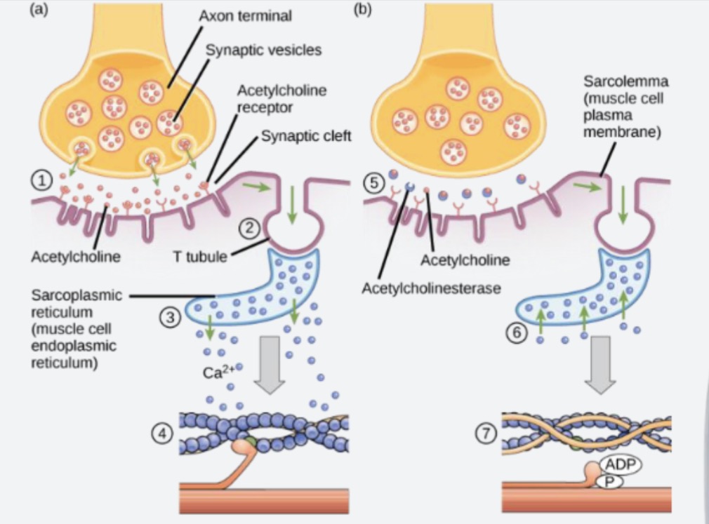
Jacobs EC Coupling Explanation
Ch 1) The Neuron
• A stimulus strong enough to reach threshold triggers an AP to travel from the motor cortex, spinal cord, to motor neuron.
• Sodium VGCs open. Na+ enters axon causing membrane potential to increase/depolarize. This occurs in a series along the length of the membrane.
• Na+ travels to buton/axon terminals. Causes Ca2+ VGCs to open. Triggers exocytosis
• Vesicles storing neurotransmitter ACh attach to T-snares on membrane with V-snares.
• ACh is released from neuron (exocytosis) into the neuromuscular junction/synaptic cleft
• Behind the AP, K+ channels open, repolarizing the cell. Na/K pump restores resting potential
Ch 2) The Muscle Fiber
• ACh attaches to ACh receptors on the sarcolemma in the synaptic cleft. These open, allowing sodium to enter the cell. This raises membrane potential, causing the AP to be transferred to the muscle fiber.
• Na+ Channels open in a series along the length of the sarcolemma(depolarization), down the length of the sarcolemma and down the T Tubule
• As AP propagates down T Tubule, triggers Ca2+ VGCs to open which connect the T tubule to the SR. Calcium now is released from cisternae into cytosol
Ch 3) Sarcomere Shortening
• Calcium binds to troponin which causes sarcomeres to shorten
• Thin filaments slide past thick filaments
• I Bands decreases in length
• H zone decrease in length
• Z- discs decrease in length
Ch 4) Cross Bridge Cycling
Calcium binds to troponin C on the troponin complex, this causes troponin T to pull on on tropomyosin exposing the myosin binding sites on actin.
Myosin heads in a high energy state attach, release ADP + Pi and pull on the actin thin filaments towards the M line
ATP attaches to myosin, releasing it from actin
ATP is hydrolyzed by ATPase to ADP and Pi. Restores myosin to high energy
"cocked" position.Cycle repeats in presence of calcium
Epilogue) Relaxation
Stimulus ends, AP ends. Neuron restored back to resting. ACh no longer released. ACh in synaptic cleft is broken down by enzymes or sequestered.
Membrane potential similarly restored in muscle cell via Na/K pump.
Calcium is sequestered via calcium pumps on the terminal cisternae back into the SR. Pumps are called SERCAs
ATP detaches myosin from actin
Muscle relaxes

Action Potentials

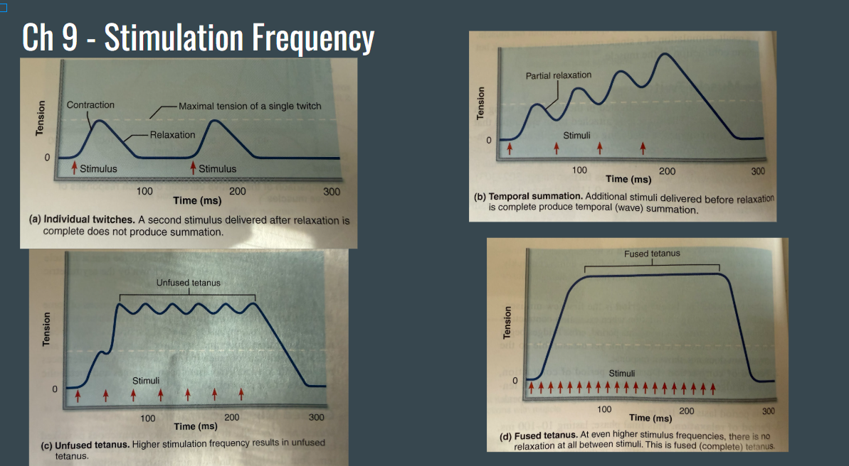
Stimulation Frequency
Neural Recruitment

Length-Tension Relationship

Powerstroke
Movement of the myosin head, bound to actin, towards the M-Line of the sarcomere
This pulls actin towards the center of the sarcomere, shortening the I-Band, H-zone, and the entire sarcomere

Cross-bridge Cycle

Arrange the following structures from largest to smallest: myofibril, whole muscle, muscle fiber, fascicle, and sarcomere.
Whole muscle → fascicle → muscle fiber → myofibril → sarcomere. A muscle fiber is one muscle cell containing many myofibrils, each composed of repeating sarcomeres.
What structures mark the boundaries of a sarcomere?
Describe what happens to the distance between these boundaries during contraction.
A sarcomere extends from one Z disc to the next.
During contraction, the Z discs move closer together, shortening the sarcomere.
Identify the main protein in each type of filament.
How do these proteins interact to cause muscle contraction?
Thick filaments consist primarily of myosin, while thin filaments consist primarily of actin. Myosin heads bind to actin and pull the thin filaments toward the center of the sarcomere.
What does the sarcoplasmic reticulum store, and how does releasing this substance help initiate muscle contraction?
It stores calcium ions (Ca?*). Released calcium binds to troponin, causing tropomyosin to move and expose myosin-binding sites on actin, allowing contraction to begin.
During contraction, do the thick and thin filaments become shorter? Explain how their movement changes the length of the sarcomere.
Thin filaments slide past thick filaments toward the sarcomere center, increasing their overlap and bringing the Z discs closer together. H-Zone shortens
Cardiac Muscle
Specialized striated muscle developed from cardiomyocytes and only found in walls the heart. Mononucleated
Cardiomyocytes are small
Automaticity - Action potentials are created spontaneously by the heart, allowing it to depolarize and beat on its own without need for external stimuli
Cardiac Muscle Neural Control
Autonomic nervous system
Parasympathetic - relaxes
Sympathetic - stimulates
No neuromuscular junction
Intercalated Discs
Spread action potentials throughout the heart, which promotes the rhythmic contraction of the heart
Rich in Desmosomes - Membrane junction between cardiomyocytes swell suited for dealing with the high stress connection from contraction of heart
Rich in Gap Junctions - Allow communication between cardiomyocytes, so that if one depolarizes they all depolarize
Smooth Muscle
Non-striated, unvoluntary, mononucleated
Smaller than skeletal muscle, thin & thick filaments are arranged diagonally
Found in the walls of hollow visceral organs (stomach, bladder, etc.)
Peristalsis – waves of smooth muscle contraction followed by waves of relaxation that move substances through the GI tract
Smooth muscle has SLOW & SMOOTH sustained contractions
Smooth Muscle Neural Control
Autonomic Nervous System
Parasympathetic
Sympathetic
No neuromuscular junction, i.e. no synaptic cleft
Instead, innervated with varicosities -Swollen bulbs of nerve bundles that release neurotransmitters
Smooth Muscle Structure
Cytoskeleton: criss-cross network of intermediate filaments, attached together via dense bodies
Caveolae: Ca2+ VGCs that diffuse calcium across plasma membrane
SR - poorly developed
One nucleus
Thick and thin filaments: more thick to thin, 1:13 (skeletal is about 1:6)
No troponin or tropomyosin
Varicosities - smooth muscle innervation
Smooth Muscle EC Coupling
AP travels from brain, spinal cord, autonomic axon, to varicosity. Neurotransmitter releases, binds to receptors. Increased membrane potential cause Ca2+ VGC to open at caveolae. Some is also released by SR.
Calcium binds to and activates calmodulin.
Calmodulin activates myosin light chain kinase (MLCK)
MLCK breaks down ATP to ADP + Pi, attaches Pi to myosin
Myosin now activated and forms cross bridge. Actin thin filaments slide across myosin thin filaments

Single Unit Type Smooth Muscle
“Visceral” muscle
Contraction rhythmically
Coupled via gap junctions
May have spontaneous potentials
Physic contractions - contraction to relaxation
Ex: peristalsis
Multi Unit Type Smooth Muscle
Gap Junctions Rare
Infrequent action potentials
Structurally independent (work on their own)
Lots of nerves
Graded contractions (force output varied rather than the entire muscle contracted/relaxed)
Tonic contractions (little to no energy needed) - contraction, hold, relaxation
Ex: Large arteries, arrector pili muscles
Smooth Muscle Response to Stretch/Stress Relaxation Response
When you stretch smooth muscle, it impulsively contracts
If you hold it, it will relax and adapt to its new length. Allows organs to store contents that change in volume. Ex: Your bladder
Sliding Filament Theory
During contraction the thin filaments slide past the thick filaments so that actin and myosin overlap to a greater degree
Cross-Bridge Cycling

Know this stuff about twitches

Fast (Unique) Twitches
Very oxidative
Uses carbs (glucose, aerobic metabolism)
Ex: extraocular muscle
Medium (considered fast if fast unique is not included) Twitch
Power (short sprint)
Relaxes relatively quickly
Uses carbs (glycogen / anaerobic metabolism)
Ex: Gastrocnemius
Slow Twitch
Endurance
Uses fates (aerobic metabolism)
Uses fats
Relaxes slowly
Postural
Ex: soleus
Explain why ATP is necessary for both muscle contraction and relaxation. How would a complete lack of ATP affect myosin attachment to actin and calcium transport, and how does this explain rigor mortis?
ATP allows myosin to detach from actin. As ATP binds to the myosin head, causing it to release actin and break the cross-bridge.
ATP is hydrolyzed into ADP and inorganic phosphate (Pi), energizing and repositioning (“cocking”) the myosin head.
The energized myosin head binds to exposed sites on actin. Release of Pi triggers the power stroke, pulling actin toward the center of the sarcomere; ADP is then released. Another ATP is needed for detachment and repetition. Mention cross-bridge cycling
Calcium binds to troponin, moving tropomyosin away from the myosin-binding sites on actin.
ATP powers calcium pumps (SERCA) that move calcium from the cytosol back into the sarcoplasmic reticulum.
Calcium dissociates from troponin, allowing tropomyosin to cover actin binding sites again.
No ATP, Cross-bridges become locked, and normal cycling stops.
No ATP, calcium cannot be actively pumped back into the sarcoplasmic reticulum. Calcium remaining in or leaking into the cytosol keeps actin binding sites exposed.
After death, ATP production stops and remaining ATP is depleted. Calcium accumulates in the cytosol, and myosin–actin cross-bridges become locked, producing muscle stiffness.
Decomposition breaks down muscle proteins and the locked cross-bridges; ATP production does not resume.
Mouth (Buccal Cavity
Stratified squamous epithelium
Site of mastication
Saliva turns food into a bolus
Some chemical digestion and absorption
Important structures
Tongue
Teeth
Hard palate
Soft palate
Frenulum - attached lips and tongue to mouth
Palatine ridge - divides food laterally

Tongue
Controlled by skeletal muscle
Intrinsic muscles - only muscle in body with no origin or insertion. Change shape, length, width
Extrinsic muscles - inserts on mandible. Change tongue position
Secretes lipase (fat breakdown)
Filiform papillae - inc surface area, holds things
Tastebuds: fungiform, vallate, and foliate
Lingual tonsils: Immune system surveillance, has WBCs

Teeth
Masticate (chew)
20 baby (deciduous) teeth
32 adult (permanent) teeth
4 types
Incisors: cut/snip
Canines: tear
Premolars (bicuspid): grind, crush
Molars: grind, crush
Two regions, crown and root

Crown
Enamel - Hydroxyapatite, hard
Dentin - Calcified protein
Pup Cavity - Blood vessels and nerves
Crown
Enamel - Hydroxyapatite, hard
Dentin - Calcified protein
Pup Cavity - Blood vessels and nerves
Root
In jaw bone, fixed via cement and ligament
Canines, premolars, incisors - 1 root
Molars - 2-3 roots
Salivary Glands
3 Glands: parotid (associated with mumps), submandibular, sublingual
Secrete saliva with 1-2 cell types: Serous cells, Mucus Cells (not in parotid)
Functions: cleanse mouth, dissolve food chemicals for taste, aides in bolus formation, contains amylase for starch digestion
Composition: 99% water, amylase, mucin, IgA antibodies, lysozymes, and defensins
Neural Control: Parasympathetic control of autonomic nervous system, Activated by chemoreceptors (taste receptors) and pressoreceptors (pressure receptors)

Esophagus
Pharynx to upper third is skeletal muscle (voluntary - swallowing)
Short transition zone - mix of skeletal and smooth
Rest is smooth muscle
Leads to stomach
Collapses when not in use
No serosa. Has adventitia instead
2 sphincters
Upper esophageal sphincter
Lower esophageal sphincter (cardiac sphincter)

How do the teeth, tongue, and saliva work together to begin digestion and form a bolus?
Teeth primarily aide in mastication. The tongue helps with mastication but also secretes lipase chemically break down triglycerides. Saliva moistens food and secretes amylase to chemically break down starches.
What are the two regions of a tooth? What role does each play?
Crown: hard part of tooth, mastication
Root: insertion into bone, innervation/blood supply
After picking up/delivering nutrients from digestive organs, where must blood go before returning to the heart to be reoxygenated? What is this circuit called?
The liver. Splanchnic portal system/hepatic portal system
A person begins chewing a flavorful piece of food.
Which receptors stimulate salivation, and which division of the autonomic nervous system promotes saliva secretion?
Chemoreceptors. Parasympathetic
What is the deepest membrane of the alimentary canal called when found in the abdominal cavity. What is it called when found outside the abdominal cavity?
Peritoneum/Serosa. Adventitia
T/F, the esophagus is made up of entirely smooth muscle
False
Types of Mechanical Breakdown
Mastication (chewing), churning (stomach), segmentation (small and large intestine food mixing)
Types of Chemical Breakdown
The different enzymes.
Mouth - lipase from salivary glands breaks down fats
Stomach: cell ty
Small intestine: cell types in crypt that secrete enzymes
Enterocytes - intestinal juice
Paneth cells - lysozyme and defensin to destroy bacteria
Goblet cells - secrete mucus
Enteroendocrine cells - secretin and cholecystokinin