Respiratory System/ Test 2
 Call Kai
 Learn
Learn Practice Test
 Spaced Repetition
 Match
Match Flashcards
 Knowt Play
1/52

There's no tags or description
Looks like no tags are added yet.
Name | Mastery | Learn | Test | Matching | Spaced | Call with Kai | Chat |
|---|
No analytics yet
Send a link to your students to track their progress
53 Terms
Nose and Nasal Cavity
Filters, warms, and humidifies incoming air.
Vibrissae (nose hairs) trap larger particles.
Nasal conchae create turbulence, helping air contact the mucous membranes.
The external nose is the visible portion; the nasal cavity is inside.

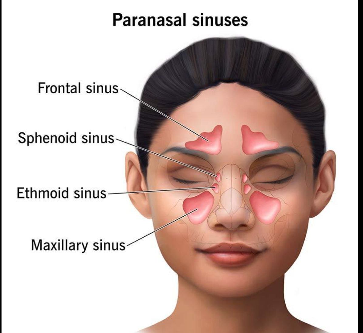
Paranasal Sinuses
Air-filled spaces in the frontal, maxillary, ethmoid, and sphenoid bones.
Help lighten the skull, produce mucus, and add resonance to the voice.
Inflammation of the nasal mucosa is called rhinitis. Inflammation of the sinuses is sinusitis.
Pharynx
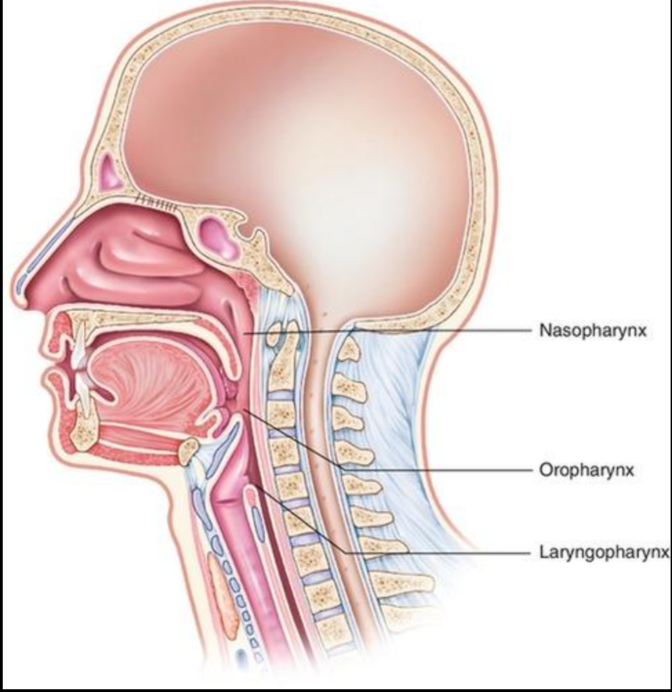
Muscular passageway shared by the respiratory and digestive systems.
Nasopharynx: behind the nasal cavity.
Oropharynx: behind the mouth.
Laryngopharynx: lower region leading toward the larynx and esophagus.
Enlarged adenoids may obstruct nasal airflow and contribute to mouth breathing.
Larynx- Voice box
Keeps an open airway.
Produces sound through the vocal folds.
Helps protect the lower airway during swallowing.
The epiglottis is elastic cartilage that helps keep food and liquid out of the airway.

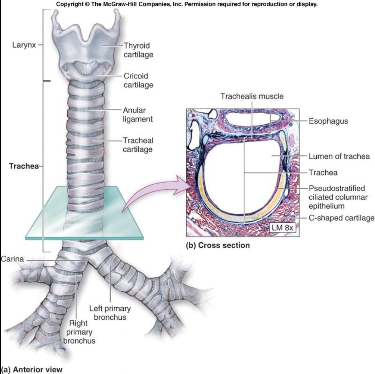
Trachea (windpipe)
Conducts air toward the lungs.
C-shaped cartilage rings help prevent collapse.
Mucus traps particles, and cilia move mucus toward the throat.
Smoking damages this clearance system, contributing to smoker’s cough.
Bronchial Tree
Main bronchi branch into smaller bronchi, then bronchioles.
Terminal bronchioles are the smallest airways of the conducting zone.
Respiratory bronchioles lead into the gas-exchange portions of the lungs.
Main bronchi enter the lungs through the hilum.

Alveoli
Tiny air sacs where oxygen and carbon dioxide are exchanged.
Type I alveolar cells form the thin walls for gas diffusion.
Type II alveolar cells produce surfactant, which reduces surface tension and helps prevent collapse.
Alveolar macrophages engulf particles and microorganisms.
An alveolar duct leads into clusters of alveoli; an alveolar sac is a common chamber opening into multiple alveoli.

Lungs and pleurae
Right lung: three lobes — superior, middle, inferior.
Left lung: two lobes — superior and inferior; it is smaller to accommodate the heart.
Pulmonary circulation carries blood to the lungs for gas exchange.
Bronchial circulation supplies oxygenated blood to lung tissues.
Parietal pleura lines the chest wall; visceral pleura covers the lungs.
Pleural fluid reduces friction during breathing.

Pleurisy
inflammation of the pleura, often causing pain when breathing
Pleural effusion
excess fluid in the pleural cavity
Pneumothorax
air in the pleural cavity that can cause lung collapse; treatment may require a chest tube
Atelectasis
collapse of alveoli or part of a lung
Conducting zone
moves, filters, warms, and humidifies air
Ex: nose, nasal cavity, pharynx, larync, trachea, bronchi, bronchioles
Respiratory zone
participates in gas exchange
Tracheal obstruction
a life-threatening emergency requiring the Heimlich Maneuver
Internal Respiration
Between the blood and tissue
systemic capillaries and tissue
External Respiration
blood and alveoli in the lungs
pulmonary capillaries and alveoli
Forced inspiration
scalenes, sternocleoidomastoid, pectoralis minor
Forced expiration
oblique and transverse abdominal muscles, internal intercostals
Blood that just left tissue
low in O2, high in CO2, low in nutrients
Surfactant
Break up surface tension from water lining the inner alveoli
Disrupting the surface tension stops the alveoli from collapsing and allows inflating during inspiration
Infant Respiratory Distress Syndrome: \Premature infants are born with the inability to make surfactant, resulting in alveolar collapse that does not allow lungs to take in air to obtain oxygen
How does O2 move through the body
Enters body from the atmosphere, reaches alveoli, diffuses to bloodstream, released to systemic circulation, unloaded at tissues
How does CO2 move through the body
Produced as a byproduct of cell activity, picked up by bloodstream from tissues, taken to lungs, diffused to alveoli, expired out
What can cause a reduction in lung compliance
Scar tissue, frequent infections, low surfactant, muscle paralysis
In terms of ventilation-perfusion coupling, arteriolar diameter is dependent on the partial pressure of which gas and bronchiolar diameter is dependent on which gas?
Partial pressure of oxygen impacts arteriolar diameter while partial pressure of carbon dioxide influences bronchiolar diameter.
Arteriolar diameter:
- Decrease in PO2 ⇒ arteriolar constriction
- Increase in PO2 ⇒ arteriolar dilation
Bronchiolar diameter:
- Decrease in PCO2 ⇒ bronchiolar constriction
- Increase in PCO2 ⇒ bronchiolar dilation
Why is ventilation-perfusion coupling important?
Ventilation-perfusion coupling bridges the ability of the body to bring in air and the ability to transport gases in/out of the body. Ventilation allows for oxygen to come in and carbon dioxide to go out. Perfusion allows for oxygen to leave the lung and into the tissues, and carbon dioxide to leave the body. If ventilation is good but perfusion is not, air is getting in/out of the lungs but carbon dioxide is not getting to the lungs to leave, and oxygen is not getting to the tissues. If perfusion is good but ventilation is not, carbon dioxide is getting to the lungs and oxygen is able to get to the tissues but the body is not bringing enough air in/out of the lungs.
What is the Bohr effect
As cells metabolize, they produce carbon dioxide and free hydrogen ions (lowers pH). The presence of these chemical substances weakens the bond between oxygen and hemoglobin, resulting in reduced affinity. This leads to increased unloading of oxygen at those tissues.
Eupnea
normal breathing pattern (12-15 breaths per min)
Hypercapnia
too much CO2 in the body
Hyperventilation
loss of too much CO2, dizziness
Hypoxia
tissues are not receiving enough oxygen
Hypoemic hypoxia- low O2 in blood due to less oxygen making it into the body
Ischemic hypoxia- blockage of vessels
Anemic hypoxia- Low RBCs, little or abnormal hemoglobin
Asthma
acute episodes and symptom-free periods.
Sleep apnea
momentary pause in breathing while sleeping
structural deformity causing obstruction of the airways
Cystic Fibrosis
A genetic disorder that causes the production of abnormally thick and sticky mucus that obstructs airways and ducts.
Treatment: To loosen or thin out mucus (there is no cure)
Chronic bronchitis
long term inflamattion of airways
mucus production, airway obstruction, and fibrosis of tissue
Where does respiration occur?
Alveoli and capillaries at tissues
Emphysema
destruction of alveolar and lung tissue
permanent enlargement of the alveoli due to trapped air
Breathing becomes difficult as trapped air maintains higher intra-alveolar pressure, reducing the ability to inspire.
Functions of Digestive System
Ingest
Digest
Absorb
Eliminate
What is the alimentary canal?
Long muscular tube that includes the exact pathway and structures that the food travels through.
Pathway: Mouth (buccal cavity), pharynx (throat), esophagus, stomach, small intestine, large intestine, rectum, anus.
Small intestine
Duodenum, jejunum, ileum
structures of the large intestine in order of pathway
Cecum, ascending colon, transverse colon, descending colon, sigmoid colon, rectum, anal canal.
Which glands produce secretions to aid in the breakdown of food and what do they each secrete?
Salivary glands secrete saliva
gastric glands in stomach secrete gastric juices
pancreas secretes digestive juices with enzymes that help break down food
gallbladder secretes bile
Saliva functions
cleanses and moistens the mouth,
assists bolus formation
and helps taste food
Enteric nervous system
Enteric neurons communicate exclusively with digestive organs/activities
operate independently of CNS. It is known as the second brain
is the 3rd branch of the ANS dedicated to the digestive system
Where does peristalsis occur?
all throughout the entire alimentary canal to propel food forward
Xerostomia
dry mouth
Nutrient rich blood is directed towards which organ and why
Nutrient rich blood is directed towards the liver to process, filter, and package nutrients so that they are fit to enter systemic circulation.
Celiac/Gluten Sensitive Enteropathy
no gluten
GERD
Gastroesophageal Reflux Disease; chronic heartburn
What is the significance of minerals in the body?
Essential for body functions:
● Calcium, phosphorus, magnesium = bone health
● Iron = important for hemoglobin
● Iodine = activation of thyroid hormone
● Sodium + chloride = maintain BP
What maintains blood glucose in postabsorptive? state
Glucagon triggers:
● Glycogenolysis in liver
● Glycogenolysis in skeletal muscle
● For gluconeogenesis (if needed): lipolysis in adipose tissue + liver, protein catabolism
What mechanisms increase core body temperature
Constriction moves blood to core
● Shivering (skeletal muscle activity)
● Increase BMR ⇒ thermogenesis
● Thyroxine increases BMR
What mechanisms lower core body temperatures
Dilation moves blood away from core
● Sweating
● Voluntary measures: clothing, stop activity