Muscles of the Trunk
 Call Kai
 Learn
Learn Practice Test
 Spaced Repetition
 Match
Match Flashcards
 Knowt Play
1/27

There's no tags or description
Looks like no tags are added yet.
Name | Mastery | Learn | Test | Matching | Spaced | Call with Kai | Chat |
|---|
No analytics yet
Send a link to your students to track their progress
28 Terms
Diaphragm
ventilation
Action: lowers the floor of the thoracic cavity during inspiration
Origin: sternum, lower ribs, and lumbar vertebrae
Insertion: central tendon of diaphragm
Dome-shaped muscle that separates the thoracic and abdominopelvic body cavities
contracts and flattens and pulls the thoracic cavity and lungs with it, increasing the size and volume of the lungs causing Inspiration
when diaphragm relaxes, the volume of the thoracic cavity decreases, causing Passive Expiration

External & internal intercostals
ventilation
External intercostals raise and spread the ribs to assist with inspiration
Internal intercostals depress the rib cage, causing forced expiration

Sternocleidomastoid & scalenes
ventilation
Assist with forced inspiration by elevating the rib cage

Rectus abdominis
Action: flexes the trunk and compresses the abdomen
Origin: superior part of pubic bones
Insertion: costal cartilage of ribs 5–7
Superficial muscle of the anterior abdomen
powerful flexor of the trunk
enclosed by the Rectus Sheath, formed by fibers from other abdominal muscles with a central tendon along the midline called the Linea Alba
muscle is divided into sections by bands of dense regular collagenous connective tissue that create “six-pack abs” look

Transversus abdominis
Action: compresses the abdominal cavity
Origin: lower six costal cartilages
Insertion: pubic bone
Compresses the abdominal cavity, which raises intra-abdominal pressure
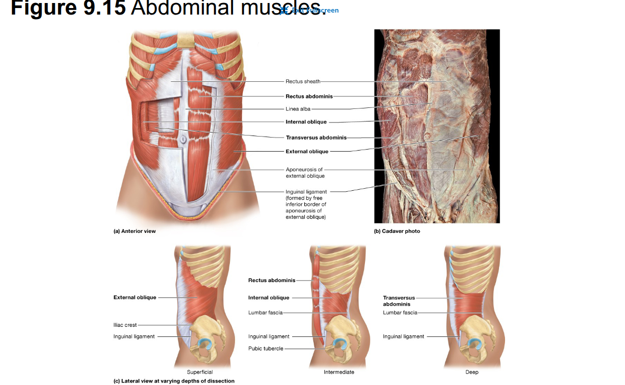
Internal oblique
Action: flexes and rotates the vertebral column and compresses the abdomen
Origin: iliac crest
Insertion: lower four ribs
rotate the trunk and flex it laterally

External oblique
Action: flexes and rotates the vertebral column and compresses the abdomen
Origin: lower eight ribs
Insertion: pubic and iliac crests
rotate the trunk and flex it laterally
Trapezius
Action: extends the head and elevates the scapula
Origin: occipital bone and cervical vertebrae
Insertion: clavicle
Rhomboid major & minor
Action: retract the scapula
Origin: spinous processes of C7–T1 and T1–T5
Insertion: scapula
Teres major
Action: adducts, extends, and rotates the arm laterally
Origin: posterior and inferior part of scapula
Insertion: medial sulcus of humerus
Erector spinae muscle group
Nine muscles arranged in three columns along each side of the vertebral column
important postural muscles and powerful extensors of the vertebral column

What are the 3 columns of the erector spinae?
Iliocostalis = lateral column
Longissimus = middle column
Spinalis = medial column

Iliocostalis muscles
lateral column
Iliocostalis lumborum( in the lower back), iliocostalis thoracis (in the mid back), iliocostalis cervicis (in the neck)

Longissimus muscles
middle column
Longissimus thoracis (in the lower back) , longissimus cervicis (in the neck), and longissimus capitis (inserts into the mastoid process in the head)

Spinalis muscles
medial column
Spinalis thoracis, spinalis cervicis, and spinalis capitis

Transversospinal group
Semispinalis, multifidus, and rotatores
actions are similar to the those of the erector spinae muscle group
generally extend the head, neck, and vertebral column

Quadratus lumborum
Helps extend and laterally flex the vertebral column

Broad, flat sheet of muscle that supports the organs within the pelvis
formed by the levator ani muscle group
Levator ani muscle group
Includes the anterior pubococcygeus, middle iliococcygeus, and posterior ischiococcygeus muscles
supports the pelvic floor, anal canal, and genitourinary organs
Pubococcygeus, iliococcygeus & ischiococcygeus
Three muscles of the levator ani group
They help support the pelvic floor
the anococcygeal ligament extends from the rectum to the coccyxserves as an insertion site for these muscles
Perineum
Diamond-shaped region inferior to the pelvic diaphragm, between the proximal thighs
contains the external genitalia and opening of the anus
Contains the external urethral sphincter and deep transverse perineal muscle
The urethra passes through it in males
the urethra and vagina pass through it in females
External urethral sphincter
Surrounds the urethra and provides voluntary control of urination
Circular muscle that surrounds the vagina in the female
Supports the genitourinary/pelvic organs and helps fix the perineal body (central tendon)
External anal sphincter
Surrounds the anus and provides voluntary control of defecation
attaches to the perineal body/central perineal tendon
Help stabilize the perineal body
Bulbospongiosus & ischiocavernosus
Aid erection of the external genitalia during sexual excitement
The bulbospongiosus also helps expel semen and urine from the urethra in males
constricts the vaginal opening in females
the ischiocavernosus helps maintain erection

Pelvic floor exercises involving repeated contraction and relaxation of the levator ani muscle
used to strengthen the pelvic floor and help treat urinary incontinence