[LAB] Epithelial Tissue And Connective Tissue

0.0(0)
Studied by 15 people
call kaiCall Kai
learnLearn
examPractice Test
spaced repetitionSpaced Repetition
heart puzzleMatch
flashcardsFlashcards
GameKnowt Play
Card Sorting

1/11

encourage image

There's no tags or description

Looks like no tags are added yet.

12 Terms

1
New cards
<p>Simple squamous epithelium</p>

Simple squamous epithelium

  • Simple - single-layered


  • Epithelium - covers or lines free, unattached surfaces (free space)


  • Squamous - flatter, disk shaped cells


  • Oval shaped nucleus


<ul><li><p>Simple - single-layered</p></li></ul><p></p><ul><li><p>Epithelium - covers or lines free, unattached surfaces (free space)</p></li></ul><p></p><ul><li><p>Squamous - flatter, disk shaped cells</p></li></ul><p></p><ul><li><p>Oval shaped nucleus</p></li></ul><p></p>
2
New cards
<p>Simple cuboidal epithelium</p>

Simple cuboidal epithelium

  • Simple - single-layered


  • Epithelium - covers or lines free, unattached surfaces (free space)


  • Cuboidal - shaped like rounded squares


  • Nucleus is near the center of the cell


<ul><li><p>Simple - single-layered</p></li></ul><p></p><ul><li><p>Epithelium - covers or lines free, unattached surfaces (free space)</p></li></ul><p></p><ul><li><p>Cuboidal - shaped like rounded squares</p></li></ul><p></p><ul><li><p>Nucleus is near the center of the cell</p></li></ul><p></p>
3
New cards
<p>Simple columnar epithelium - <span style="color: red;"><strong>NO CILIA</strong></span></p>

Simple columnar epithelium - NO CILIA

  • Simple - single-layered


  • Epithelium - covers or lines free, unattached surfaces (free space)


  • Tall and narrow


  • Nucleus is oval-shaped and is near the base of the cell


  • Contains goblet cells that hold mucus


NO CILIA

<ul><li><p>Simple - single-layered</p></li></ul><p></p><ul><li><p>Epithelium - covers or lines free, unattached surfaces (free space)</p></li></ul><p></p><ul><li><p>Tall and narrow</p></li></ul><p></p><ul><li><p>Nucleus is oval-shaped and is near the base of the cell</p></li></ul><p></p><ul><li><p>Contains goblet cells that hold mucus</p></li></ul><p></p><p><span style="color: red;"><strong>NO CILIA</strong></span></p>
4
New cards
<p>Pseudostratified columnar epithelium - <span style="color: red;"><strong>YES CILIA</strong></span></p>

Pseudostratified columnar epithelium - YES CILIA

  • Pseudostratified - appears to have many layers but is actually single-layered


  • Epithelium - covers or lines free, unattached surfaces (free space)


  • Tall and narrow


Contains goblet cells that hold mucus and DOES CONTAIN CILIA


<ul><li><p>Pseudostratified - appears to have many layers but is actually single-layered</p></li></ul><p></p><ul><li><p>Epithelium - covers or lines free, unattached surfaces (free space)</p></li></ul><p></p><ul><li><p>Tall and narrow</p></li></ul><p></p><p>Contains goblet cells that hold mucus and <span style="color: red;"><strong>DOES CONTAIN CILIA</strong></span></p><p></p>
5
New cards
<p>Stratified squamous epithelium - <strong>keratinized</strong></p>

Stratified squamous epithelium - keratinized

  • Stratified - multi-layered


  • Squamous - flatter, disk shaped cells


  • Epithelium - covers or lines free, unattached surfaces (free space)


  • Keratinized - exposed to the outside world; palms of hand and soles of feet


These cells can be found on the skin in the form of several layers of dead skin cells which keeps us hydrated


No nuclei present in the dead cells


The cells get flatter towards the surface of the skin
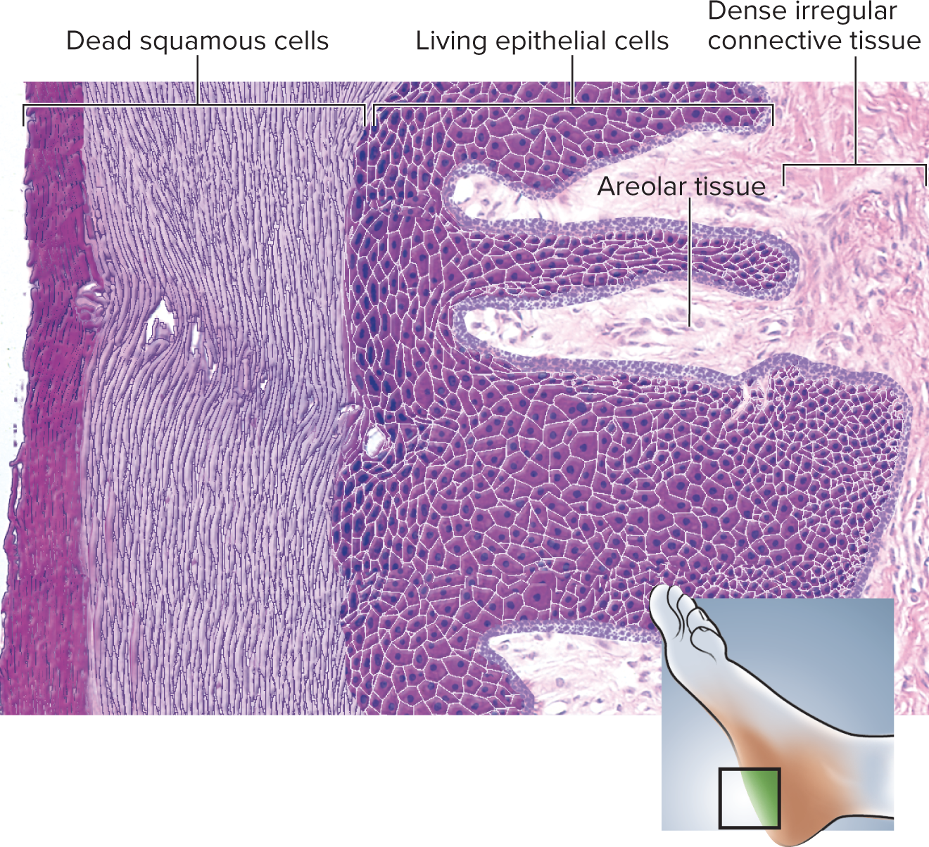
<ul><li><p>Stratified - multi-layered</p></li></ul><p></p><ul><li><p>Squamous - flatter, disk shaped cells</p></li></ul><p></p><ul><li><p>Epithelium - covers or lines free, unattached surfaces (free space)</p></li></ul><p></p><ul><li><p><strong>Keratinized </strong>- exposed to the outside world; palms of hand and soles of feet</p></li></ul><p></p><p>These cells can be found on the skin in the form of several layers of dead skin cells which keeps us hydrated</p><p></p><p>No nuclei present in the dead cells</p><p></p><p>The cells get flatter towards the surface of the skin</p>
6
New cards
<p>Stratified squamous epithelium - <strong>nonkeratinized</strong></p>

Stratified squamous epithelium - nonkeratinized

  • Stratified - multi-layered


  • Squamous - flatter, disk shaped cells


  • Epithelium - covers or lines free, unattached surfaces (free space)


  • Nonkeratinized - exposed to the outside world, but in a moist environment; cheek cells in the mouth, esophagus


These cells are living and nuclei are present

<ul><li><p>Stratified - multi-layered</p></li></ul><p></p><ul><li><p>Squamous - flatter, disk shaped cells</p></li></ul><p></p><ul><li><p>Epithelium - covers or lines free, unattached surfaces (free space)</p></li></ul><p></p><ul><li><p><strong>Nonkeratinized </strong>- exposed to the outside world, but in a moist environment; cheek cells in the mouth, esophagus</p></li></ul><p></p><p>These cells are living and nuclei are present</p>
7
New cards
<p>Areolar connective tissue - <strong>FOUND IN THE DERMIS</strong></p>

Areolar connective tissue - FOUND IN THE DERMIS

  • Areolar - contains lots of fibers


  • Connective tissue - no free space


  • Makes up stretchy parts of the body like skin


  • Cells usually do not come into contact with each other


  • The dark red spots are the nuclei of the cells that are responsible for creating all the messy fibers visible in the images


<ul><li><p>Areolar - contains lots of fibers</p></li></ul><p></p><ul><li><p>Connective tissue - no free space</p></li></ul><p></p><ul><li><p>Makes up stretchy parts of the body like skin</p></li></ul><p></p><ul><li><p>Cells usually do not come into contact with each other</p></li></ul><p></p><ul><li><p>The dark red spots are the nuclei of the cells that are responsible for creating all the messy fibers visible in the images</p></li></ul><p></p>
8
New cards
<p>Adipose tissue - <strong>FOUND IN THE HYPODERMIS</strong></p>

Adipose tissue - FOUND IN THE HYPODERMIS

  • Adipose - cells containing fat molecules


  • Connective tissue - no free space


  • The cells contain very little amounts of cytoplasm, but many fat molecules


  • The darker purple dots are the nuclei of the adipocytes (cloud-like images)


<ul><li><p>Adipose - cells containing fat molecules</p></li></ul><p></p><ul><li><p>Connective tissue - no free space</p></li></ul><p></p><ul><li><p>The cells contain very little amounts of cytoplasm, but many fat molecules</p></li></ul><p></p><ul><li><p>The darker purple dots are the nuclei of the adipocytes (cloud-like images)</p></li></ul><p></p>
9
New cards
<p>Dense <strong>regular </strong>connective tissue</p>

Dense regular connective tissue

  • Wave-like pattern that is very uniform


  • Slender nuclei


  • Found in tendons and ligaments

    • Tendons - connect muscle to bone

    • Ligaments - connect bone to bone


<ul><li><p>Wave-like pattern that is very uniform</p></li></ul><p></p><ul><li><p>Slender nuclei</p></li></ul><p></p><ul><li><p>Found in tendons and ligaments</p><ul><li><p>Tendons - connect muscle to bone</p></li><li><p>Ligaments - connect bone to bone</p></li></ul></li></ul><p></p>
10
New cards
<p>Dense <strong>irregular </strong>connective tissue - <strong>FOUND IN THE DERMIS</strong></p>

Dense irregular connective tissue - FOUND IN THE DERMIS

  • Less wave-like in appearance; much more messy, with fibers running in random directions


  • Few visible cells


<ul><li><p>Less wave-like in appearance; much more messy, with fibers running in random directions</p></li></ul><p></p><ul><li><p>Few visible cells</p></li></ul><p></p>
11
New cards
<p>What tissues are present in this photo?</p>

What tissues are present in this photo?

Top layer - stratified squamous epithelium; dead skin cells on the epidermis


Middle layer - areolar connective tissue found in the dermal papilla


Bottom layer near the arrow - dense irregular connective tissue, making up the layer of skin known as the dermis


Skin layers from superficial to deep: epidermis » dermis » hypodermis

<p>Top layer - <strong>stratified squamous epithelium</strong>; dead skin cells on the epidermis</p><p></p><p>Middle layer - <strong>areolar connective tissue</strong> found in the dermal papilla</p><p></p><p>Bottom layer near the arrow - <strong>dense irregular connective tissue</strong>, making up the layer of skin known as the dermis</p><p></p><p>Skin layers from superficial to deep: epidermis » dermis » hypodermis</p>
12
New cards
<p>What tissues are present in this photo?</p>

What tissues are present in this photo?

Blue arrow - stratified squamous epithelium; dead skin cells on the epidermis


Red arrows - areolar connective tissue found in the dermal papilla


Green arrow - dense irregular connective tissue, making up the layer of the skin known as the dermis


Skin layers from superficial to deep: epidermis » dermis » hypodermis

<p>Blue arrow - <strong>stratified squamous epithelium</strong>; dead skin cells on the epidermis</p><p></p><p>Red arrows - <strong>areolar connective tissue</strong> found in the dermal papilla</p><p></p><p>Green arrow - <strong>dense irregular connective tissue</strong>, making up the layer of the skin known as the dermis</p><p></p><p>Skin layers from superficial to deep: epidermis » dermis » hypodermis</p>



