Anatomy and Physiology
 Call Kai
 Learn
Learn Practice Test
 Spaced Repetition
 Match
Match Flashcards
 Knowt Play
1/356

There's no tags or description
Looks like no tags are added yet.
Name | Mastery | Learn | Test | Matching | Spaced | Call with Kai | Chat |
|---|
No analytics yet
Send a link to your students to track their progress
357 Terms
A system always involves____
Multiple elements that are connected in specific ways.
A system always displays ____
Novel properties that none of the parts possess.
Intelligence of a biological system is embedded in:
The design of the elements and the connections between the elements.
Anatomy
Structural connections
Physiology
Functional connections
Types of Proteins
Enzymes
Receptors
Channels
Transporters
Motors
Highways
Adhesives
Building blocks
Protein structure is sensitive to the physical and chemical factors in their environment including:
Detergents
pH
Temperature
Pressure
Solutes
Homeostasis
A stable internal environment that supports the structure and function of proteins. Essential for survival.
Protein function depends entirely on ___
3-D structure
Thermoregulation
Maintaining homeostasis in body temperature

Heat receptor examples
TRPV1, TRPV2, TRPV3, TRPV4

Cold receptor examples
TRPA1, TRPM8, TRPC5
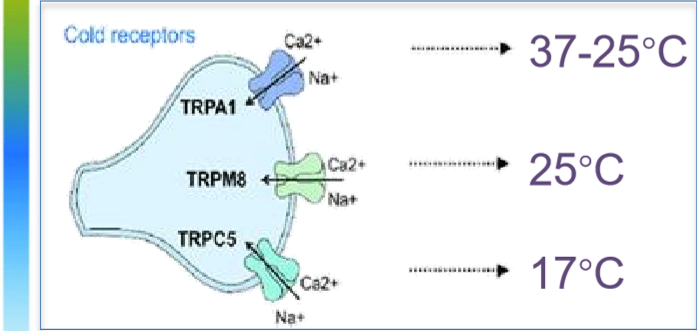
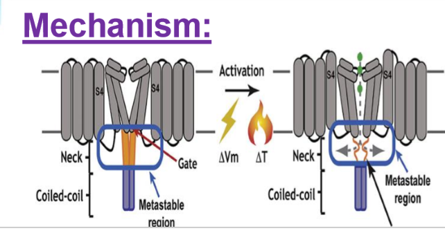
Mechanism of temperature receptor activation
Temperature changes alter ion channel structure => opening or closing
Effectors for changing temperatures
Skeletal muscles
Brown fat
Blood vessels
Sweat glands
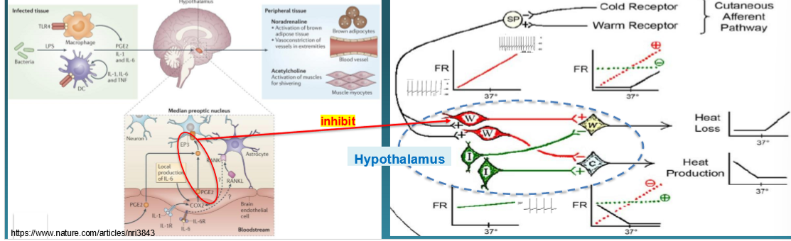
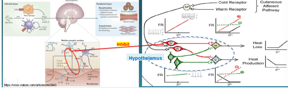
Thermoregulatory center
In the hypothalamus

W neurons
Stimulated by warm sensitive receptors
Inhibited by cold sensitive receptors
Stimulate W efferent neurons
Inhibits C efferent neurons

I neurons
Insensitive to temp
Auto fire at a stable frequency (stabilizes the thermoregulatory network by providing steady background activity)
Stimulates C efferent neurons
Inhibits W efferent neurons
Number of steps in fever mechanism
6
Step 1 of Fever mechanism
Step 1: Trigger
Exogenous pyrogens activate immune cells

Example of exogenous pyrogen
LPS
Example of immune cells (fever mechanism)
Macrophages
Dendritic cells
Step 2 of Fever mechanism
Step 2: Release of Endogenous Pyrogens
Activates immune cells release cytokines
Example of cytokines (Fever mechanism)
IL-1β
IL-6
TNF-α
Step 3 of Fever Mechanism
Step 3: Signal to the Hypothalamus
Cytokines to to hypothalamus and stimulate expression of COX-2

Step 4 of Fever Mechanism
Step 4: Cox-2 converts arachidonic acid to prostaglandins especially prostaglandin E2(PGE-2)

Step 5 of Fever mechanism
Step 5: PGE2 Action on the hypothalamus
PGE2 binds to EP3 receptors on warm sensitive neurons in the hypothalamus
Leading to reduced firing of warm sensitive neurons

Step 6 of Fever mechanism
Step 6: Physiological response (Set-point is raised)
Vasoconstriction
Shivering
Behavioral changes
=> Fever develops
Positive feedback loop
Response to a stimulus amplifies the original stimulus, moving the system further from homeostasis

Examples of positive feedback loop
Blood clotting
Childbirth
Lactation

Negative feedback loop
Response reduces or counteracts the original stimulus helping maintain homeostasis

Example of negative feedback loops
Thermoregulation
Blood glucose regulation
Blood pressure regulation
Thyroid hormone regulation

LECTURE 2- TISSUES EPITHELIUM
Four types of Human Tissues
Epithelial tissue: Forms boundaries
Muscle tissue: Enables contraction
Nervous tissue: Facilitates communication and control
Connective tissue: Provides connection and support
Epithelial tissue/Epithelium
A sheet of cells that covers a body surface or lines a body cavity
1 Cell Layer Epithelium
Simple Epithelium
More than 1 Cell Layer Epithelium
Stratified
Scalelike Cell shape
Squamous
Cubelike Cell shape
Cuboidal
Column-shaped Cell
Columnar
Structure supports___
Function
Simple squamous epithelia function
For exchange of material by diffusion and filtration
In sites where protection is not important
Secretes lubricating substances in serosae

Simple squamous epithelia location
Kidney glomeruli, air sacs of lungs (alveoli), lining of heart, blood vessels, lymphatic vessels, lining of ventral body cavity (serosae).

Simple cuboidal epithelia function
Secretion and absorption
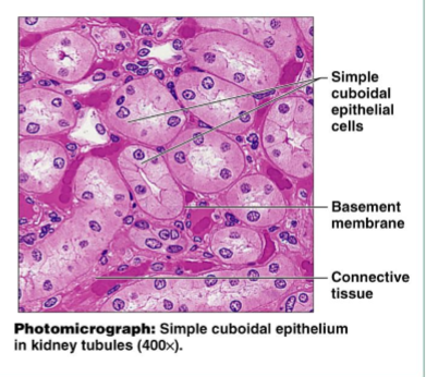
Simple cuboidal epithelia location
Kidney tubules, ducts and secretory portions of small glands, ovary surface

Simple columnar epithelia function
Absorption, secretion of mucus, enzymes, and other substance
Ciliated type propels mucus (or reproductive cells) by ciliary action

Simple columnar epithelia location
Non-ciliated type lines most of the digestive tract (stomach to anal canal), gallbladder, and excretory ducts of some glands
Ciliated type lines small bronchi, uterine tubes and some regions of the uterus

Pseudostratified columnar epithelia function
Secretion, particularly of mucus
Propulsion of mucus by ciliary action
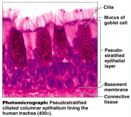
Pseudostratified columnar epithelia location
Non-ciliated type in male’s sperm-carrying ducts and ducts of large glands
Ciliated variety lines the trachea, most of the upper respiratory tract

Stratified squamous epithelia function
Protects underlying tissues in areas subject to abrasion

Stratified squamous epithelia location
Nonkeratinized type forms the moist linings of the esophagus, mouth, and vagina
Keratinized variety forms the epidermis of the skin (a dry membrane)

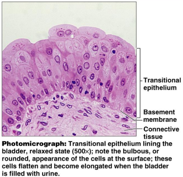
Transitional epithelia function
Stretches readily and permits distension of urinary organs by contained urine

Transitional epithelia location
Lines the ureters, bladder, and part of the urethra
Common functions of epithelial tissue
Forms barriers
Skin
Endothelium of blood vessels
Blood brain barrier
Secretion
Absorption
Sensation
External boundaries
Separate the body from external environment

Internal boundaries
Separate one part of the body from another

Exocrine glands: secretory epithelium surrounds a ___
Duct, which empties their secretion out of the body
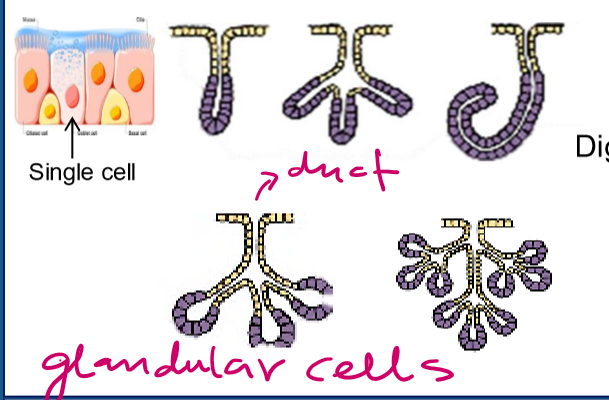
Exocrine gland secretion examples
Sweat glands
Salivary glands
Sebaceous glands
Lacrimal glands
Mammary glands
Exocrine pancreas
Stomach and intestinal glands

Endocrine glands: are ____
Ductless, they release their secretion (hormones) into the blood- circulatory system.
Endocrine glands example
Pituitary gland
Adrenal gland
Thyroid gland

Key characteristics of Epithelial tissues
Polarity: apical and basal
Specialized junctions
Supported by connective tissue: basement membrane (an acellular material secreted partly by the epithelial cells and connective tissue cells)
Avascular but innervated: depend on diffusion of nutrients from underlying connective tissue. (The epidermis is Aneural)
Regeneration: epithelial cells can easily divide to regenerate the tissue
Cell junction types
Tight Junctions
Adherens junctions
Desmosomes
Hemidesmosomes
Gap junctions
Tight junctions Function
Seal space between cells to prevent paracellular leakage
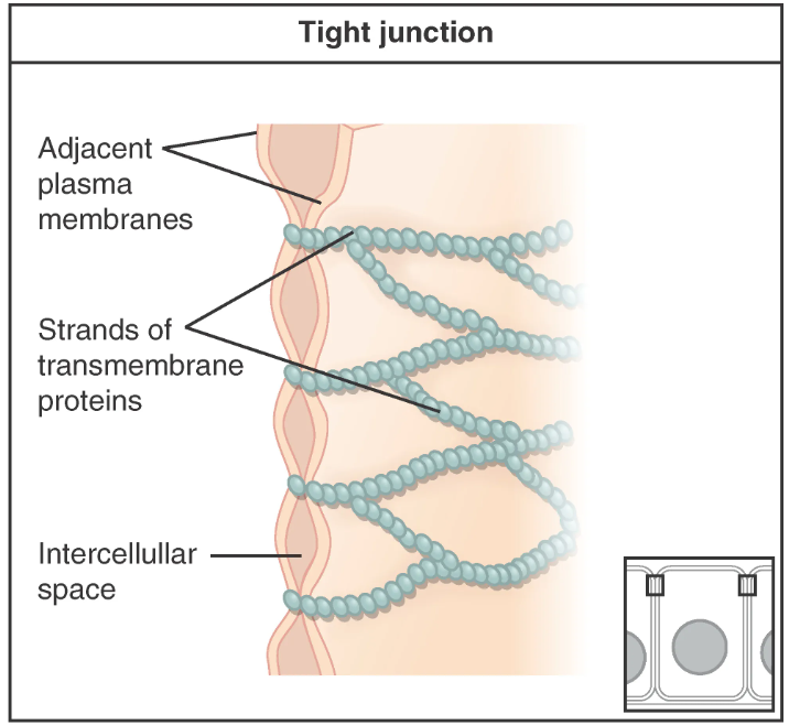
Tight junctions key proteins
Claudins, occludins
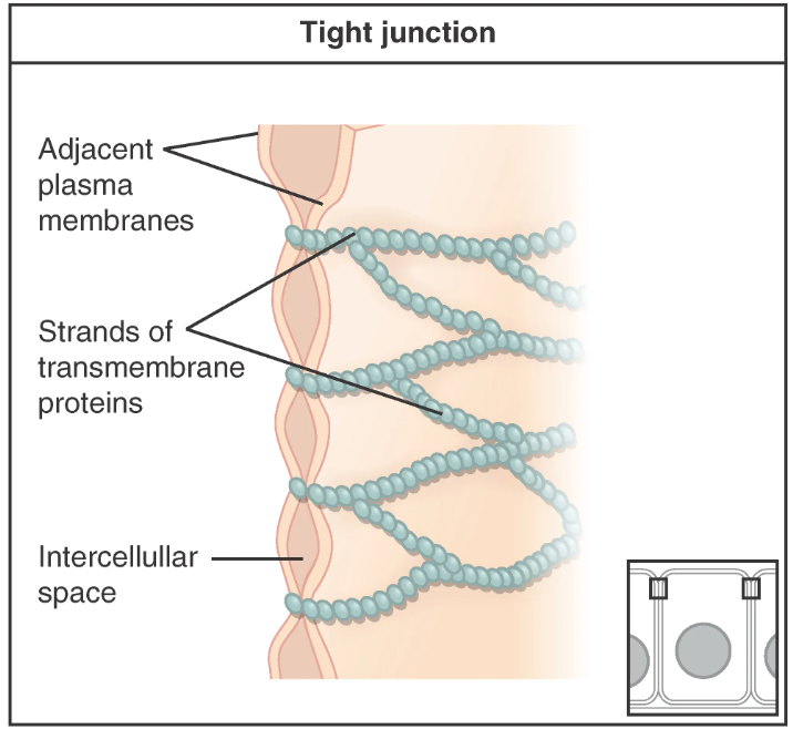
Tight junctions typical distributions in epithelium
Apical region of absorptive epithelia to maintain polarity
E.g. intestinal lining, kidney tubules

Adherens junctions Function
Mechanical link between actin cytoskeletons of adjacent cells. Belt like

Adherens junctions key proteins
Cadherins (E cadherins), catenins

Adherens junctions typical distribution in epithelium
Just below tight junctions in most simple epithelia to maintain tissue integrity
eg. intestinal epithelium
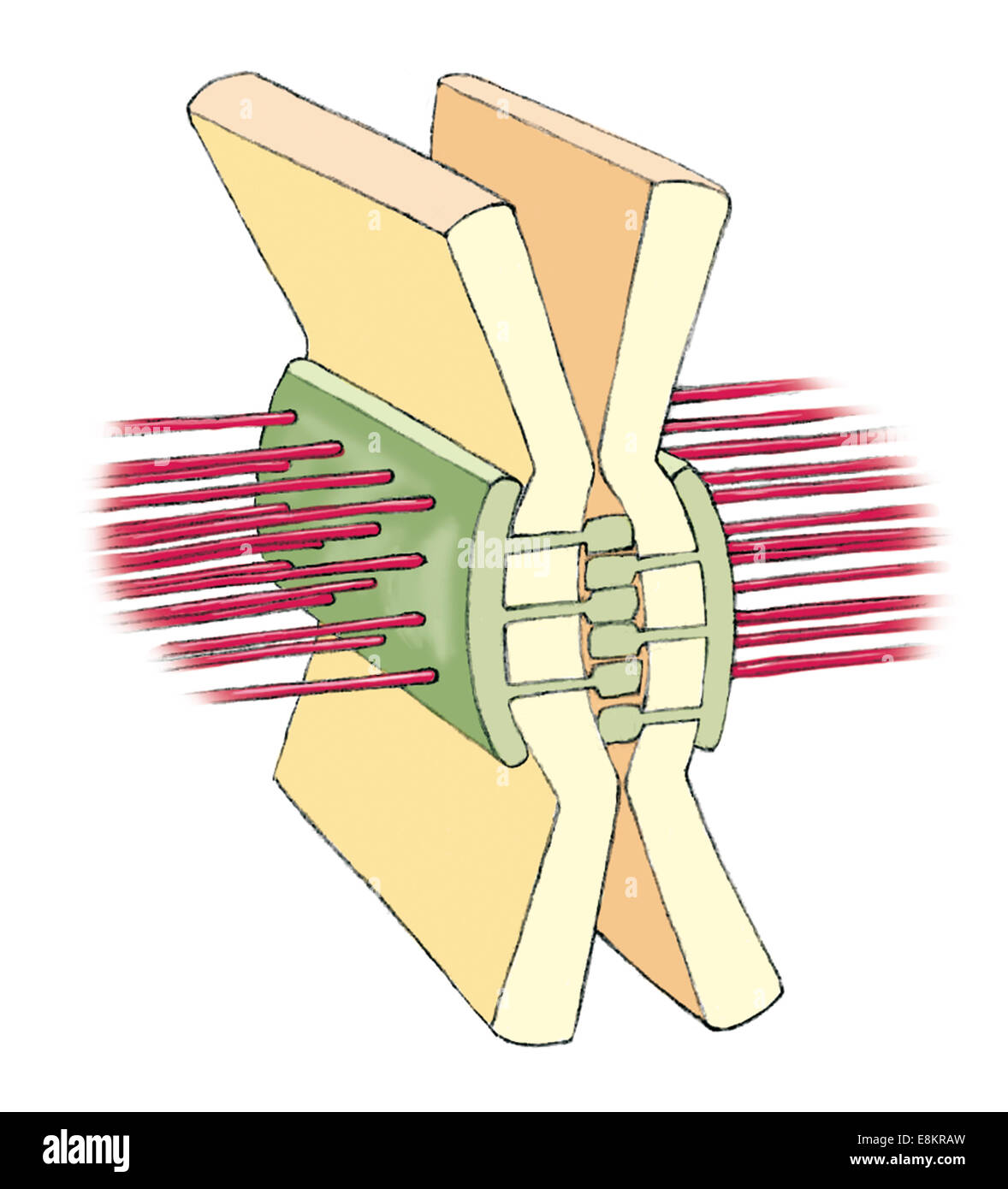
Desmosomes function
Strong spot-like adhesion, resist mechanical stress. Spot adhesion.

Desmosomes key protein
Desmoglein, desmocollin (cadherins)
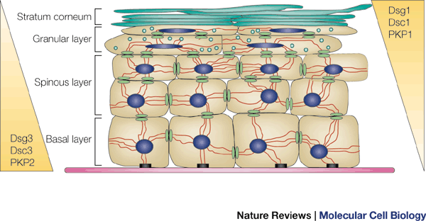
Desmosomes typical distribution in epithelium
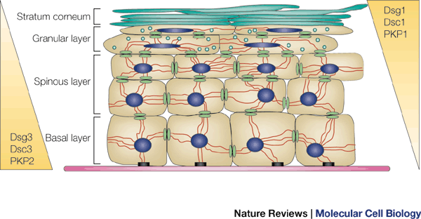
Abundant in stratified squamous epithelium
Hemidesmosomes function
Anchor basal surface of epithelial cells to basement membrane

Hemidesmosomes key protein
Integrins, BP180, BP230

Hemidesmosomes typical distribution in epithelium
Basal surface of epidermis and other epithelia under mechanical stress

Gap junctions function
Direct communication via ion and small molecule exchange

Gap junctions key proteins
Connexins
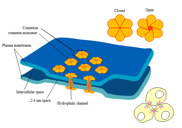
Gap junctions typical distribution in epithelium
Found in epithelia that coordinate activity
E.g. ciliated epithelium of respiratory tract, secretory epithelium of glands.

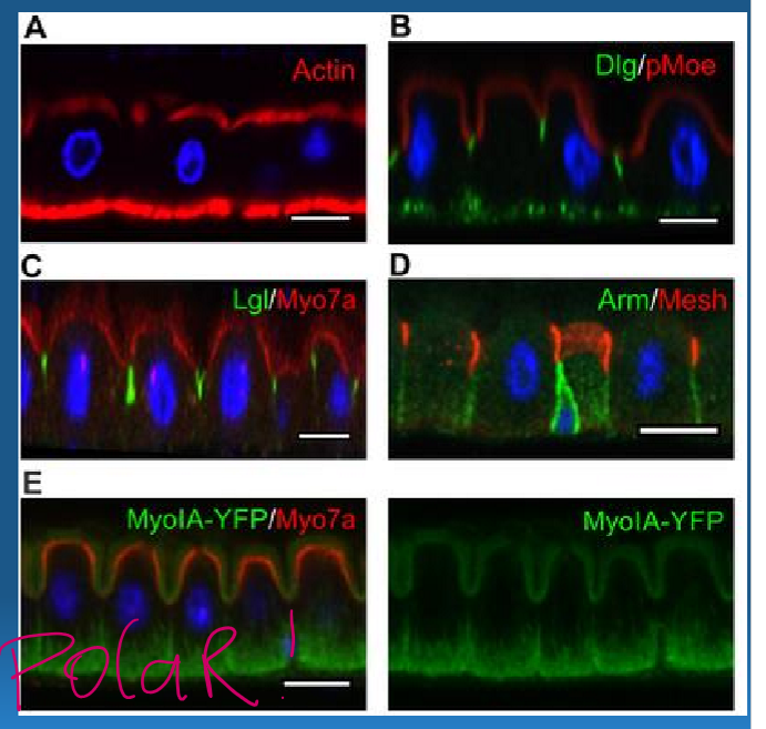
Cell polarity
A structural and functional asymmetry exists within the cell.
Three surfaces of a cell
Apical surfaces: faces the lumen of external space
Lateral surface
Basal surface: adheres to basement membrane (layers of extracellular matrix)
Epithelial tissue in the small intestine have specialized protein transporter that _____
Recognize and absorb various nutrients.
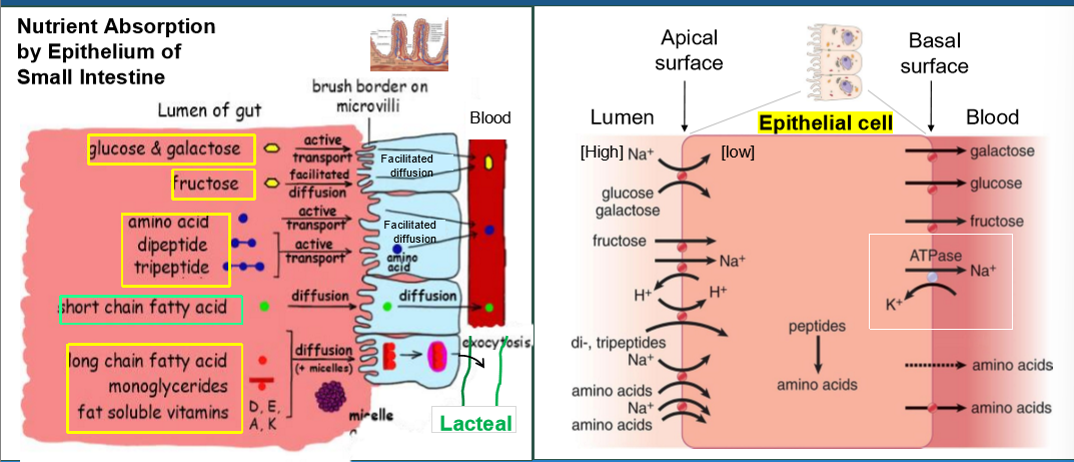
Apical-Basal Polarity
Protein machinery is localized to different surfaces, ensuring the proper execution of specific functions.

Villi+Microvilli increase the surface area by ___ x compared to a smooth tube
600
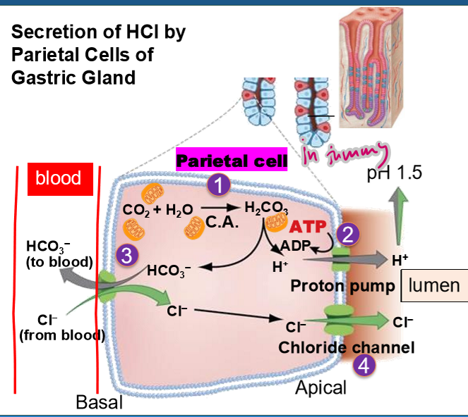
Epithelial tissue in the gastric gland have specialized protein tools for _____
generating acids and secretion.
To ensure the proper execution of specific function, there is Apical-basal polarity.
(4 steps) Secretion of HCl by parietal cells of gastric gland
The enzyme carbonic anhydrase catalyzes the formation of carbonic acid (H2CO3) from CO2 and H2O. They H2CO3 dissociates and HCO3- (bicarbonate ion), and H+
The protons are actively (using ATP) pumped into the stomach lumen
HCO3- transported to blood in exchange for Cl- from the blood. This is called Chloride Shift.
Cl- is then transported into the lumen via a Cl- channel.
Endothelium main functions
Regulates vascular tone by releasing signaling molecules.
Involved in angiogenesis (formation of new blood vessels)
Facilitates immune cell trafficking
Regulate blood clotting
In capillaries, its a selective barrier between blood and tissues
Three types of capillaries
Continuous capillaries
Fenestrated capillaries
Sinusoidal capillaries

Continuous capillaries characteristics
Have tight junctions, forming barrier as in the blood brain barrier (BBB)
E.g. in the brain, lungs

Fenestrated capillaries characteristics
Have small pores within endothelial cells to allow greater permeability.
E.g. in kidneys, intestines

Sinusoidal capillaries characteristics
Have large gaps between cells to allow the passage of larger molecules like proteins and cells
E.g. in liver, spleen

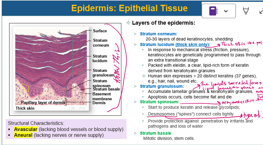
Layers of the epidermis
Stratum corneum
Stratum lucidum (thick skin only)
Stratum granulosum
Stratum spinosum
Stratum basale

Stratum corneum
20-30 layers of dead keratinocytes, shedding
Stratum lucidum
In response to mechanical stress (friction, pressure), keratinocytes are genetically programmed to pass through an extra transitional stage
Packed with eleidin, a clear, lipid-rich form of keratin derived from keratohyalin granules
Stratum granulosum
Accumulate lamellar granules & keratohyalin granules (makes skin waterproof)
Apoptosis occurs, cells become flat and die
Stratum spinosum
Start to produce keratin and release glycolipids (makes skin water proof)
Desmosomes (“spines”) connect cells tightly
Provide protection against penetration by irritants and pathogens and loss of water
Stratum basale
Mitotic division, stem cells
CONNECTIVE TISSUE
Lecture 3
Two main parts of connective tissue
Cells
Matrix (abundant)
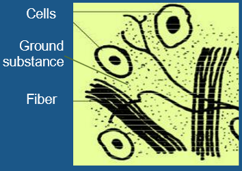
Two main parts of connective tissue matrix
Fibers
Ground substance
Three main types of connective tissue fibers
Collagen fiber (collagen proteins)- resist stretch
Elastic fiber (elastin + fibrillin proteins)- stretch and recoil
Reticular fiber (collagen proteins)- delicate mesh
Two main types of connective tissue ground substance
Polysaccharide- negatively charged so fills space with H2O
Proteoglycans- provides cushioning