CH 6 BIO 110
 Call Kai
 Learn
 Practice Test
 Spaced Repetition
 Match
 Flashcards
 Knowt Play
1/120

There's no tags or description
Looks like no tags are added yet.
121 Terms
Muscle Types
skeletal muscle, cardiac muscle, smooth muscle
“myo” and “mys”
refer to muscle
“sarco”
refers to flesh
skeletal and smooth muscle cells are
elongated
contraction of muscle is due to
the movement of microfilaments
Skeletal Muscle Characteristics
most are attached by tendons to bones
cells are multinucleated
striated (have visible banding)
voluntary- subject to conscious control
Connective Tissue Wrappings
cells are surrounded and bundled by connective tissue
Endomysium
enclosed a single muscle fiber

Perimysium
wraps around a fascicle (bundle) of muscle fibers

Epimysium
covers the entire skeletal muscle

Fascia
on the outside of the epimysium
Skeletal Muscle Attachments
epimysium blends into tendons
Tendons
cord-like structures that connect muscle to bone; most parallel collage fibers
Smooth Muscle Characteristics
lacks striations
spindle-shaped cells
single nucleus
involuntary - no conscious control
found mainly in walls of hollow organs
Cardiac Muscle Characteristics
striations
usually has a single nucleus
branching cells
joined to another muscle cell at an intercalated discs
involuntary
Muscle Functions
produce movement
maintain posture
stabilize joints
generate heat
Muscle Fiber
thin, elongated cylindrical cell with rounded ends; extends length of muscle

Isotonic Contractions
muscle shortens and movement occurs with contraction
Isometric Contractions
muscle cells do not shorten even though muscle tension increases
Flexion
decreases the angle of the joint, brings two bones closer together, typical of hinge joints like knee and elbow

Extension
opposite of flexion, increases angle between two bones
hyperextension

Rotation
movement of a bone around its longitudinal axis, common in ball-and-socket joints, example is when you move atlas around the dens of axis
lateral and medial rotation

Abduction
movement of a limb away from the midline

Adduction
opposite of abduction- movement of a limb toward the midline

Circumduction
scribing a cone in space seen with ball and socket joints

Dorsiflexion
lifting the foot so that the superior surface approaches the shin (movement at the ankle)
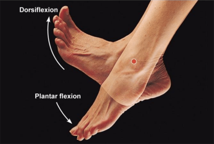
Plantar Flexion
depressing the foot, pointing the toes (movement is at the ankle)

Inversion
turn sole of foot medially

Eversion
turn sole of foot laterally

Supination
forearm rotates laterally so palm faces anteriorly (at the ulna)
can hold a cup of soup (sup)
Pronation
forearm rotates medially so palm faces posteriorly (at the ulna)
pour (pro) the soup out
Sarcolemma
plasma membrane of a skeletal muscle

Myofibrils
contractile organelles found in the cytoplasm of muscle cells; they push nuclei aside and nearly fill the cytoplasm

Transverse Tubules
invaginations (surface folds inward to create a tube) of sarcolemma, extend all the way through the fiber

Sarcoplasmic Reticulum
modified ER, sacs and tubes that surround each myofibril and stores Calcium (releases when the muscle fiber is stimulated to contract)

Striation Pattern of Skeletal Muscle
due to the arrangement of thin and thick filaments
A Band
dark band, length of the thick filament

I Band
light band, contains thin filament and anchored to the Z disc

Sarcomere
contractile unit of muscle fiber; the segment of a myofibril between the Z discs

Sarcomere Organization
consists of the h zone, m line, and myofilaments: thick and thin filaments
Thick Filaments
composed of many myosin molecules and located in the center of a sarcomere

Myosin
composed of 2 twisted protein with globular heads projecting outwards
myosin heads form cross bridges

Myosin Heads
has ATPase enzymes: which split ATP into ADP and P to release energy
energy released is used to cock myosin head so it is ready to bind to actin (a binding site exists for actin)

Thin Filament
actin is the main protein
anchored to the z disc
troponin and tropomyosin are regulatory proteins found on the thin filament

Actin
double stranded helical molecule where each actin monomer has a binding site for myosin

H Zone
region in the center of the A band that contains only myosin at rest

M Line
proteins that hold myosin molecules in place

Excitability
ability to receive and respond to a stimulus; responsivenessCC
Contractitlity
ability to shorten when an adequate stimulus is received
Extensibility
ability of muscle cells to be stretchedE
Elasticity
ability to recoil and resume length after stretching
Motor Unit
one neuron and all the skeletal muscle fibers it stimulates
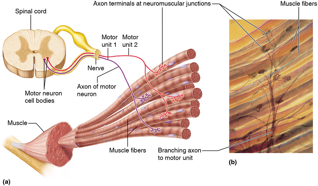
Axon
long, thread-like extension of a neuron

Axon Terminal
where the axon reaches the muscle and branches out into axon terminals

Neuromuscular Junctions
connection between the axon terminal of the motor neuron and the sarcolemma of a muscle fiber

Motor Neuron
the axon terminal contains synaptic vesicles which contain acetylcholine, ACh
ACh
neurotransmitter- chemical that transmits signal from neuron to the next cell
Motor End Plate
folded region of the sarcolemma under the axon terminal that contains ACh receptors
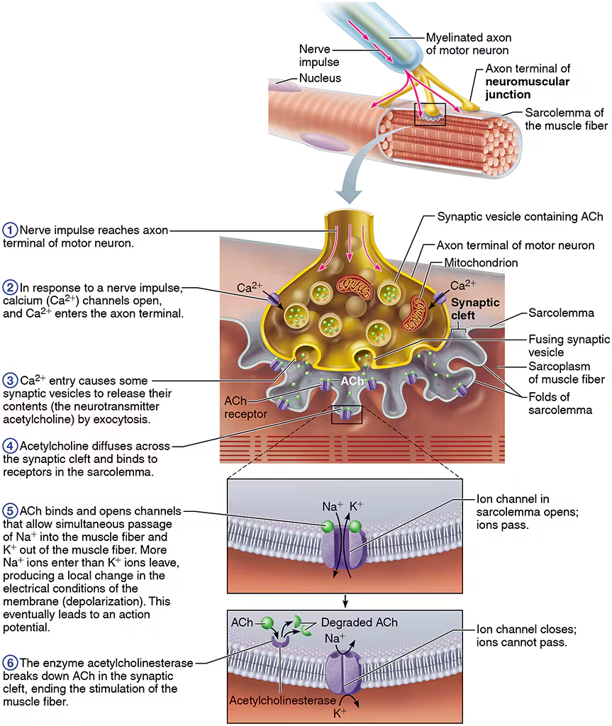
Synaptic Cleft
the gap between neuron and muscle fiber- they do not make contact and this area is filled with interstitial fluid

Transmission of Nerve Impulses to Muscle
Nerve impulse reaches the axon terminals of the motor neuron
Calcium channels open and Calcium enters the axon
The calcium entry causes some synaptic vesicles to release ACh by exocytosis
ACh diffuses across the synaptic cleft and binds and binds to receptors on the motor end plate (sarcolemma)
ACh opens channels increasing Sodium entry into the muscle fiber and Potassium out; more Na ions enter than K ions leave which causes a change in electrical conditions
Local depolarization leads to an action potential (electrical signal) on sarcolemma
Action potential travels on sarcolemma and stimulates muscle fiber to contract
ACh is degraded by acetylcholinesterase (AChE) and muscle contraction ends
Head and Neck Muscles
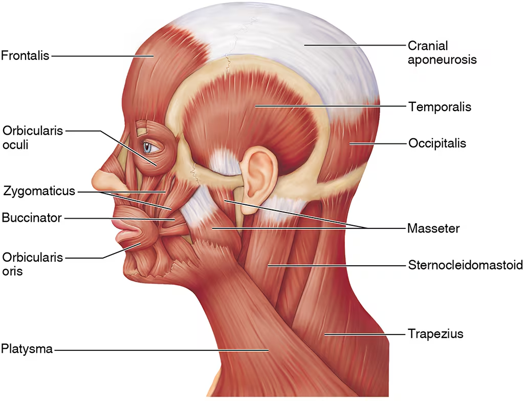
facial muscles (orbicularis oculi, orbicularis oris, zygomaticus major), chewing muscles (masseter, temporalis), and neck muscles (sternocleidomastoid)
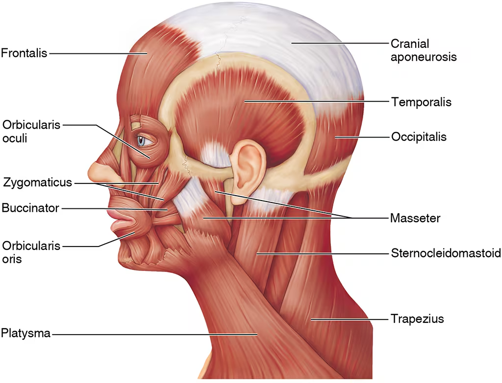
Facial Muscles
orbicularis oculi, orbicularis oris, zygomaticus major

Orbicularis oculi
closes eyes, squints, blinks, winks

Orbicularis oris
closes mouth and protrudes the lips

Zygomaticus major

Chewing Muscles
masseter, temporalis

Masseter
elevates mandible

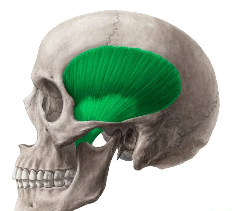
Temporalis
elevates mandible
Neck muscles
Sternocleidomastoid
Sternocleidomastoid
flexes the neck, rotates the head

Muscles of the Trunk
anterior muscles (pectoralis major), muscles of abdominal girdle (rectus abdominis, external oblique, internal oblique, transversus abdominis), and posterior muscles (trapezius, latissimus dorsi, deltoid)

Anterior Muscles
Pectoralis major

Pectoralis major

Muscles of the Abdominal Girdle
rectus abdominus, external oblique, internal oblique, transverse abdominis

Rectus abdominus
flexes vertebral column; compresses abdominal content

External oblique

Internal oblique
deep to the external oblique

Transversus abdominis
deep to internal oblique

Posterior Muscles
trapezius, latissimus dorsi, deltoid

Trapezius

Latissimus dorsi

Deltoid

Muscles of the Upper LImb
biceps brachii, brachialis, brachioradialis, triceps brachii

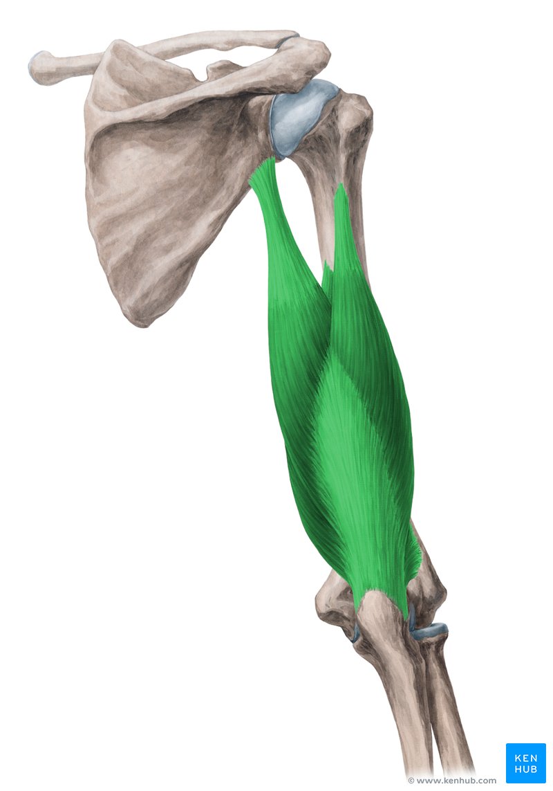
biceps brachii
flexes elbow, supinates forearm

Brachialis

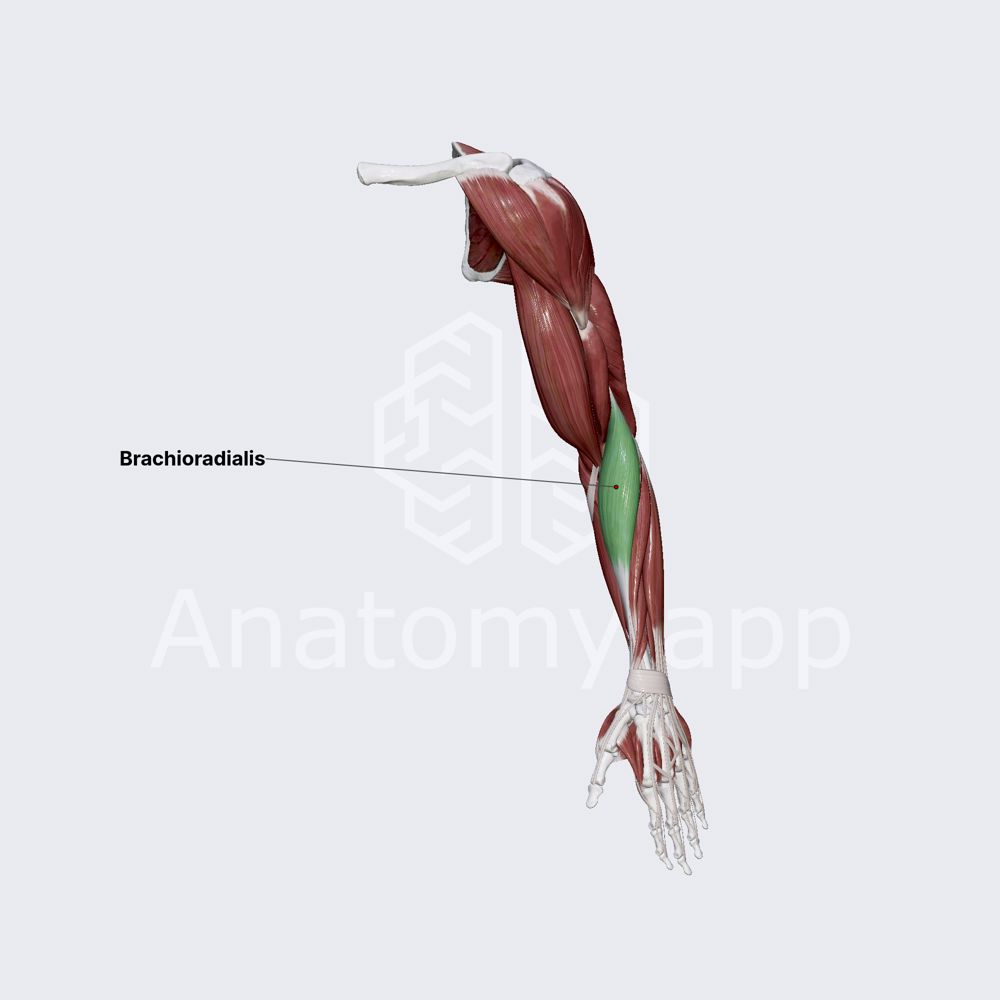
Brachioradialis
Triceps brachii
elbow extension
Muscles of the Lower Limb
gluteus maximus, adductor muscles, hamstring group (biceps femoris, semimembranosus, semitendinosus), sartorius, and quadriceps group (rectus femoris, vastus lateralis, vastus medialis, vastus intermedius)
Gluteus maximus
extends thigh at hip, abducts thigh

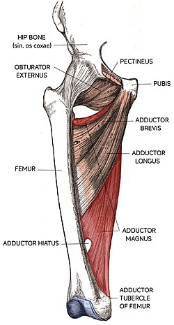
Adductor muscles
several muscles on medial side of thigh, adducts thigh, seen in both anterior and posterior views
Hamstring Group
thigh extension and knee flexion
biceps femoris, semimembranosus, semitendinosus

Biceps femoris
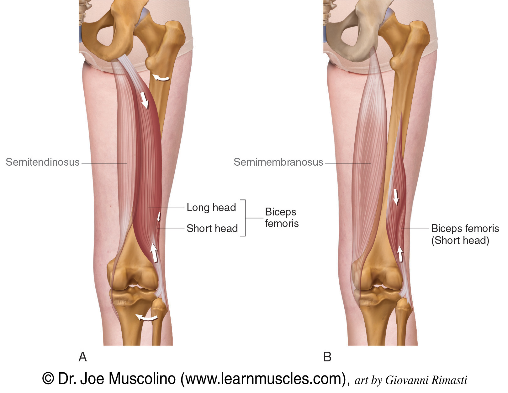
Semimembranosus

Semitendinosus
slightly superficial to semimembranosus

Sartorius

Quadriceps Group
rectus femoris, vastus lateralis, vastus medialis, vastus intermedius

Rectus femoris
extends the knee, flexes hip

Vastus lateralis
extends the knee

Vastus medialis
extends the knee

Vastus intermedius
