A&P Integumentary System
 Call Kai
 Learn
 Practice Test
 Spaced Repetition
 Match
 Flashcards
 Knowt Play
1/141

There's no tags or description
Looks like no tags are added yet.
142 Terms
Structure included in the Integumentary System
Skin, Hair, Nails, Sweat Glands, and Sebaceous (oil) glands
How many distinct regions are in the skin
2: Epidermis and Dermis
What is the third region that is not a part of the skin but shares some functions
Hypodermis, or subcutaneous tissue.
Epidermis
The superficial region consisting of epithelial tissue. Avascular
Dermis
Underlies the epidermis. Made of mostly fibrous connective tissue, vascular.
Hypodermis (superficial fascia)
Subcutaneous layer deep to skin. Made of mostly adipose tissue that absorbs shock and insulates. Anchors skin to underlying structures: mostly muscles.

1
Dermal Papillae
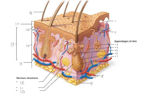
2
Sweat Pore

3
Eccrine Sweat Gland
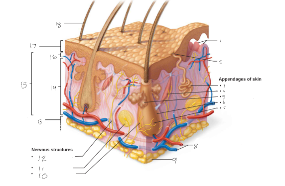
4
Arrector Pili Muscle
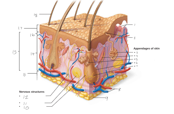
5
Sebaceous (oil) gland

6
Hair follicle

7
Hair root

8
Dermal vascular plexus

9
Adipose Tissue

10
Hair follicle receptor (root hair plexus)

11
lamellar corpuscle
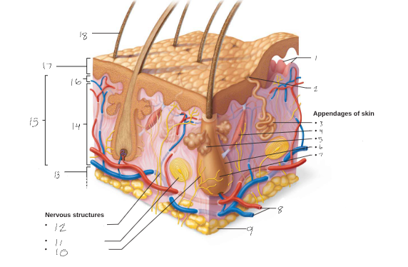
12
Sensory nerve fiber with free nerve endings

13
Subcutaneous Tissue (Hypodermis: not part of Skin)

14
Reticular Layer

15
Dermis

16
Papillary Layer

17
Epidermis

18
Hair Shaft
What is the epidermis mostly made up of
Keratinized Stratified squamous epithelium
Four cell types found in the epidermis
Keratinocytes, Melanocytes, Dendritic (Langerhans) cells, Tactile (Merkel) cells
Keratinocytes
Produce fibrous keratin (protein that gives skin its protective properties)
Major cells of epidermis
Tightly connected by desmosomes
Millions slough off every day
Melanocytes
Spider-shaped cells located in deepest epidermis
Produce pigment melanin, which is packaged into melanosomes
Melanosomes are transferred to keratinocytes, where they protect nucleus from UV damage
Dendritic (Langerhans) cells
Star-shaped macrophages that patrol deep epidermis
Are key activators of immune system
Tactile (Merkel) cells
Sensory receptors that sense touch
In thin skin epidermis contains how many layers?
4: Stratum basale, stratum spinosum, stratum granulosum, stratum corneum
How many layers are found in thick skin?
5: the stratum lucidum is found between the stratum granulosum and the stratum corneum
Where is thick skin found
High-abrasion areas such as hands and feet
Which layer is the deepest of all epidermal layers
Stratum basale (basal layer): is firmly attached to the dermis
What is the make up of thee stratum basale
Consists of a single row of stem cells that actively divide (mitotic), producing two daughter cells each time
One daughter cell journeys from basal layer to surface, taking 25–45 days to reach surface
Cell dies as it moves toward surface
Other daughter cell remains in stratum basale as stem cell
What is the stratum basale also know as
Layer also known as stratum germinativum because of active mitosis
What type of cell makes up 10-25% of the stratum basale
melanocytes
Characteristics of the stratum spinosum
Several cell layers thick
Cells contain weblike system of intermediate prekeratin filaments attached to desmosomes
Allows them to resist tension and pulling
Keratinocytes in this layer appear spikey, so they are called prickle cells
Scattered among keratinocytes are abundant melanosomes and dendritic cells
How many cells thick is the stratum granulosum?
Four to six cells thick but these cells are flattened making the layer thin
What changes occur in the appearance of cells found in the stratum granulosum?
Cells flatten, nuclei and organelles disintegrate
Keratinization begins
Cells accumulate keratohyaline granules that help form keratin fibers in upper layers
Cells also accumulate lamellar granules, a water-resistant glycolipid that slows water loss
Why do cells found above the stratum granulosum die?
They are too far from dermal capillaries to facilitate access of nutrients from the bloodstream
What is the stratum lucidum?
Found only in thick skin
Consists of thin, translucent band of two to three rows of clear, flat, dead keratinocytes
Lies superficial to the stratum granulosum
What is the stratum corneum?
20–30 rows of flat, anucleated, keratinized dead cells
Accounts for three-quarters of epidermal thickness
What functions do the dead cells making up the stratum corneum maintain?
Protect deeper cells from the environment
Prevent water loss
Protect from abrasion and penetration
Act as a barrier against biological, chemical, and physical assaults
How do cells change?
Cells change by going though apoptosis (controlled cell death)
Dead cells slough off as dandruff and dander
Humans can shed ~50,000 cells every minute
What type of tissue and cells make up the dermis?
Strong, flexible connective tissue
Cells include fibroblasts, macrophages, and occasionally mast cells and white blood cells
Fibers in matrix bind body together
Makes up the “hide” that is used to make leather
What structures can be found in the dermis?
Contains nerves, blood vessels, and lymphatic vessels
Contains epidermal hair follicles, oil glands, and sweat glands
How many layer make up the dermis (what are they called)
Two layers: Papillary and Reticular
What is the papillary layer?
Superficial layer of areolar connective tissue consisting of loose, interlacing collagen and elastic fibers and blood vessels
Loose fibers allow phagocytes to patrol for microorganisms
Dermal papillae
What are the dermal papillae found in the papillary layer
superficial region of dermis that sends fingerlike projections up into epidermis
Projections contains capillary loops, free nerve endings, and touch receptors (tactile corpuscles, also called Meissner’s corpuscles)
In thick skin, dermal papillae lie on top of dermal ridges, which give rise to epidermal ridges
Collectively ridges are called friction ridges
Enhance gripping ability
Contribute to sense of touch
Sweat pores in ridges leave unique fingerprint pattern
What is the Reticular Layer?
Makes up ~80% of dermal thickness
Consists of coarse, dense fibrous connective tissue
Many elastic fibers provide stretch-recoil properties
Collagen fibers provide strength and resiliency
Bind water, keeping skin hydrated
Cutaneous plexus
Extracellular matric
What is the cutaneous plexus in the reticular layer
network of blood vessels between reticular layer and hypodermis
What is contained in the reticular layer’s extracellular matrix
Pockets of adipose cells
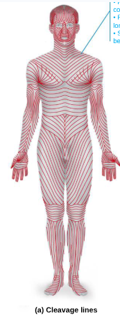
What causes cleavage (tension) lines in reticular layer
many collagen fibers running parallel to skin surface
Externally invisible
Important to surgeons because incisions parallel to cleavage lines heal more readily
What are flexure lines in the reticular layer
Dermal folds at or near joints
Dermis is tightly secured to deeper structures
Skin’s inability to slide easily for joint movement causes deep creases
Visible on hands, wrists, fingers, soles, toes

What causes striae or stretch marks
Extreme stretching of skin can cause dermal tears that leave silvery white scars

What causes blisters?
Acute, short-term traumas to skin can cause
What are blisters
fluid-filled pockets that separate epidermal and dermal layers
What are the three pigments that contribute to skin color?
Melanin, Carotene, Hemoglobin
Key characteristics about Melanin
Only pigment made in skin; made by melanocytes
Made from amino acid tyrosine by tyrosinase
Packaged into melanosomes that are sent to shield DNA of keratinocytes from damaging UV sunlight
The more sun, the more need for protective shield, the more melanin will be produced
Two forms: reddish yellow to brownish black
Skin color differences are due to amount and form of melanin
Freckles and pigmented moles are local accumulations of melanin
How does excessive sun exposure damage skin
Elastic fibers clump, causing skin to become leathery
Can depress immune system and cause alterations in DNA that may lead to skin cancer
UV light destroys folic acid
Necessary for DNA synthesis, so insufficient folic acid is especially dangerous for developing embryos
Photosensitivity is increased reaction to sun
Some drugs (e.g., antibiotics, antihistamines) and perfumes cause photosensitivity, leading to skin rashes
What is the pigment carotene?
Yellow to orange pigment
Most obvious in palms and soles
Accumulates in stratum corneum and hypodermis
Can be converted to vitamin A for vision and epidermal health
What is the pigment Hemoglobin?
Pinkish hue of fair skin is due to lower levels of melanin
Skin of Caucasians is more transparent, so color of hemoglobin shows through
Examples of alterations in skin cancer that can indicate disease
Cyanosis
Blue skin color: low oxygenation of hemoglobin
Pallor (blanching or pale color)
Anemia, low blood pressure, fear, anger
Erythema (redness)
Fever, hypertension, inflammation, allergy
Jaundice (yellow cast)
Liver disorders
Alterations in skin color that can indicate disease
Bruises (black-and-blue marks)
Also referred to as ecchymoses or hematomas, are a result of clotted blood beneath skin
As clot is broken down, color of bruise changes
Brown or black “necklace” or bruises
Hyperpigmented dark areas in axillae and around neck may be a sign of insulin resistance and elevated blood glucose levels
What is hair/pili made of?
flexible strands of dead, keratinized cells
Produced by hair follicles
Contains hard keratin, not like soft keratin found in skin
Hard keratin is tougher and more durable, and cells do not flake off
Where is hair not located?
None located on palms, soles, lips, nipples, and portions of external genitalia
What are the functions of hair?
Warn of insects on skin
Hair on head guards against physical trauma
Protect from heat loss
Shield skin from sunlight
Regions of the hair
Shaft: area that extends above scalp, where keratinization is complete
Root: area within scalp, where keratinization is still going on
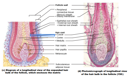
What are the three parts of hair shaft?
Medulla: central core of large cells and air spaces
Cortex: several layers of flattened cells surrounding medulla
Cuticle: outer layer consisting of overlapping layers of single cells

How are hair pigments mad by melanocytes in hair follicles
Combinations of different melanins (yellow, rust, brown, black) create all the hair colors
Red hair has additional pheomelanin pigment
Gray/white hair results when melanin production decreases and air bubbles replace melanin in shaft

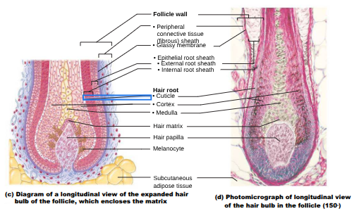
What is the structure of a hair follicle?
Extends from epidermal surface to dermis
Hair bulb: expanded area at deep end of follicle
Hair follicle receptor (or root hair plexus): sensory nerve endings that wrap around bulb
Hair is considered a sensory touch receptor

What is the wall of a follicle composed of?
Peripheral connective tissue sheath
Derived from dermis
Also called fibrous sheath
Glassy membrane: thickened basal lamina
Epithelial root sheath
Derived from epidermis
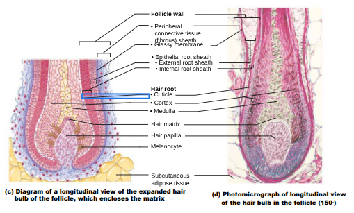
What is the hair matrix
actively dividing area of bulb that produces hair cells
As matrix makes new cells, it pushes older ones upward
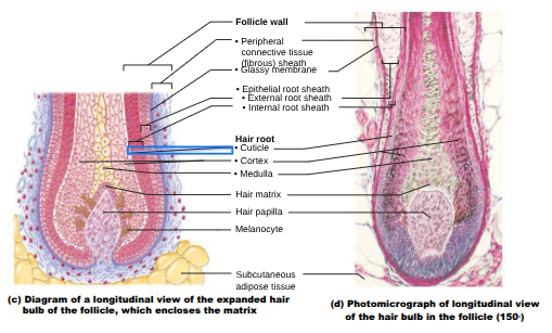
What is the arrector pili
small band of smooth muscle attached to follicle
Responsible for “goose bumps”

What is the hair papilla?
Dermal tissue containing a knot of capillaries that supplies nutrients to growing hair
Vellus Hair
pale, fine body hair of children and adult females
Terminal Hair
coarse, long hair
Found on scalp and eyebrows
At puberty
Appear in axillary and pubic regions of both sexes
Also on face and neck of males
Hair growth
Nutrition and hormones affect hair growth
Follicles cycle between active and regressive phases
Average 2.25 mm growth per week
Lose 90 scalp hairs daily
Homeostatic imbalance of androgens in women
In women, ovaries and adrenal glands produce small amounts of androgens (male sex hormones), but tumors on these organs can cause abnormally large amounts of androgens
Ex: polycystic ovary syndrome
Can result in excessive hairiness, called hirsutism, as well as other signs of masculinization
In many cases, no clinical problem is found
Alopecia
hair thinning in both sexes after age 40
True baldness
Genetically determined and sex-influenced condition
Male pattern baldness caused by follicular response to DHT (dihydrotestosterone)
Telogen effluvium (TE)
is abrupt hair thinning caused by an abundance of hair follicles entering resting phase at same time
Can be due to stress-induced factors such as: hormonal changes, surgery, severe emotional trauma, or crash dieting
Other causes or thinning and/or texture change:
Drugs (ex: antidepressants, chemotherapeutic drugs)
Hypothyroidism
Protein-deficient diets
Nail key information
Scalelike modifications of epidermis that contain hard keratin
Act as a protective cover for distal, dorsal surface of fingers and toes
Consist of free edge, nail plate, and root
Nail bed is epidermis underneath keratinized nail plate
Nail matrix
thickened portion of bed responsible for nail growth
Nail folds:
skin folds that overlap border of nail
Eponychium
nail fold that projects onto surface of nail body
Also called cuticle
Hyponychium
Area under free edge of plate that accumulates dirt
Nail Color
Nails normally appear pink because of underlying capillaries
Lunule: thickened nail matrix, appears white
Abnormal color or shape can be an indicator of disease
Instances when nail appearance can help in diagnosing some diseases
Yellow-tinged may indicate respiratory or thyroid gland disorder
Thickened yellow nails can be due to fungal infection of nail
Koilonychia, also called “spoon nail” – an outward concavity of nail may signal iron deficiency
Beau’s lines – horizontal lines across nails may indicate severe illnesses such as uncontrolled diabetes, heart attack or cancer chemotherapy
Basic Information of Sweat Glands
Also called sudoriferous glands
All skin surfaces except nipples and parts of external genitalia contain sweat glands
About 3 million per person
Two main types
Eccrine (merocrine) sweat glands
Apocrine sweat glands
Contain myoepithelial cells
Contract upon nervous system stimulation to force sweat into ducts
Multicellular exocrine glands mode of secretion
Merocrine: most secrete products by exocytosis as secretions are produced (sweat, pancreas)
Holocrine: accumulate products within, then rupture (sebaceous oil glands)
Apocrine: accumulate products within, but only apex ruptures; whether this type exists in humans is controversial (maybe mammary cells?)
Eccrine (Merocrine) Sweat Glands
Most numerous type
Abundant on palms, soles, and forehead
Ducts connect to pores
Function in thermoregulation
Regulated by sympathetic nervous system
Their secretion is sweat
99% water, salts, vitamin C, antibodies, dermcidin (microbe-killing peptide), metabolic wastes

Apocrine Sweat Glands
Confined to axillary and anogenital areas
Secrete viscous milky or yellowish sweat that contains fatty substances and proteins
Bacteria break down sweat, leading to body odor
Larger than eccrine sweat glands with ducts emptying into hair follicles
Begin functioning at puberty
Function unknown but may act as sexual scent gland
Modified Apocrine Glands
Ceruminous glands: lining of external ear canal; secrete cerumen (earwax)
Mammary glands: secrete milk
Sebacious (Oil) Glands
Widely distributed, except for thick skin of palms and soles
Most develop from hair follicles and secrete into hair follicles
Relatively inactive until puberty
Stimulated by hormones, especially androgens
Secrete sebum
What is sebum
Oily holocrine secretion
Bactericidal (bacteria-killing) properties
Softens hair and skin

What causes acne?
Acne is usually an infectious inflammation of the sebaceous glands, resulting in pimples (pustules or cysts)
Associated with Propionibacterium acne infection
Whiteheads are blocked sebaceous glands
If secretion is oxidized, whitehead becomes a blackhead
What causes “cradle cap”?
Overactive sebaceous glands in infants can lead to seborrhea, known as “cradle cap”
Begins as pink, raised lesions on scalp that turn yellow/brown and flake off
