Basics of Folding and Structure Prediction
 Call Kai
 Learn
 Practice Test
 Spaced Repetition
 Match
 Flashcards
 Knowt Play
1/19

There's no tags or description
Looks like no tags are added yet.
20 Terms
Peptides
R is the side chain that gives the characteristic to the amino acid for eg. hydrophobic, hydrophilic, basic, acidic etc…
The amide group must remain planar and in the trans configuration. Hence, rotations are only possible about the two bonds adjacent to the alpha-carbon in each amino acid residue
There are also two rotational parameters:
1. Psi represents the angle about the C(alpha)-C bond
2. Phi represents the angle about the C(alpha)-N bond
Hence, the entire path of the peptide will be known only if all Psi and Phi angles are known
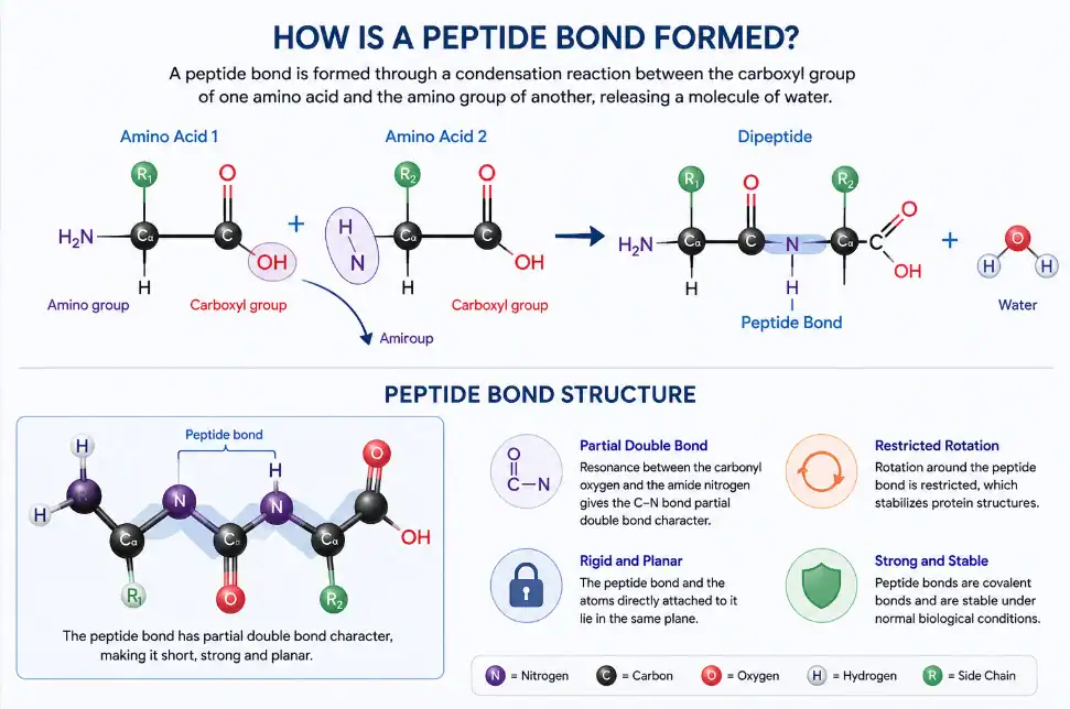
The Ramachandran Plot (1963):
2D graphical map (using torsion angles φ and ψ) to visualize and analyze allowed and disallowed conformations of protein backbones
Displays ψ-angles as a function of φ-angles
All theoretically possible secondary structures are shaded in blue
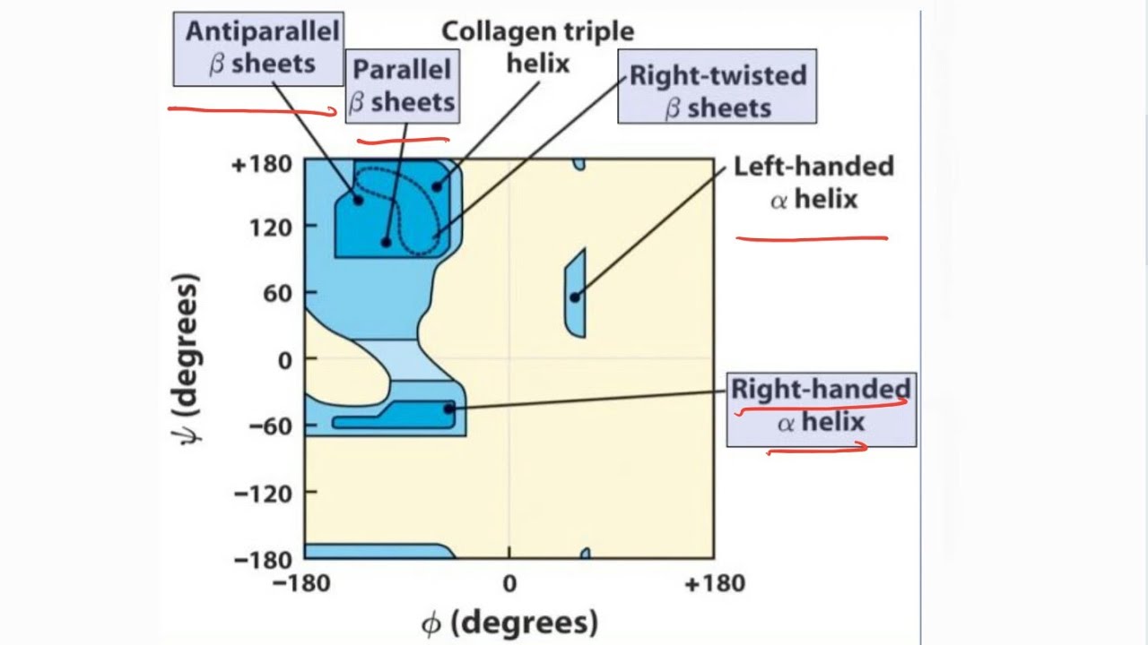
Alpha Helix
Polypeptide chain twists into a right-handed spiral or coil
It is stabilized by a regular network of hydrogen bonds.
The carbonyl oxygen (C=O) of each amino acid (residue i) forms a hydrogen bond with the amide hydrogen (N-H) of the amino acid four residues ahead (i + 4)

Beta Hairpin (Super Secondary Structure)
two antiparallel beta strands connected by loop/turn

Helix-turn-helix (Super Secondary Structure)
Two helices connected by loop/turn found in DNA binding proteins (~20 residues)
Second helix involved in DNA binding-enters in major groove

Beta-alpha-beta (Super Secondary Structure)
The helix axis is parallel to the beta sheets and helix is always above the plane of the beta sheets
alpha/beta horseshoe/barrel

Zinc Finger Motif (Super Secondary Structure)
Zinc atom stabilizes the structure / fold by coordinate bond
Each motif contains 30-50 residues
All other residues positions may not be conserved except cysteine and histidine
Regular patterns of Cys and His coordinate zinc atom

Greek Key Motif (Super Secondary Structures)
Three/Four adjacent antiparallel beta-sheet

EF Hand Motif
The 5th -e- and 6th -f - helices binding to calcium
Carboxyl containing residues are common.
(Aspartic acid, Glutamic acids); Present in all calcium signaling proteins: Calmodulin, Troponin

Alpha-alpha/Coiled Coils:
Alpha helix packed together via hydrophobic interactions
Stable in solution, parallel or antiparallel helices
Can form up to six helix-bundles
Transcription factors, intermediate filaments
Specific heptad repeat sequences (7 residues)

Omega Loop
The beginning and the end of the loop come together
Globular compact structure
No repeating hydrogen bonds
Side chains pack to the center of the loops
Eg. phosphate binding loops of nucleotide binding protein

Domains
Different combinations of secondary structure/super-secondary structure/motifs, connected by loops and turns.
Conserved part of protein structure (evolution),
Can exist independent of the protein structure.
(30-500 residues, with 90% < 200 residues)
4 classes:
All beta domain
All alpha domain
Alpha-beta domain
Alpha + beta domain
Can be classified into more than 800 groups
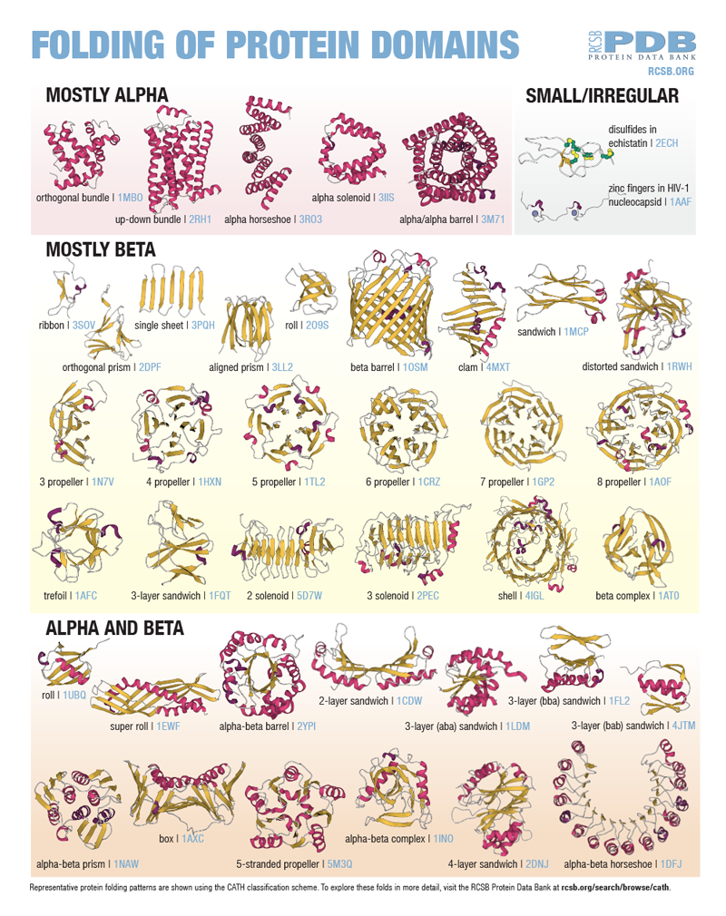
Motif vs. Domain
Both: A common three-dimensional structure that appears repeatedly in different proteins
Motif:
Smaller in size (10-20 a.a.)
May or may not fold on its own
May or may not have a function (other than being a part of a bigger structure)
Evolutionary relationship is not considered
Domain:
Larger in size (50-250 a.a.)
Folds into a specific shape independently of the rest of the polypeptide
Often associated with a specific function
Serves as an evolutionary module
Multiple motifs can combine to form a single domain
There is also an overlap: sometimes a motif is also a domain

B-form DNA
10-10.5 base pairs per turn
Bases perpendicular to the axis
Compact right-handed structure
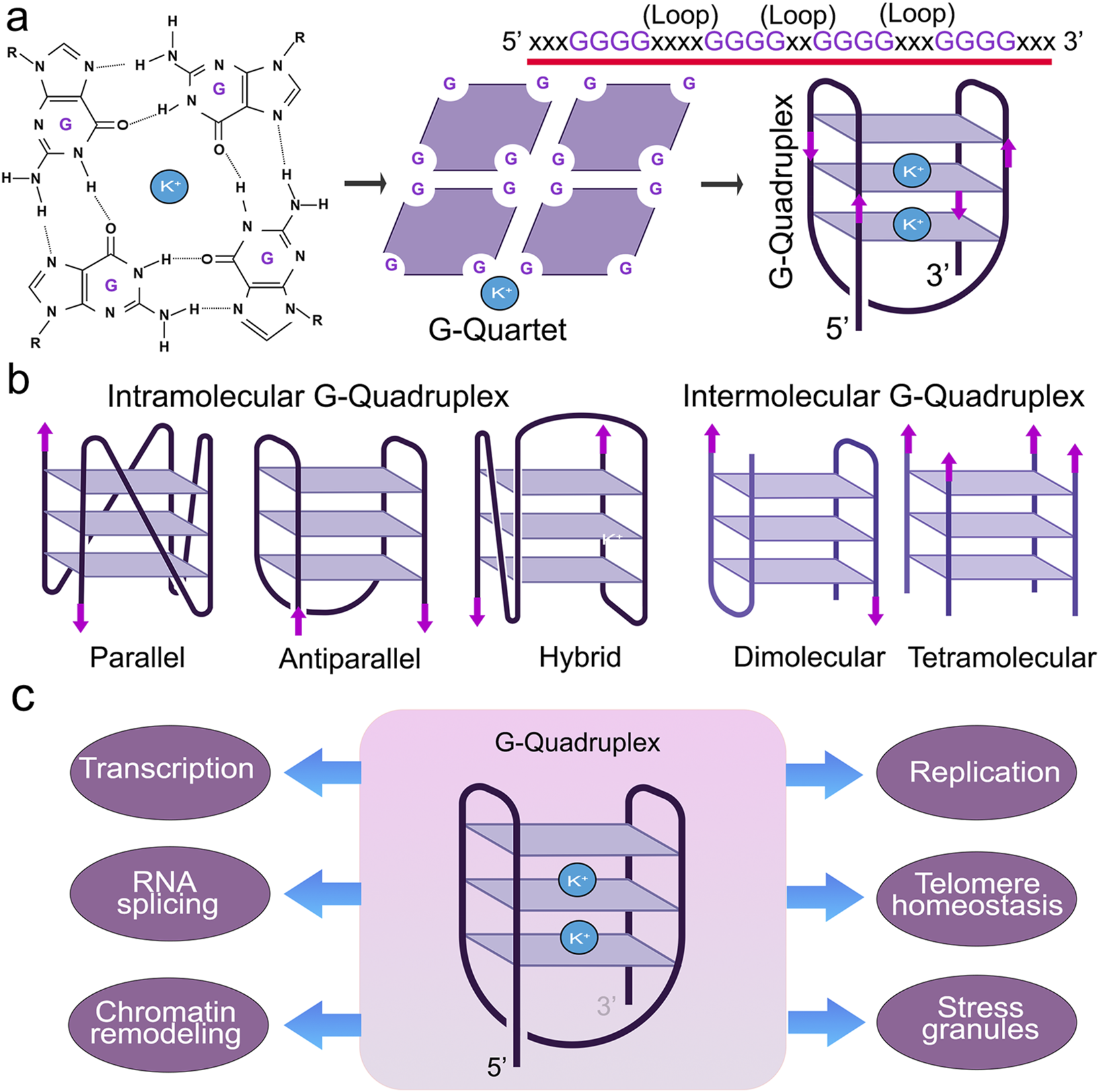
Undergoes a conformation transition to G-quadruplex
Inhibit telomerase, DNA polymerase
A-form DNA
11 base pairs per turn
Bases are 200 to helical axis
Wider (2.3 nm) than B-form
20% shorter than B-form
Compact right-handed structure
Z-form DNA
Elongated
Rigid
Unfavorable under normal conditions (occurs- high salt, cesium ions GC rich)
Left-handed, zig-zag structure
DNA folds in very precise and complex ways:
About 2 meters (6 feet) of DNA must fit inside a microscopic cell nucleus.
DNA wraps around protein spools called histones to form chromatin, which then twists, coils, and loops into thousands of specific configurations (such as topologically associating domains and chromosomes).
This folding helps control which genes turn on or off.

How does DNA folding work?
At the simplest level, chromatin is a double-stranded helical structure of DNA
DNA is completed with histones to form nucleosomes
Each nucleosome consists of eight histone proteins around which the DNA wraps 1.65 times
A chromosome consists of a nucleosome plus the H1 histone
The nucleosomes fold up to produce a 30-nm fibre
That forms loops averaging 300 nm in length
The 300-nm fibres are compressed and folded to produce a 250-nm-wide fibre
Tight controlling of the 250-nm fibre produces the chromatid of a chromosome
Human Telomeric i-Motifs
Four-stranded, non-canonical secondary DNA structures formed in cytosine (C)-rich single-stranded telomeric repeats (CCCTAA)
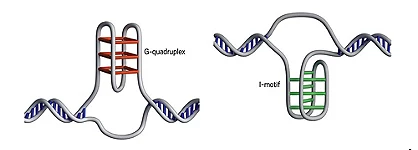
ATP-binding protein that resolves and unfolds G-quadruplex (G4) structures:
DHX36 (also known as RHAU or G4 resolvase 1)
