Chapter 7: Bone tissue
 Call Kai
 Learn
Learn Practice Test
 Spaced Repetition
 Match
Match Flashcards
 Knowt Play
1/93

There's no tags or description
Looks like no tags are added yet.
Name | Mastery | Learn | Test | Matching | Spaced | Call with Kai | Chat |
|---|
No analytics yet
Send a link to your students to track their progress
94 Terms
Skeletal system
composed of bones, cartilages, and ligaments joined to form a framework for the body
Osteology
the study of bones
Cartiladge, ligaments, and tendons
Cartilage is the forerunner of most bones and covers
many joint surfaces
Ligaments hold bones together at joints
Tendons attach muscle to bone.
7 Roles/functions of the skeletal system
Support—limb bones and vertebrae support body; jaw bones support teeth; some bones support viscera.
Protection—of brain, spinal cord, heart, lungs, and more.
Leverage—limb movements, breathing, and other movements depend on bone.
Storage of nutrients, mineral and electrolyte balance—calcium and phosphate levels.
pH (Acid–base) balance—buffers blood against large pH changes
by altering phosphate and carbonate salt levels.Blood formation—red bone marrow is the chief producer of blood cells.
Hormone secretion—bone cells secrete hormones that affect the action of insulin and moderate the stress response.
Osseous (bone) Tissue
connective tissue with the matrix hardened by calcium phosphate and other minerals
Mineralization or calcification
the hardening process of bone
What tissue types are found in individual Bones (the organ)
Bone tissue, bone marrow, cartilage, adipose tissue, nervous tissue, and fibrous connective tissue.
The word bone can refer to the organ or just the osseous tissue.
What are flat bones?
Thin, curved plates; protect soft organs. Examples: parietal bones of skull, sternum, scapula, ribs, hip bones
What are long bones?
Longer than wide; rigid levers acted upon by muscles; crucial for movement. Examples: humerus, radius, ulna, femur, tibia, fibula, metacarpals, metatarsals, phalanges
What are short bones?
approximately equal in length and width; carpals, tarsals, meta carpals, metatarsals
What are irregular bones?
elaborate shapes; vertebrae and some skull bones.
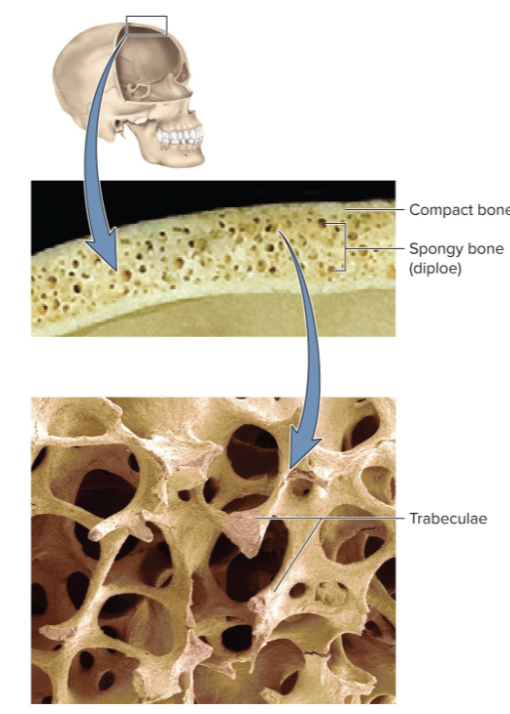
Compact bone
AKA dense or cortical bone has a dense outer shell of bone;
encloses the medullary cavity
Spongy (AKA cancellous) bone
Loosely organized bone tissue. Found in center of ends and center of shafts of long bones and in middle of nearly all others. Covered by more durable compact bone. Skeleton is three-fourths compact and one-fourth spongy bone by
weight
Diaphysis
shaft that provides leverage.

Epiphysis
Enlarged end of a long bone. Strengthens the joint and anchors ligaments and tendons
Epiphyseal line
Remnant of childhood growth zone, epiphyseal plate
Articular cartilage
Layer of hyaline cartilage that covers the joint surface; allows the joint to move more freely.

Nutrient foramina
Minute holes in the bone surface that allow blood vessels to penetrate
Periosteum
Periosteum—external sheath covering most of bone made of dense regular connective tissue. Outer fibrous layer of collagen and inner osteogenic layer of bone-forming cells. Perforating fibers of the periosteum penetrate the underlying bone matrix.

Endosteum
thin layer of reticular connective tissue lining marrow cavity and all internal bone surfaces
Dipole
Called dipoler in cranium. Inner and outer tables of compact bone enclosing layer of spongy bone in between. Flat bones have this type of snadwich like structure.
What are the four types of bone cells?
Osteogenic cells, osteoblast, osteocytes, and osteoclasts
Osteogenic cells
stem cells found in endosteum and inner layer of periosteum. Arise from embryonic mesenchyme; multiply continuously and give rise to osteoblasts.
Osteoblasts
bone-forming cells in endosteum and inner layer of periosteum. Perform osteogenesis—synthesize soft organic matter of matrix and promote its mineralization. Stress stimulates osteogenic cells to multiply rapidly and increase the number of osteoblasts which reinforce bone.
Osteocytes
Former osteoblasts become trapped in the matrix they deposited, forming lacunae—tiny cavities where osteocytes reside. Gap junctions allow for the passage of nutrients, wastes, and signals. Some osteocytes reabsorb bone matrix while others deposit it. Act as strain sensors—when stressed, they produce biochemical signals that regulate bone remodeling (shape and density changes that are adaptive).
Canaliculi
Little channels that protrude from the lacunae and connect to neighboring lacunae. Cytoplasmic processes of osteocytes reach into canaliculi and contact processes of neighboring cells;
Osteocalcin
Osteoblasts and osteocytes secrete hormone osteocalcin. Part of body’s acute stress response, stimulates pancreatic secretion of insulin, increases insulin sensitivity, promotes energy availability
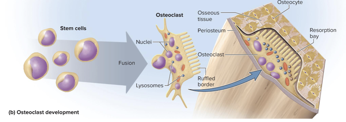
Osteoclasts
Bone-dissolving cells found on bone surface.
Perform osteolysis, the breakdown of bone, as part of bone remodeling.
Develop from same bone marrow stem cells that give rise to blood cells (different origin from other bone cells).
Very large cells formed from fusion of several stem cells; have multiple (usually3-4, but up to 50) nuclei in each cell.
Infoldings increase surface area and form ruffled border facing bone. Often reside in pits called resorption bays that they etch into the
bone surface
Osteocyte development

Osteoclast development
Composition of osseous tissue matrix:
By dry weight, averages one-third organic and two-thirds inorganic matter.
Organic matter—synthesized by osteoblasts. Collagen and carbohydrate–protein complexes, such as glycosaminoglycans, proteoglycans, and glycoproteins. The protein portion gives some flexibility.
Inorganic matter—mineral component. 85% hydroxyapatite, a crystallized calcium phosphate salt. 10% calcium carbonate. Many inorganic ions. The mineral portion allows the bone to support body weight without sagging
Combination forms a composite material that provides flexibility and strength
Rickets
A disease caused by mineral deficiency and resulting in
soft, deformed bones
Osteogenesis imperfecta (brittle bone disease)
Results from a defect in collagen deposition. Collagen molecules contain sacrificial bonds that break under stress and dissipate shock under load
Histology Bone: concentric lamellae
Concentric lamellae—layers of matrix surrounding a
central (Haversian) canal running longitudinally
• A central canal and its lamellae constitute and osteon (haversian
system); central canals connected by transverse perforating
canals
• Collagen corkscrews down each lamella; helical arrangement in
one lamella is opposite of adjacent lamella—enhances strength
• Osteons separated by a cement line, which prevents spread of
microfractures.
Circumferential lamellae
Found encircling the outer region of dense bone just underneath the periosteum.
Interstitial lamellae
Fill irregular regions between osteons
Spongy bone anatomy: Spicules
Bone slivers that make up the lattice of spongy bone
Spongy bone anatomy: trabeculae
plates that make up the lattice of psongy bone
Spongy bone anatomy and physiology
Anatomy:
Lattice of bone slivers (spicules) and plates (trabeculae), covered with endosteum. Sponge-like appearance. Spaces filled with red bone marrow.
Matrix arranged in lamellae, but few osteons and no
central canals. All osteocytes close to bone marrow.
Physiology:
Provides strength with minimal weight. Trabeculae develop along bone’s lines of stress.
How does the spongy bone structure organize itself to mechanical stress?
Trabeculae form along lines of mechanical stress in the spongy bone to provide strength with minimal additional weight.


What type of tissue is this?
Spongy bone

Bone marrow
soft tissue occupying marrow cavities of long bones and small spaces of spongy bone
Red marrow (myeloid tissue) (anatomy, physiology, and location)
Anatomy: contains multiple tissues, including hematopoietic tissue, which is tissue that produces blood cells.
Physiology: creates blood cells
Location: (for adults) skull, vertebrae, ribs, sternum, parts of the pelvic girdle, and proximal heads of the humerus and femur. In nearly every bone in a child.

Yellow marrow
Found in adults. Fatty marrow that does not produce blood. Can transform back to red marrow in the event of chronic anemia.
Ossification or osteogenesis
the formation of bone.
What are the two ways that bone can develop?
Both begin with embryonic mesenchyme. Intramembranous ossification and Endochondral ossification.
Describe the process of Intramembranous ossification
produces the flat bones of the skull, most of the clavicle, and part of the mandible
Summary of stages:
1. Deposition of osteoid tissue into embryonic mesenchyme
2. Calcification of osteoid tissue and entrapment of osteocytes
3. Honeycomb of spongy bone with developing periosteum
4. Filling of space to form compact bone at surfaces, leaving spongy bone in middle
Intramembranous ossification also important in lifelong thickening and remodeling of long bones.

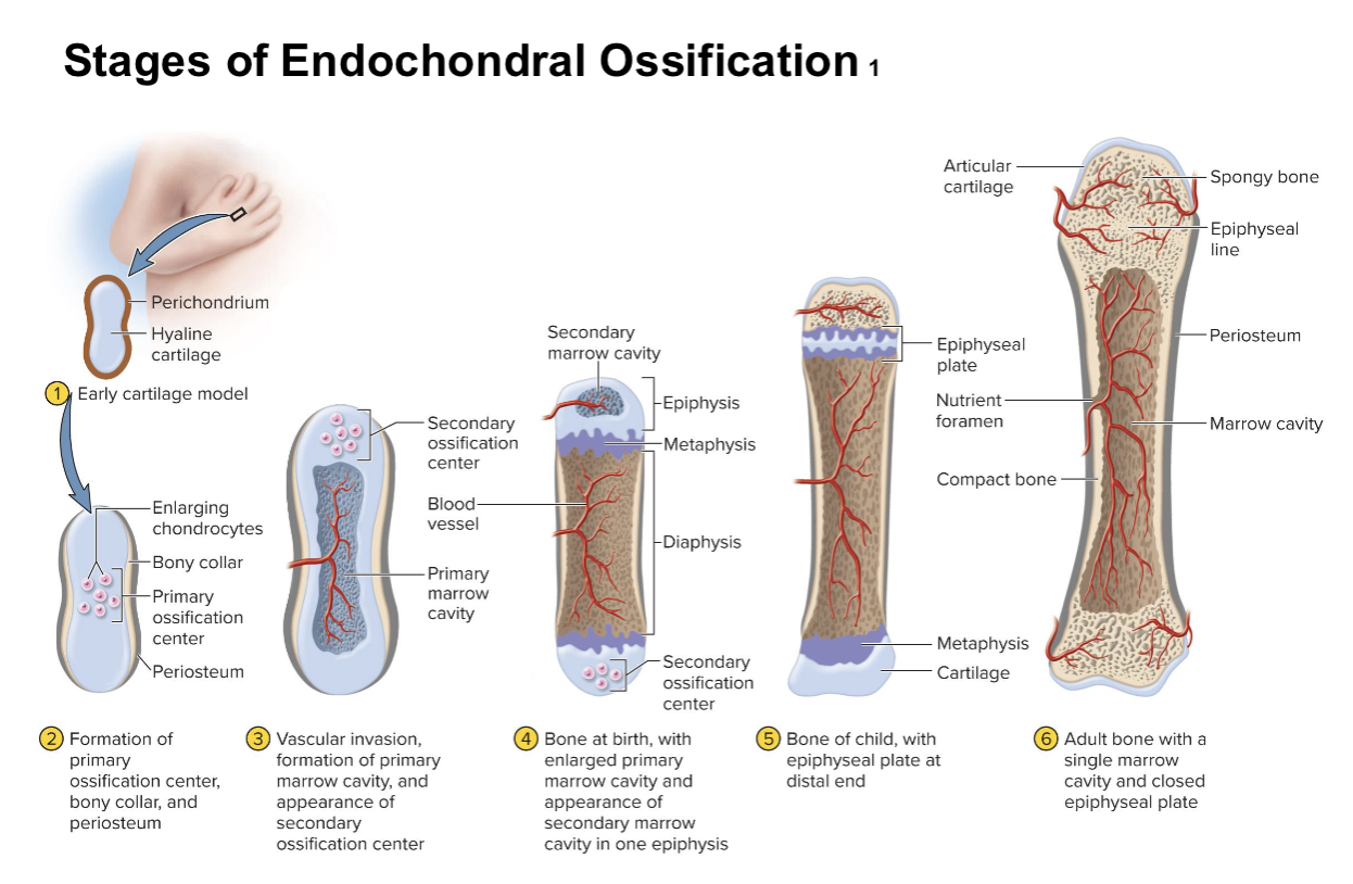
Describe the process of endochondral ossification
Bone develops from hyaline cartilage model; produces most bones of body (bones of limbs, vertebrae, ribs, sternum, scapula, pelvic girdle)
Summary of stages (metacarpal example):
1. Mesenchyme forms early hyaline cartilage model
2. Formation of primary ossification center, bony collar, and periosteum
3. Vascular invasion, formation of primary marrow cavity, and appearance of secondary ossification center
4. Bone at birth contains enlarged marrow cavity and secondary marrow cavity in one epiphysis.
5. Bone of child contains epiphyseal plate
6. Adult bone contains a single marrow cavity and close
epiphyseal plate (becomes epiphyseal line).
Epiphyseal plate
serves as growth zone growht plate
Metaphysis
transinal zone in between epiphyseal plate and diaphysis on each side where cartilage is replaced by bone.
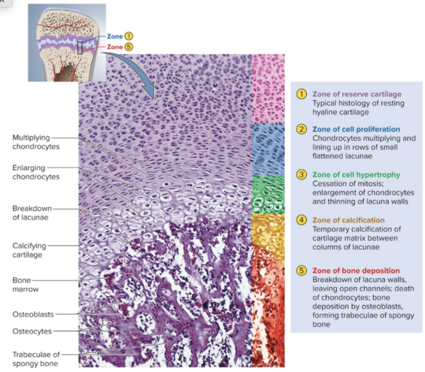
5 Zones of metaphysis (steps of cartiglage replacement)
Zone of reserve cartilage typical hiusology of resting hyaline cartlage.
Zone of cell proliferation. chondrocytes multiplying and lining up in rows of small flatted lacunae.
Zone of cell hypertrophy. Chrondrocytes engage, lacuna walls thin.
Zone of calcification. Calcification of cartilage matrix between columns of lacuna.
Zone of bone deposition. Lacuna walls breadown. Chondrocytes are replaced by osteoblast that deposit bone matrix to form trabiculae in lines of mechanical stress forming spongy bone.
Growth in height
Chondrocyte multiplication in zone 2 and hypertrophy in
zone 3 push the zone of reserve cartilage toward ends of the
bone, and the bone elongates. This is interstitial growth.
Interstital growth
cartilage growth from within the epiphyseal plate.
When does cartilage of epiphyseal plate deplete?
Late teens, early 20s. Primary and secondary marrow cavities unite into one cavity. Junctions are filled with spongy bone. Site of original epiphyseal plate is now epiphyseal line.
Epiphyseal line
Present on adults shows where the epiphyseal plate was.
Achondroplastic dwarfism
Long bones stop growing in childhood; results in normal torso, short limbs. Failure of cartilage growth in metaphysis. Spontaneous mutation produces mutant dominant allele.
Pituitary dwarfism
Caused by a lack of growth hormone. Normal proportions with short stature.
Appositional growth (definition, mechanism
The deposition of new tissue at bone surface that cause the growth of bone in diamter and thickness.
Mechanism: occurs by intramembranous ossifcation at bone surface:
Osteoblasts of the inner layer of periosteum deposit osteoid tissue, become trapped as tissue calcifies.
This lays down the matrix in layers parallel to surface, forming circumferential lamellae.
Osteoclasts of the endosteum enlarge the marrow cavity.
Bone remodeling ( what percent of the skeleton is replaced epr year?)
Bones continually remodeled (absorption and deposition) throughout life, replacing 10% of skeleton per year.
Bone remodeling repairs microfractures, releases minerals into blood, reshapes bones in response to use and disuse/mechanical stress
Wolff’s law of bone
architecture of bone determined by mechanical stresses placed on it. Remodeling is a collaborative and precise action of osteoblasts and
osteoclasts.
Bony processes grow larger in response to mechanical stress
Mineral depositon (mineralization)
crystallization process in which calcium, phosphate, and other ions are taken from blood and deposited in bone.
Osteoblasts produce collagen fibers that spiral the length of the osteon; fibers become encrusted with minerals. First few crystals act as seed crystals that attract more calcium and phosphate from solution
Ectopic ossification
abnormal calcification of tissues, such as a lung, brain, eye, muscle, tendon, or artery (arteriosclerosis) (atherosclerosis)
Calculus
a calcided mass in an otherwise soft tissue organ.
Mineral reabsorption
process of dissolving bone; releases minerals into blood. Performed by osteoclasts at ruffled border. Hydrogen pumps in the membranes pump hydrogen into space between osteoclast and bone surface. Chloride ions follow by electrical attraction.
Hydrochloric acid (pH 4) dissolves bone minerals. Acid-tolerant protease enzyme digests collagen.
Bone remodeling example: braces.
Orthodontic appliances (braces) reposition teeth through resorption and deposit.
Tooth moves because osteoclasts dissolve bone ahead of
tooth; osteoblasts deposit bone behind the tooth.
Calcium homeostasis: other than boen where is calcium also need in the body?
Calcium needed for bone structure, neuron communication, muscle
contraction, blood clotting, and exocytosis
• Minerals deposited in the skeleton and withdrawn when they are
needed for these other purposes
• About 18% of skeletal calcium is exchanged with blood each year.
What is the normal calcium conentration in blood plasma?
9.2–10.4 mg/dl
Hypocalcemia
calcium deficiency less than 9.2 mg/dl; causes excessive excitability
of nervous system and muscles
Hypercalcemia
calcium excess over 10.4 mg/dl; causes nerve and muscle cells to be less excitable than normal. Hypercalcemia is rare, but hypocalcemia results from a variety of causes (low vitamin D, diarrhea, thyroid tumors, underactive parathyroid glands).
What factors affect calcium homeostasis
Calcium homeostasis depends on a balance between dietary intake, urinary and fecal losses, and exchanges between osseous tissue
What three hormones rgulate calcium homeostasis?
Calcitriol, Calcitonin, and Parathyroid hormone
General overview of hormonal control of calcium balance
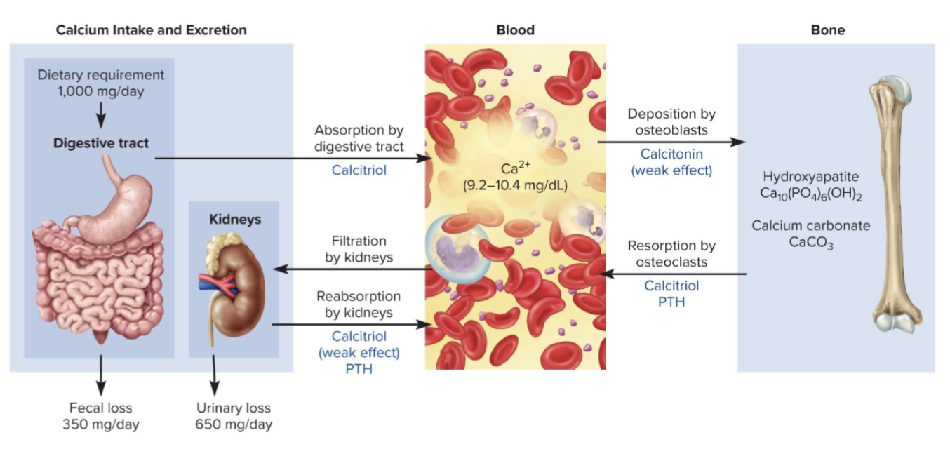
How is calcitriol produced?
Form of vitamin D produced by sequential actions of skin, liver, and kidneys
1. Epidermal keratinocytes use UV radiation to convert 7-
dehydrocholesterol to previtamin D3; warm sun on skin
converts this to vitamin D3 (cholecalciferol)
2. Liver adds hydroxyl group converting that to calcidiol
3. Kidney adds hydroxyl group converting that to calcitriol
What does Calcitrol do in the body? (3 mechanisms)
Calcitriol acts to raise blood calcium in three ways
• Increase calcium absorption by small intestine
• Increase calcium resorption from skeleton (stimulates formation of new osteoclasts)
• Weakly promotes reabsorption of calcium from the kidneys.
Calcitriol is necessary for bone deposition—helping
provide adequate calcium and phosphate
Bone resorption, reduced excretion of Ca by the kidneys, and increased absorption of Ca by the digestive tract.

Rickets
Inadequate calcitriol results in abnormal softness of bones
in children
Osteomalacia
Inadequate calcitriol results in abnormal softness of bones
in adults
Calcitonin (2 mechanisms)
produced by parafollicular (clear, C) cells of thyroid gland; secreted when blood calcium levels are too high.
Lowers blood calcium concentration in two ways:
Osteoclast inhibition, thereby reducing bone resorption.
Osteoblast stimulation, to deposit calcium into bone.
Important in children with more active osteoclast and osteoblast, weak effect in adults. Osteoclasts more active in children due to faster remodeling.
May inhibit bone loss in pregnant and lactating women.

Parathyroid hormone (PTH)
Secreted by the parathyroid glands on the posterior surface of the thyroid, in response to low calcium in the blood.
PTH raises blood calcium level by four mechanisms:
Binds to receptors on osteoblasts, which then stimulate osteoclasts to resorb bone.
Promotes calcium reabsorption by kidneys, so less lost in urine. Less urinary calcium excretion, more urinary phosphate excretion.
Promotes the final step of calcitriol synthesis in the kidneys, enhancing the calcium-raising effect of calcitriol.
Inhibits collagen synthesis by osteoblasts, inhibiting bone
deposition.

How much phosphaste doe sthe average adult have? What percentage of that is in the bones?
500 to 800 g phosphorus with 85 to 90% of it in the bones.
What is the normal plasma concentration of phosphorus
Normal plasma concentration is 3.5 to 4.0 mg/dl
How are phosphate levels regulated by PTH and Calcitriol?
Phosphate levels are not regulated as tightly as calcium
levels.
Calcitriol raises phosphate levels by promoting its
absorption by small intestine.
PTH lowers blood phosphate levels by promoting its urinary excretion.
What are the other factors that affect bone?
At least 20 or more hormones, vitamins, and growth factors affect
osseous tissue.
Growth hormone, estrogen, and testosterone occur
and promote ossification. These hormones stimulate multiplication of osteogenic cells, matrix deposition by osteoblasts, and chondrocyte multiplication and hypertrophy in the metaphysis
How does gender affect bone growth?
Girls grow faster than boys and reach full height earlier—estrogen has stronger effect than testosterone on bone growth.
Males grow for a longer time and also taller
Orthopedics
Branch of medicine dealing with the prevention and correction of injuries and disorders of bones, joints, and muscles.
The name implies its origin as a field treating skeletal deformities
in children.
Includes the design of artificial joints and limbs and the treatment of athletic injuries.
Stress fracture
break caused by abnormal trauma to a bone (example: in a fall)
Pathological fracture
Break in a bone weakened by disease (such as bone cancer or osteoporosis), usually caused by a stress that would not break a healthy bone
What strcutral charcteristics are used to classify fractures?
Direction of fracture line, such as nondisplaced, displaced.
Cracked or in multiple pieces, such as greenstick (cracked) and comunicated (in multiple pieces) .

How logn does it take for an uncomplicated fracture to heal?
about 8-12 weeks; longer for more complex fractures
What are the 4 stages of fracture healing?
Hematoma formation—blood clot (fracture hematoma) converted to granulation tissue by invasion of cells an blood capillaries.
Soft callus formation—deposition of collagen and fibrocartilage converts granulation tissue to soft callus.
Hart callus formation—osteoblasts deposit temporary bony collar to unite broken pieces while ossification occurs.
Bone remodeling—small bone fragments are removed by osteoclasts; osteoblasts deposit spongy bone and convert it to compact bone.

Closed reduction
procedure in which bone fragments are manipulated into their normal positions without surgery
Open reduction and internal fixation (ORIF)
involves surgical exposure of the bone and the use of plates, screws, or pins to realign the fragments
How is electrical stimulation used to heal a fracture?
Fractures taking more than 2 months to heal may be treated with electrical stimulation, which suppresses the effects of parathyroid hormone, including bone resorption. PTH also inhibits collagen production, so when it is suppressed, bone production can increase.
Osteopenia (defnition, age, and contributing factors)
Measurable decline in bone density; may advance to osteoporosis.
Beginning around age 40, net loss of bone mass as resorption outpaces deposition.
Contributing factors:
• Sex—women more affected than men
• Build—small, light frames affected more than heavier people
• Ancestry—White women of Asian and European origin
especially vulnerable
• Age—older individuals are more vulnerable
Osteoprosis (definition, assessment, treatments and prevention)
Severe loss of bone density; bones easily fracture.
Common assessment procedure: Bone densitometry, or DEXA scan (dual energy X-ray absorptiometry)—scans the spine and hips with low-dose X-rays.
Treatment strategies center around drugs that stimulate
bone deposition or slow the rate of resorption.
Preventative methods include weight-bearing exercise throughout life, even old age.