ANS 710 28 Male Reproductive Anatomy
 Call Kai
 Learn
 Practice Test
 Spaced Repetition
 Match
 Flashcards
 Knowt Play
1/65

There's no tags or description
Looks like no tags are added yet.
66 Terms
Male genital apparatus
Paired testes
Paired duct systems
Epididymis
Ductus deferens
Urethra
Penis
Skin adaptations
Prepuce
Scrotum
Testes exocrine function
Gamete production (spermatocytes)
Testes endocrine function
Testosterone, estrogen, etc
Temperature regulations for testes
Pampiniform plexus
Network of testicular veins that allows countercurrent exchange (hot arterial blood cooled down by venous blood)
Cremaster muscle (poorly developed in toms)
Strip of internal abdominal oblique that helps to raise and lower the testicle within the scrotum
Tunica dartos
Layer of smooth muscle beneath scrotal skin that helps raise and lower testicles
Scrotal skin
Sweat glands
Where are testes developed?
In abdomen
Testes Composition
Thick capsule = tunica albuginea
Lobules
Seminiferous tubules: Produce sperm cells
Mediastinum testis
Rete testis
Efferent ductules
Leads to epididymis

Superficial inguinal ring
Slit in the aponeurosis of the external abdominal oblique → on the outside of abdominal wall
2 crura (borders), 2 commissures (craniolateral & caudomedial)
Deep inguinal ring
On the inside of abdominal wall
Boundary → NOT a distinct structure
Caudal border of the internal abdominal oblique M
Arcus inguinalis (inguinal ligament)
Thickened caudal portion of aponeurosis of EAO M
Inguinal canal
Between superficial and deep rings
What is the developing testicle invested in?
Visceral peritoneum
Gubernaculum testis
Mesenchymal tissue from testis through inguinal canal to eventual scrotum
As the gubernaculum testis retracts through the inguinal canal and rings, the ________ _____ will also surround the testicle
parietal peritoneum
Vaginal tunic
Visceral peritoneum → visceral vaginal tunic
Parietal peritoneum → parietal vaginal tunic
Virtual space between tunics
Vaginal canal: Within spermatic cord
Vaginal cavity: Within scrotum

What becomes the internal spermatic fascia?
Endoabdominal fascia (aka transverse abdominal fascia)
External spermatic fascia
Layer of loose connective tissue deep to scrotal skin
True or False: The vaginal cavity/canal directly communicates with the peritoneal cavity
True
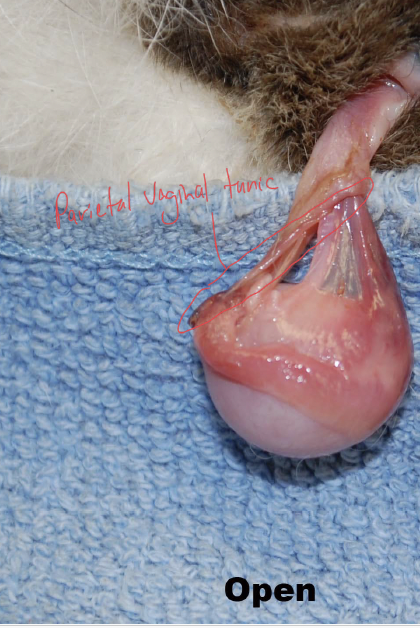
Risk of open neuter
If the parietal layer of the vaginal tunic is penetrated during castration, then a channel to the peritoneal cavity is opened with the potential for an ascending infection to cause peritonitis

Benefit of closed neuter
If the parietal layer of the vaginal tunic is not penetrated, then no pathway to the peritoneal cavity exists
Scrotal ligament
Between tail of epididymis and tunica dartos
Ligament of the tail of the epididymis
Absent in cats and dogs
Proper ligament of the testis
Tail of epididymis to the testis
Cryptorchidism timeline
If not resolved by 7 months of age, testicle(s) will need to be removed abdominally (like a spay)
Cryptorchidism: Temporary inguinal retention
Palpable, likely to eventually descend with growth
Cryptorchidism: Permanent inguinal retention
Has passed through the deep inguinal ring but is partly within inguinal canal
Cryptorchidism: Incomplete abdominal retention
Cremaster muscle, epididynmal tail; partially through vaginal ring
Cryptorchidism: Complete abdominal retention
Testis and epididymis
Cryptorchidism significance
No sperm production (too warm) but still produces testosterone
Higher risk for cancer
Head of epididymis
Attached firmly to testis
Efferent ductules enter here
Body of epididymis
Only part not firmly adhered to testis
Testicular bursa: space between the body and the testis
Tail of epididymis
Adhered to internal spermatic fascia
Picture of Epididymis

Ductus deferens
Continuation of epididymis
Connects to urethra
Colliculus seminalis
Ejaculatory ducts
Prostate
Invested in mesentery
Spermatic cord
Testicular artery, vein, and nerve
Pampiniform plexus
Lymphatics
Ductus deferens
Visceral vaginal tunic (remnant of visceral peritoneum)
Mesorchium
Covers vessels and nerves of spermatic cord
Mesoductus deferens
Covers the ductus deferens and attached to mesorchium
Mesofuniculus
Between origin of mesorchium and mesoductus deferens
Dog mesenteric structures picture

Accessory reproductive glands
Help sperm cells live longer in female reproductive tract with secretions (forms semen)
Dog accessory reproductive glands
Ampulla of the ductus deferens (small in dog)
Ampullary glands
Prostate gland
Right and left lobes with dense capsule
Bulk is around the urethra
Small disseminate part within the urethral mucosa
Cat accessory reproductive glands
Prostate - small body
Prostatic glands within walls of pelvic urethra, not just in body of prostate
Small bulboeurethral glands
No ampullae present
Male urethra
Divided into pelvic and penile parts
Pelvic part of male urethra
Preprostatic - very short
Prostatic part - urethral crest in dorsal wall
Colliculus seminalis: Openings for deferent ducts (aka deferent duct orifices)

Penile part of male urethra
Terminates at external urethral ostium
Root of the penis
2 crura + bulb of the penis
Crura
Proximal end of each corpus cavernosum penis, that attaches to ischiatic arch
Each crus has a core of cavernous tissue
Corpus cavernosum penis
Tunica albuginea
Covered by ischiocavernosus muscle
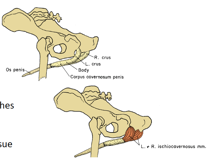
Bulb of the penis
The caudal expansion of the corpus spongiosum penis
Corpus spongiosum penis surrounds the caudal part of pelvic urethra and all of the penile urethra
Do not confuse with bulbus glandis
Covered by bulbospongiosus muscle

Muscles of penis
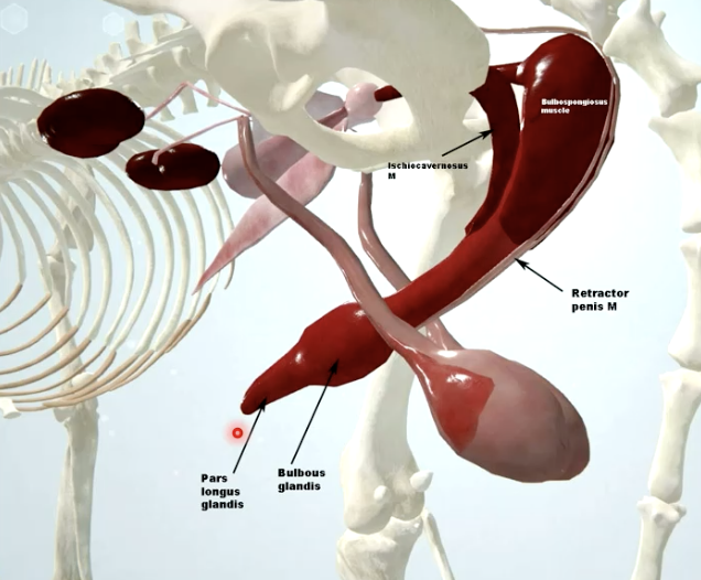
Ischiocavernosus
Bulbospongiosus
Retractor penis

How far can the body of the dog penis bend during tie?
180 degrees
Os penis (dogs)
Located within the glans penis
Attached to terminal CSP
Urethral groove
A place where urinary stones often block urethra

Glans penis in dogs
The part of the penis that will be introduced into the female repro tract to create the tie
Parts:
Bulbus glandis
Large thickening
Pars longa glandis
Corona glandis
Tip of penis
Glans penis in cats
Penis directed caudoventrally
Glans with keratinized spines
Stimulates ovulation

Prepuce
External lamina
Internal lamina
Paired preputial muscles
External lamina of prepuce
Continuous with the abdominal skin
Internal lamina of prepuce
Reflects onto penis at the level of the preputial fornix
Lined by glands and lymphoid tissue
Midventral incision through prepuce

Phimosis
Constriction of preputial ostium
Penis can’t come out
Paraphimosis
Inability to retract penis due to swelling or preputial constriction
Ex. hair wrapped around penis
Priapism
Persistent abnormal erection
Penile frenulum
Thin band of connective tissue joining ventral glans penis to corpus of penis or to the prepuce
Persistent penile frenulum
Clinical signs
Excessive licking
Urine scald
Phallocampsis (curvature of the erect penis)
Treatment
Thin: transect
Thick: local anesthesia then cut

Scrotum
Median raphe (aka groove) → scrotal septum
Sebaceous and sudoriferous (sweat) glands
Tunica dartos
Dog testis orientation in scrotum
Dorsocaudal
Cat testis orientation in scrotum
Obliquely toward anus
Dog scrotum
Mostly hairless
Cat scrotum
Haired