Biol 225 (A&P) - Muscular System Objectives #1 - #9 - Dr. Steele
 Call Kai
 Learn
 Practice Test
 Spaced Repetition
 Match
 Flashcards
 Knowt Play
1/85

There's no tags or description
Looks like no tags are added yet.
86 Terms
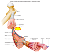
Epimysium
This connective tissue covering surrounds and bundles all of the fascicles bound by the perimysium

Perimysium
This layer of connective tissue surrounds each fascicle (bundle of muscle fibers/cells) - it extends inward from the epimysium and separates the muscle tissue into small section (fascicles).

Endomysium
This layer of connective tissue surrounds each muscle cell/fiber within a fascicle - Consists of individual connective fibrous cells that cannot conduct an impulse
Periosteum
This is a continuous covering over bones that eventually "oozes" off and connects muscle to bone (tendons)
Aponeuroses
These connect muscle to muscle and are found in the connection between the muscles of the occipitofrontalis (epicranius) and the obliques
Tendons
These connect muscle to bone
Understand this diagram and the different levels of a muscle cell

Muscle fibers
Muscle cells are also known as these
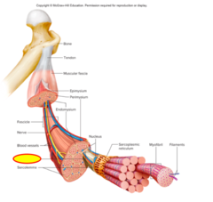
Sarcolemma
This term is used to describe the cell membrane of a muscle cell

Sarcoplasm
This is the internal fluid of muscle cells
Multinucleate
The cells that form muscles keep fusing together and have many nuclei. They are formed by smaller cells (myoblasts) that join together - Myoblasts fuse to form myocytes
Myofibrils
These are what give muscle its stripes (seen where proteins do/do not overlap) - Actin and myosin - produce alternating light and dark striations

Sarcomeres
This is the area from one z-line to another - a repeating pattern of units creating striations - a lot of light passes through this and is referred to as the I-band
Striations
These patterns are created due to the filaments actin and myosin
Smaller
The size of filaments (actin and myosin), relative to a myofibril, is described as what?
Myosin
This filament is thicker and bulkier - considered a globular protein
Actin
This filament is longer and thinner, described as filamentous
The A-band
In a sarcomere, Which band's length is the exact same length as myosin?
I-band
This striation pattern (light band) in a sarcomere is composed of thin actin filaments held by direct attachment to z-lines, which appear in the center of these
A-band
This striation pattern (dark band) in a sarcomere is composed of thick myosin filaments overlapping thin actin filaments
Z-line
In a sarcomere, this appears in the center of I-bands and are shown by a zig-zag pattern - their job is to hold myosin filaments in place
H-zone
In a sarcomere, this is a slightly lighter central region of the A band consisting of only thick filaments
M-line
In a sarcomere, this is a thickening of the A band that consists of proteins that help hold the thick filaments in place
Tropomyosin and troponin
What are the two regulatory proteins that exert control over when actin and myosin can physically touch - Job is to control muscle contraction by blocking the binding of actin and myosin (prevent them from touching)?
Ca2+
What ion allows the binding of actin and myosin and overpowers the regulatory proteins?
Sarcoplasmic reticulum (SR)
This is a membranous cell organelle in a muscle cell that fills all available space around the myofibrils and is capable of the transfer of impulses around the entire myofibril
Transverse tubules
These connect the outside of a muscle cell to the SR and surround myofibril - this is continuous with the sarcolemma
Terminal cisternae
These are storage containers that surround the transverse tubules and contain Ca2+ due to active transport - contain voltage-gated ion channels, and when open, move Ca2+ with its concentration gradient (stores Ca2+ aside of a cell organelle)
Triad
This is formed when a t-tubule is combined with the surrounding cisternae
Motor unit
This is the entire system of a motor neuron and its connections to other cells
Neuromuscular junction
This consists of a motor neuron, a motor end plate, and a synaptic cleft - it is where the terminal axon/knob "attaches" to the motor end plate at the synapse
However many synaptic knobs are on the presynaptic neuron
How many muscle fibers can a neuromuscular junction connect to?
Motor neuron
This part of the neuromuscular junction controls effectors and causes the contraction of a skeletal muscle fiber when stimulated by its neuron
Motor end plate
This part of a neuromuscular junction is the area of the muscle fiber that is specialized to create a connection with the presynaptic neuron knobs - abundant in nuclei and mitochondria as well as having very folded sarcolemma
Synaptic cleft
This part of a neuromuscular junction is the small gap that separates the membrane of the neuron (knob) and the membrane of the muscle fiber (motor end plate) - this is where neurotransmitters are released into
1. There is a release of acetylcholine (ACh)
2. ACh binds to ligand-gated ion channels which allow the transfer of Na+ to stimulate the post synaptic cell due to the influx of positive charge, creating a second set of Na+ channels to open as an impulse/action potential - end plate potential and muscle action potential/impulse
3. Calcium is released form the SR and causes muscle contraction by freeing the binding sites of actin and myosin
How is a skeletal muscle fiber stimulated to contract?
Acetylcholine (ACh)
What is the only neurotransmitter used in skeletal muscle?
At the motor end plates
Where is the only place a neurotransmitter can bind?
1. Myofilaments do not shorten but slide over one another
2. Sarcomeres shorten, causing myofibrils to shorten
3. Areas of actin/myosin overlap increase
Describe the sliding filament model for skeletal muscle fiber contraction
Filaments do not change length, but their orientation to each other changes
In the sliding filament model for skeletal muscle fiber contraction, do filaments change in length? What changes?
Z-lines
As sarcomeres shorten, what becomes closer together?
The motor neuron releases acetylcholine as the neurotransmitter that binds to motor end plates and causes ligand-gated channels on the sarcolemma to open. This starts the transfer of an impulse as Na+ channels open following it
What is acetylcholine's role in excitation-contraction coupling?
Calcium has to be present for actin to bond to myosin and actually contract the muscle - if calcium is not present, contraction will not occur
What is Calcium's role in excitation-contraction coupling?
1. Motor neuron releases acetylcholine
2. Ligand-gated channels on the sarcolemma open due to presence of ACh
3. Na+ and K+ channels open (Na+ influx predominates)
4. End-plate potential results, threshold achieved, voltage-gated Na+ channels open
5. Sarcolemma and t-tubules depolarize
6. Voltage-gated channels on SR open - cisternae of SR store calcium - T-tubule and surrounding cistern = triad membranes
7. Ca2+ is released into the sarcoplasm by diffusion
8. Ca2+ binds to troponin, shifts regulatory proteins
9. Actin and myosin change position relative to one another
Explain excitation-contraction coupling
ACh
In excitation-contraction coupling, what opens ligand-gated ion channels on motor end plates?
Voltage-gated Na+ channels
In excitation-contraction coupling, if excitation is great enough and brings in enough Na+, what can be triggered just outside of the motor end plate to create an action potential?
Voltage-gated Na+ channels
These channels are located just outside of the motor end plate and can use the extra excitation from the neurotransmitter action at the motor end plate to stimulate them
The opening of Ca2+ channels
In excitation-contraction coupling, the impulse eventually travels along and depolarizes the sarcolemma and t-tubules, what does this lead to?
Comes from the sarcoplasmic reticulum (including cisternae) and the extracellular environment
When Ca2+ flows into the sarcoplasm by diffusion, where does it come from?
The I-band and H-zone
Z-line
In excitation-contraction coupling, when actin and myosin change position relative to one another, what two things become shorter? And, what is pulled towards the M-line?
ATP binds to myosin and hydrolyzes to provide energy to contract - ATP also binds to myosin after contraction to detach it from actin
What is the role of ATP in cross-bridge formation
1. ATP binds to myosin and hydrolyzes (ATP → ADP+P)
2. Activated myosin binds to actin in the presence of Ca2+
3. ADP+P is released at contraction , and myosin head pivots (power stroke) as it is slung forward
4. New ATP binds and myosin detaches from actin
5. Hydrolysis of new ATP returns myosin to its activated position
6. Cross-bridge cycling continues as long as Ca2+ and ATP are available
Explain cross-bridge formation
Calcium
Regulatory proteins prevent binding of actin and myosin in the absence of what ion?
One hydrolyzed ATP (ATP → ADP+P)
How many ATP/ADP+P are responsible for stimulating one myosin filament to bind to one actin filament
Once the Ca2+ supply is returned back into the SR or extracellular matrix so that the regulatory proteins can block binding sites of actin and myosin - Ca2+ pumps re-establish the gradient
How does cross-bridge cycling end?
1. Ca2+ is pumped back into the SR and extracellular fluid because voltage-gated ion channels are closed, so the pump re-establishes concentrations
2. Binding sites are blocked when Ca2+ is removed and muscles cannot contract
3. Sarcomere returns to its original length
Explain muscle relaxation
Rigor mortis
This is the contraction of dead muscle cells
- Muscle cells run out of ATP and the muscles contract due to the presence of Ca2+ (bc pumps fail and cannot reset contraction)
- Ca2+ present frees binding sites allowing energized myosin to sling forward
- Once myosin and actin sling forward in contraction, the energy is used and the cells cannot relax because there is no ATP to free the binding site
What is the cause of rigor mortis?
They are long, skinny, and covered by connective fibrous tissue
Describe muscle cells
Myofibrils
These are long and thin structures that are smaller than a cell and are located inside one muscle cell
Skeletal muscle cells are individualized (covered by dense fibrous connective tissue) and independent of one another
Describe the connectivity of skeletal muscle cells
Impulses transfer cell-to-cell in cardiac muscle so that the muscle can contract every single time and have a complete reaction
How do impulses travel in cardiac and smooth muscle muscle?
Because skeletal muscle cells do not communicate with one another since they are independent, allowing us to only perform a certain extent of an action compared to a complete action of a cardiac or smooth muscle
Why can skeletal muscle do partial contractions?
Skeletal muscle cells are independent from one another
Are skeletal muscle cells interdependent or independent of one another?
Smooth and cardiac muscle cells are interdependent on one another
In cardiac and smooth muscle, are cells interdependent or independent?
Bands
When looking at sarcomeres and striations, what do we use as reference points?
Transfer of electrical signals (impulses)
The membrane that compartmentalizes myofibrils and is connected to the outside of the cell has what function?
No, the impulse stays in the skeletal muscle fiber because those fibers are separated from neighboring cells
Does an impulse travel from one skeletal muscle fiber to another?
A nerve
Skeletal muscle requires what to tell them what to do?
Damage cannot be at the cell body because separation damages beyond repair, partially because of high nucleus concentration
The only way you can recover from nerve damage is if it is not at this part of the neuron
Pumps - the calcium there can then diffuse with its gradient
Where does all of the calcium in the synaptic cleft come from?
Highly folded
What is a characteristic of the motor end plate?
Overshoot
What allows stimulation and opening of adjacent ion channels to the motor end plate?
yes
In the sliding filament model, do myofibrils change length?
Closer together as each sarcomere becomes shorter
In the sliding filament model, in which direction do Z-lines travel?
The sarcoplasmic reticulum
What stores calcium which creates a regulatory protein shift?
I-band
This band is on either side of a z-line where there is only actin, this decreases in size during contraction
Z-lines
Which lines are pulled closer together when myosin binds to actin?
It decreases
What happens to the H-band during contraction?
A-band increases overlap but stay the same length
What happens to the A-band during contraction?
The impulse is what lets the calcium out of storage to free binding sites to start contraction
What is a reason why skeletal muscle requires an impulse?
Excitation
What is the impulse that leads to a contraction
Contraction
This occurs when filaments slide and the muscle gets shorter
Nerve impulse
You need this to cause contraction
T-tubules/cisternae
Excitation stimulates all of the __________ and __________ that are continuous with the cell membrane
During the power stroke
In cross-bridge cycling, when is energy released (ADP+P is spent)?