E&E Histology Exam2
 Call Kai
 Learn
 Practice Test
 Spaced Repetition
 Match
 Flashcards
 Knowt Play
1/17

There's no tags or description
Looks like no tags are added yet.
18 Terms
Interior of Liver Lobule

Hepatocyte
Large, round, centrally located(sometimes binucleate) nuclei. Polygonal, have microvilli, bound to each other by gap junctions, occluding junctions and desmosomes

Perisinusoidal space
space between hepatocytes and sinusoids. Microvili extend into this space

Reticular Fibers
Between hepatocytes and sinusoids. only type of CT between the two

Kupffer Cells
Filled with macropahges. Within sinusoids. Phagocytic

Perisinusoidal stellate cell
in perisinusoid space. Aids in vitamin A storage. Lipid droplet on top, nucleus underneath. Takes place in cirrhosis

Classic lobule
Central Vein in middle. Blood goes in, bile goes out

Bile Canaliculi
Formed from tight junctions between hepatocytes. Merge with bile ducts. Completely separate from sinusoids

Portal Triad


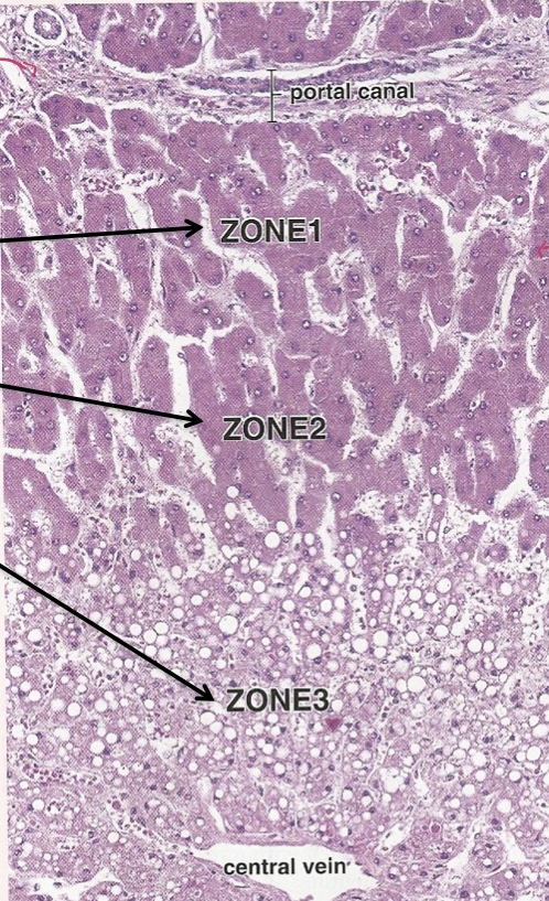
Portal Acinus
3 zones of oxygenation

Portal lobule
drains bile from hepatocytes to bile duct

Acinar Zones
Pancreatic Acini
supportive cells-capillaries, fibroblasts to secrete collagen

Cetroacinar Cells
the first cell of the intercalated duct that protrudes into the lumen of the acinus


Intralobular Ducts

Interlobular Ducts
Between Ducts

Islets of Langerhans
Endocrine Pancreas. Large, round, euchromatic nucleus. Abundant cytoplasm. Comprised of four main cell types

Posterior Pituitary
Unmyelinated axons, astrocytes, fenestrated capillaries

