Chapter 3-Sesay Lecture
 Call Kai
 Learn
Learn Practice Test
 Spaced Repetition
 Match
Match Flashcards
 Knowt Play
1/105

Earn XP
Description and Tags
Will be on test
Name | Mastery | Learn | Test | Matching | Spaced | Call with Kai | Chat |
|---|
No analytics yet
Send a link to your students to track their progress
106 Terms
Cell theory
1- All organisms are composed of cells and cell products
2-Cell is the simplest structural and functional unity of life
3- organisms structure and function are due to activities of its cells.
4- cells only come from existing cells.
what is a tissue?
a group of similar cells that work together to perform a similar function.
There are apx 200 cell types in the body. 9 common shapes are:
To know for test: Shape and thinness/thickness:
Squamous: flat tissue (like a floor tile), scaly- lungs
Cuboidal: cube/cuboidal- in kidney(absorbs and secretes)
Columnar: taller than wide- has large surface area- in stomach, small intestines
fusiform
fibrous
cuboidal-
columnar
discoidal
polygonal
stellate
spheroidal
Squamous-

squamous cell- function and where?
thin flat and scaly, often with bulge where nucleus is.
Diffusion and protection
outer layer of skin, esophogus.
cuboidal cells
secretion and absorbtion
liver, secretory portion of many glands
columnar cells- function and where?
high volume absorption and secretion
inner lining of stomach and intestines
Polygonal cells
irregular angular shapes
structural packing. allow cells to pack tightly
widespread, and many glands
stellate cells
communication
nerve cells
spheroidal cells
mobility and storage
white blood cells and egg cells
discoidal cells
gas exchange
red blood cells
fusiform cells:
sustained contraction
smooth muscle
fibrous cells
directional force and reach, long slender, and thread like
skeletal muscle cells and the axons of nerve cells
Microscope moments for the cell
Light vs. Electron (better mag and resolution)
light microscope revealed: plasma membrane, nucleus, and cytoplasm
Transmission electron microscope revealed : better resolution
Scanning electron microscope: dramatic 3d images at high mag and res but only on the surface.
Cell is surrounded by?
Plasma membrane:
Function: 1-protects/ defines cell boundary 2- controls exchange/what comes in and out of cell 3- communication-
components: made of proteins and lipids and defines cell boundaries.phospholipds, cholesterol, glycolipids
Cytoplasm
within the cell: contains organelles, cytoskeleton, inclusions (stored or foreign particles), and clear gel called cytosol or ICF (intracellular fluid).
cytosol
Intracellular fluid
Extracellular Fluid (ECF)
any fluid outside of cells including tissue (interstitial) fluid, blood plasma, lymph, cerebrospinal fluid
Membrane Lipids
98% of the cell membrane is composed of lipids, and mostly phospholipids (75%).
20% is cholesterol
5% glycolipids
Phospholipids
Primary (75%) lipid on plasma membrane.
amphipathic molecules arranged in a bilayer
Drifts lateral to keep membrane fluid
Phosphate head (hyrdophilic) with fatty acid tails (hydrophobic)
cholesterol
20% of the membrane lipids
Holds phospholipids still and can stiffen membrane, but at higher concentrations can increase membrane fluidity.
considered “parent” steroid from which other steroids are synthesized.
Glycolipids
5% of the membrane lipids
• Phospholipids with short carbohydrate chains on extracellular face
• Contribute to glycocalyx— comes in as glycolipid and comes at as glycoprotein. carbohydrate coating on cell surface
like a tag, helps to ID the cell.
Glycocalyx
carbohydrate moietie of glycoprotein and glycolipid, external to plasma membrane.
works as an ID tag.
Allows us to see foreign cells.
-protection
-immunity to infection
-cancer defense
membrane proteins % of membrane
not on test:
5% of membrane
larger than lipids, and account for 50% of weight
two classes are transmembrane and peripheral proteins.
The Plasma Membrane image
depict image
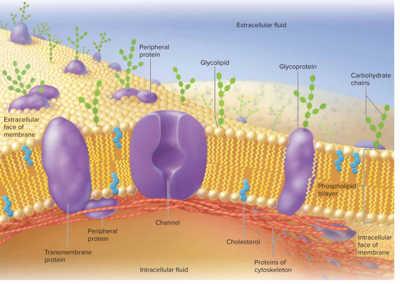
Transmembrane proteins
pass completely through
membrane
most are Glycoproteins bound to oligosaccharides on the extracellular side
• Hydrophilic regions contact the watery cytoplasm, extracellular fluid
• Hydrophobic regions pass through lipid of the membrane
Transmembrane Proteins Image
pass completely through
membrane
• Hydrophilic regions contact the watery cytoplasm, extracellular fluid
• Hydrophobic regions pass through lipid of the membrane

receptor (membrane proteins)
-bind chemical signals to trigger internal
changes
• May cause production of a second messenger within cell receiving
chemical message
Channel proteins—
allow hydrophilic solutes and water to
pass through membrane
• Some are always open, called leak channels, and some are gates
(gated channels) that open only when triggered
• Ligand-gated channels—respond to chemical messengers
• Voltage-gated channels—respond to charge changes
• Mechanically gated channels—respond to physical stress on cell
Leaky membrane channel
always open.
Ligand-gated channel
responds to chemical messengers
Voltage-gated channels—
respond to charge changes (ions)
Mechanically gated channels—respond to physical stress on cell
respond to physical stress on cell
Carriers—membrane proteins
bind solutes and transfer them across
membrane
Pump proteins—
carriers membrane proteins that consume ATP
Cell-identity markers—
glycoproteins acting as
identification tags
Functions of membrane proteins:
Receptor
enzyme
channel
gated channel
cell identity marker
cell adhesion molecule (CAM)
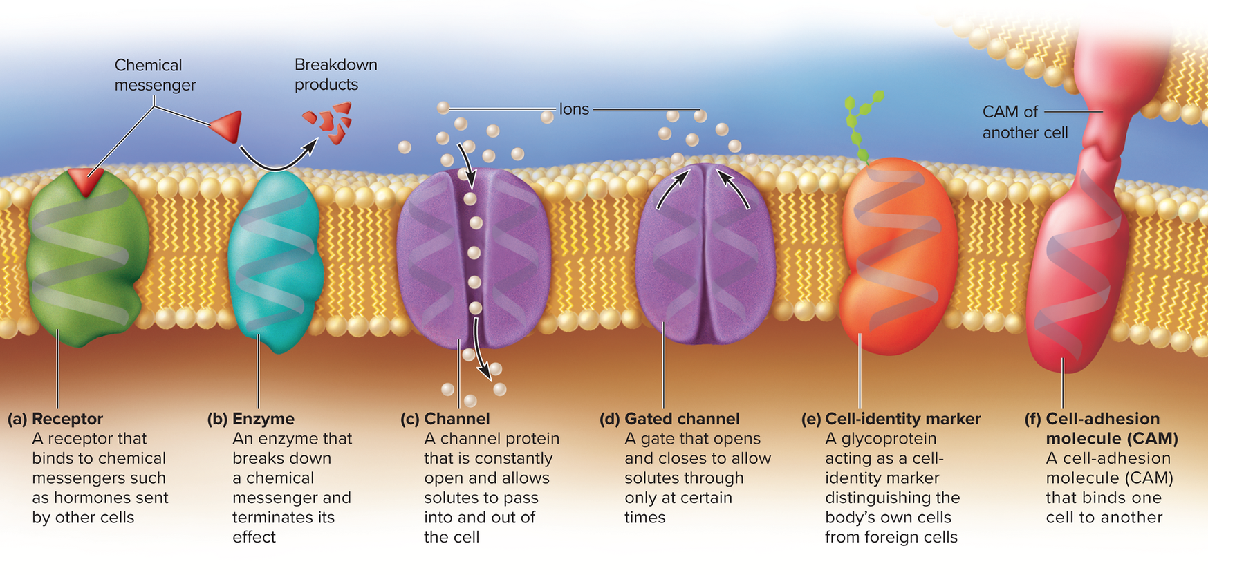
receptor protein
surface protein that receives chemical signal
enzymes in the plasma membrane
carry out final stages of starch and protein digestion in small intestine
produce secondary messengers
break down hormones and other signaling molecules
channel protein
allow water and hydrophilic molecules to pass through membrane.
Cell adhesion molecule (CAM)
how cells adhere to each other and extracellular material.
with few exceptions (such as blood cells and metastasizing cancer cells) cells need to be attached to EC material to grow properly.
Functions of Membrane Proteins image “abc”
receptor, enzyme, channel

functions of membrane proteins “cdf”
gated channel, cell identity marker, CAM cell adhesion molecule

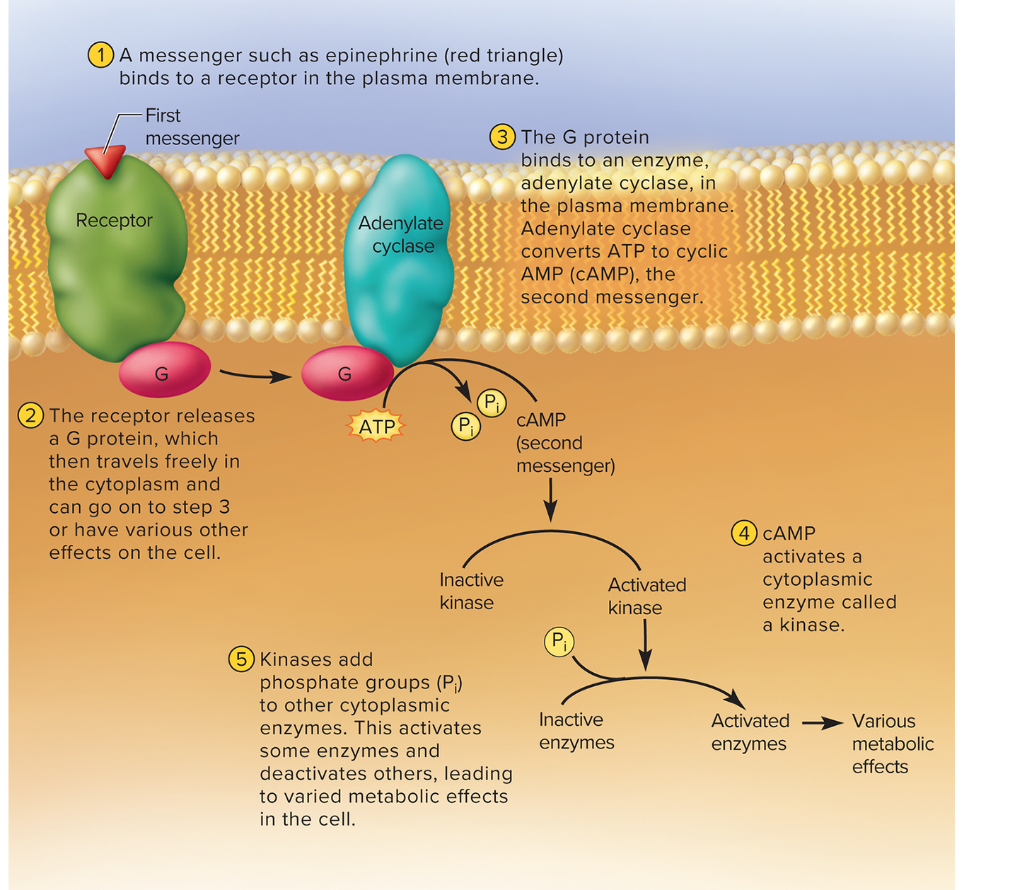
secondary messengers
example how epinephrine works:
1- epinephrine (the first messenger) binds to receptor
2-receptor is linked, on the intracellular side to a g protein (get energy from GTP)
3- G protein relays signal to another membrane molecule adenylate cyclase
4- adenylate cyclase removes two phosphate groups from ATP and converts it into cyclic AMP (cAMP)- the second messenger
5- cyclic AMP activates cytoplasmic enzymes like kinases which add phosphate groups to other cellular enzymes.
this activates some enzymes, deactivates others, and triggers a lot of physiological change in the cell.
Glycocalyx— (3.2b)
“A fuzzy coat” carbohydrate moieties of glycoproteins and
glycolipids external to plasma membrane
• Unique in everyone but identical twins. acts like ID tag, enables body to distinguish healthy cells from bad.
• Functions-
• Protection-
• Immunity to infection
• Defense against cancer
• Transplant compatibility
• Cell adhesion
• Fertilization
• Embryonic development
Microvilli- Extensions of the cell surface (3.2c)
Found in the small intestine and allows more nutrients to absorb into blood stream.
destroying microvilli will inhibit food nutrition from getting into blood stream.
extensions (1 to 2 μm) of the membrane that
serve to increase surface area
• Provide 15 to 40 times more surface area to cells that
have them
• Best developed in cells specialized in absorption (epithelial cells of kidney and intestine)
• On some absorptive cells they are very dense and appear
as a fringe called the brush border
• Some microvilli contain actin filaments that are tugged
toward center of cell to milk absorbed contents into cell

Cilia—
Motile cilia- found in the lungs, respiratory tract. lungs are a big structure. located at the top of the lungs.
cilia repels unwanted things! thats why cilia is at the top.
hair-like processes 7 to 10 μm long
•
Single, nonmotile primary cilium found on nearly every
cell; serves as “antenna” for monitoring nearby conditions
• Helps with balance in inner ear; light detection in retina
non motile cilia- helps with balance in inner ear, light detection in retina.
Brush border
Microvilli densely packed at apical cell surface
Made of actin
motile verse nonmotile cilia
Multiple nonmotile cilia found on sensory cells of nose
• Motile cilia less widespread
• Found in respiratory tract, uterine tubes, ventricles of brain, ducts of
testes
• 50 to 200 on each cell
• Beat in waves sweeping material across a surface in one direction
• Power stroke followed by recovery stroke
image of Cilia
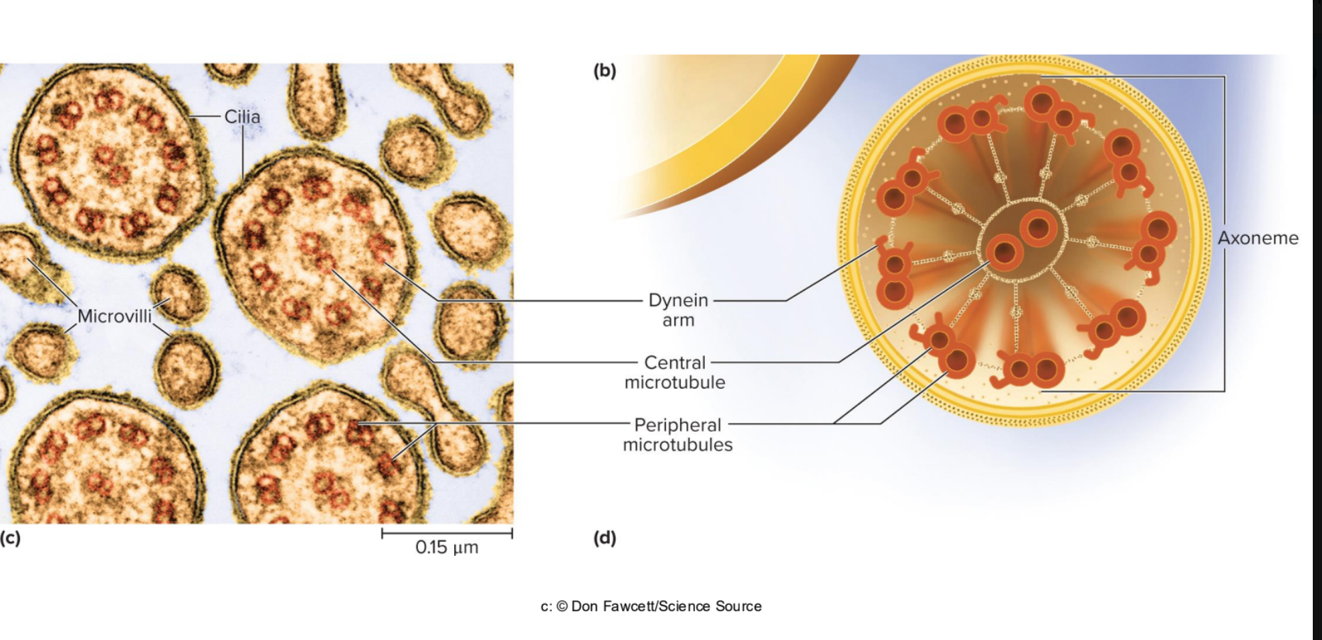
How Cilia Moves
Cilia move within saline layer at cell surface; mucus
“floats” atop this layer. this happens at the top of the lungs.
• Structure cilium:
• Dyenin arms “crawl” up adjacent microtubule, bending the cilium;
uses energy from ATP
Power stroke and recovery stroke
Kartagener syndrome
disease caused by destruction of dyenin arm
symptoms- difficulty breathing at birth, chronic cough/runny nose, frequent lung and sinus infection.
Cystic Fibrosis
problem with cilia! more respiratory issues.
hereditary disease in which cells make
chloride pumps, but fail to install them in the plasma
membrane
• Chloride pumps fail to create adequate saline layer on cell
surface
• Thick mucus plugs pancreatic ducts and respiratory tract
• Inadequate digestion of nutrients and absorption of
oxygen
• Chronic respiratory infections
• Mean life expectancy of 44
Flagellum—
whiplike structure
• Tail of a sperm is only functional flagellum in humans. sperm must travel all the way to the ovaries- flagellum helps.
Ciliary movement
1-axoneme is an array of thin protein cylinders called microtubules
2- 2 central microtubules are surrounded by a ring of nine microtubule pairs called the 9 plus 2 structure.
3- central microtubules stop at the cell surface.
4- peripheral microtubules continue a short distance into the cell as part of a basal body that anchors the cilium.
5- in each peripheral microtubules one tubule has two Dynenin arms. Dynenin is a motor protein, uses energy from ATP to crawl up the adjacent pair of microtubules.
6- on one side of the cilium, dynenin reaches out and grabs a microtubule of the adjacent pair, then pull towards them. collective movment makes the ciilium bend in one direction.
7- activation and inhibition alternate so that the back and forth of power and recovery strokes happens
Pseudopods
cytoplasm filled exensions of the cell varying in shape from fine filamentous processes to blunt fingerlike.

Filtration 3.3a
a physical pressure forces fluid through a selectively permeable membrane.
how kidneys filter waste from blood. how water, salts, nutrients, and other solutes are transferred from the blood stream to the tissue fluid.

simple diffusion 3.3b
net movement of particles from high to low concentration
affected by:
Temperature
Molecular weight
steepness of gradient
membrane surface area
membrane permeability
selectively permeable—
Plasma membrane and organelle membranes are
selectively permeable—allowing some things through, but
preventing others from passing
Passive mechanisms
require no ATP
• Random molecular motion of particles provides necessary energy
• Filtration, diffusion, osmosis
Active mechanisms
consume ATP
Active transport and vesicular transport
• Carrier-mediated mechanisms use a membrane protein to
transport substances across membrane
Osmosis
net flow of water through a selectively permeable
membrane
• Water moves from an area of higher water (lower solute)
concentration to an area of lower water (higher solute)
concentration
aquaporins
Water can diffuse through phospholipid bilayers, but osmosis is
enhanced by.————-—channel proteins in membrane
specialized for water passage
• Cells can speed osmosis by installing more ———————
• Crucial consideration for IV fluids
• Osmotic imbalances underlie diarrhea, constipation,
edema
Tonicity
1-Tonicity is describing extracellular…salt (NaCl) and glucose are major solutes..
2- water will go towards where we have more solutes
3- intracellular and extracellular fluid
Hypotonic solution—causes cell to absorb water, swell,
and possibly burst (lyse). less solute outside of cell.
• Cell Has a lower concentration of nonpermeating solutes than
intracellular fluid (ICF)
• Distilled water is an extreme example
• Hypertonic solution—causes cell to lose water and
shrivel (crenate). more solute outside of the cell.
• Has a higher concentration of nonpermeating solutes than ICF
• Isotonic solution—causes no change in cell volume
• Concentrations of nonpermeating solutes in ECF and ICF are the
same
• Normal saline (0.9% NaCl) is an example
Hypotonic solution—
causes cell to absorb water, swell,
and possibly burst (lyse). less solute outside of cell.
• Cell Has a lower concentration of nonpermeating solutes than
intracellular fluid (ICF)
• Distilled water is an extreme example
Hypertonic solution—
• causes cell to lose water and
shrivel (crenate). more solute outside of the cell.
• Has a higher concentration of nonpermeating solutes than ICF
Isotonic solution—
causes no change in cell volume
• Concentrations of nonpermeating solutes in ECF and ICF are the
same
• Normal saline (0.9% NaCl) is an example- must know!!!
There are three kinds of carrier proteins:
Uniport—carrier that moves one type of solute
• Example: calcium pump
• Symport—carrier that moves two or more solutes
simultaneously in same direction (cotransport)
• Example: sodium–glucose transporters
• Antiport—carrier that moves two or more solutes in
opposite directions (countertransport)
• Example: Sodium–potassium pump removes
+ Na
, brings in k
Three mechanisms of carrier-mediated transport
1-Facilitated diffusion,2- primary active transport,3- secondary
active transport
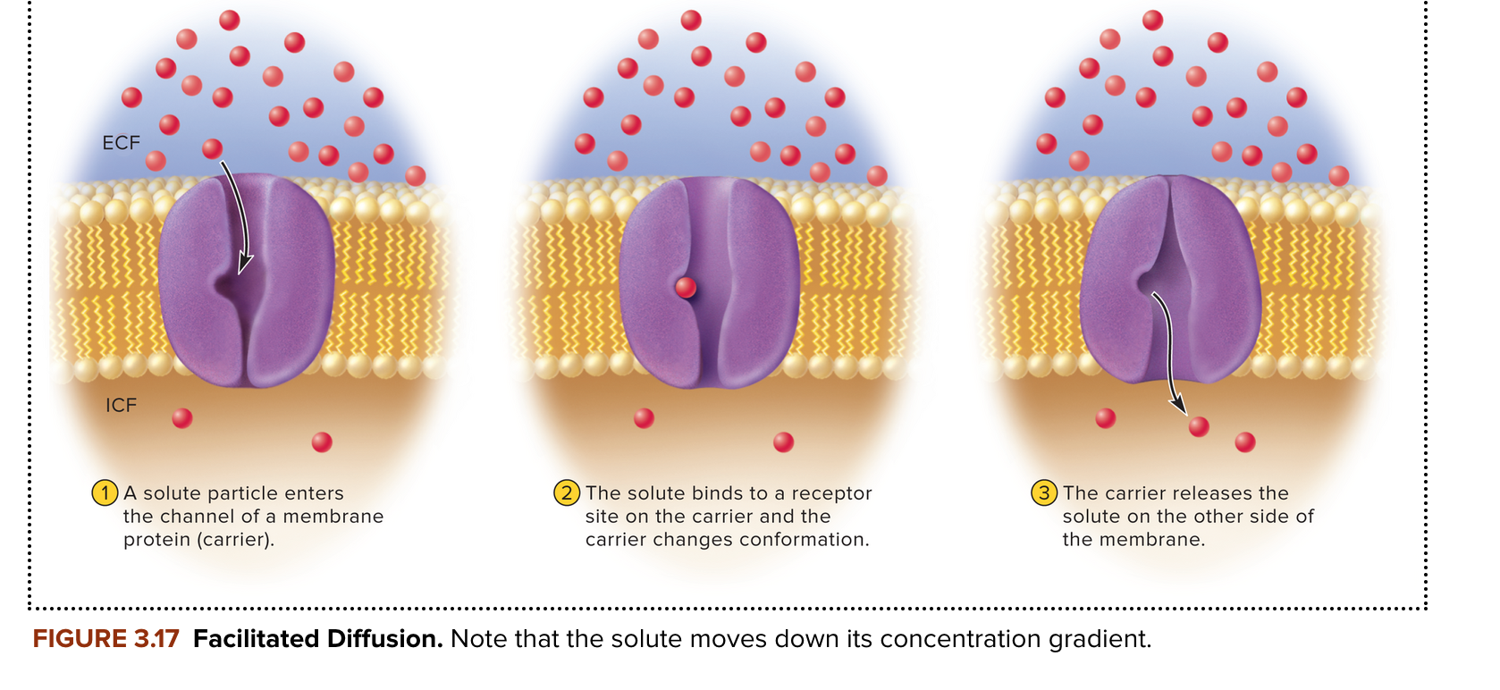
facilitated diffusion
carrier mediated transport of a solute through a membrane down its concentration gradient. require no metabolic energy (ATP) expenditure.
Primary active transport
carrier moves a substance through a cell membrane UP its concentration gradient, using ATP to do so. ATP supplies energy by transfering a phosphate group to the transport protein.
sodium potassium pump is a good example- half a day’s calories fuel this purpose!- maintains steep NA+ concetration across membrane.
regulates cell volume, maintains membrane potentials, heat production

secondary active transport
carrier moves solute
through membrane, but only uses ATP indirectly
• Example: sodium–glucose transporter (SGLT)
• Moves glucose into cell, up its concentration gradient, while
simultaneously carrying sodium down its gradient
• Depends on the primary transport performed by sodium-
potassium pump
• Does not itself use ATP
• SGLTs work in kidney cells that have sodium-potassium pump at
other end of cell; prevents loss of glucose to urine
-

Vesicular transport
moves large particles, fluid droplets, or numerous
molecules at once through the membrane in vesicles
Vesicles—
bubblelike enclosures of membrane
Endocytosis and exocytosis
brings material into cell;releases material
from cell
Three forms of endocytosis:
Phagocytosis—
• Pinocytosis—
• Receptor-mediated endocytosis—
Phagocytosis—
engulfing and destroying large particles; “cell eating”
• Pseudopods surround object, fuse to form internal phagosome, which merges
with lysosome to form phagolysosome within which object is digested
macrophages also phagocytize
Pinocytosis—
taking in droplets of ECF containing molecules useful in the
cell; pinocytic vesicles in cytoplasm; “cell drinking”
Receptor-mediated endocytosis—
particles bind to specific receptors on
plasma membrane
• Pit forms in membrane, cytosolic side covered in clathrin protein; forms clathrin-coated vesicle
coated vesicle that is directed to destination within cell by its “address label” (the clathrin coat)
Classic example is ldl (low density lipprotein) - liver has ldl receptor- that does receptor mediated endocytosis.
Receptor helps dump ldl into liver
Phagocytosis, Intracellular Digestion, and Exocytosis Image:

receptor mediated endocytosis image

Exocytosis—
discharge material from cell
• Examples:
• Release of insulin by endocrine cells
• Sperm cells release enzymes for penetrating egg
• Mammary gland cells release milk sugar
• Also functions to replace any plasma membrane lost by
endocytosis

Organelles
are the internal structures of a cell that carry out
specialized metabolic tasks
• Membranous organelles are surrounded by membranes
• Nucleus, mitochondria, lysosomes, peroxisomes, endoplasmic
reticulum, and Golgi complex
• Other organelles are without membranes
• free Ribosomes only one to know on test, centrosomes, centrioles, basal bodies
cytosol
clear vicous, watery colloid that bathes cytskelton and organelles. contains enzymes, proteins, amino acids, ATP, electrolytes, diverse ions, oxygen, CO2, and metabolic waste.
cytoskeleton
network of protein filaments and cylinders that structurally support a cell, determine its shape, organize contents, direct movement of material within cell. dense supportive scaffold within cytoplasm.
connects to transmembrane proteins, which is inturn connected to protein fibers external to the cell
cytoskeleton elements even connect to chromosomes in the nucleus!- could stimulate genetic function, timing of cell division
Made of microfilaments, intermediate filaments, and microtubules
microfilament
6nm thick and made of actin protein.
concentrated into fibrous mat called terminal web (membrane skeleton) on the cytoplasmic side of membrane.
phospholipds spread on the terminal web like butter on bread.
actin filaments form supportive core of the microvilli
intermediate filament
thicker and stiffer than microfilaments
make up strength of hair and fingernails
give cell its shape, resist stress, and form junction that attach cell to cell
microtubules
cylinders made of 13 parallel strands called protofilaments. each protofilamanet is a long chain of globular proteins called Tubulin.
these radiate from the centromeres,
hold organelles in place, maintain cell shape,
act like monorails for motor proteins to carry organelles/macromolecules around the cell.
microtubules form the axonemes of cilia and flagella
form mitotic spindle
nucleus—
usually largest organelle (5 μm in diameter);
contains cell’s genetic material
• Material within nucleus is called nucleoplasm
• Includes threadlike chromatin (DNA and proteins) and one or more
nucleoli (singular: nucleolus) where ribosomes are produced
mature red blood cells are anuclear.
some liver, urinary bladder, and heart cells have two nuclei.
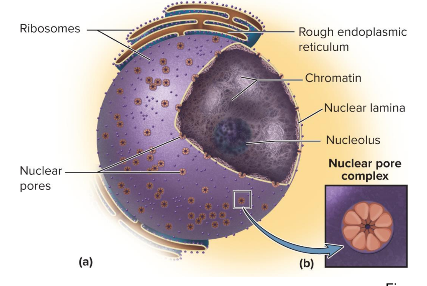
nuclear envelope
double membrane encloses nucleus, peforated with nuclear pores.
raw material for DNA/RNA come into nucleus via the pores.
nucleoplasm
material in the nucleus”
chromatin..
nucleoplasm
Material within nucleus is called —————-
• Includes threadlike chromatin (DNA and proteins) and one or more
nucleoli (singular: nucleolus) where ribosomes are produced
structure of the nucleus
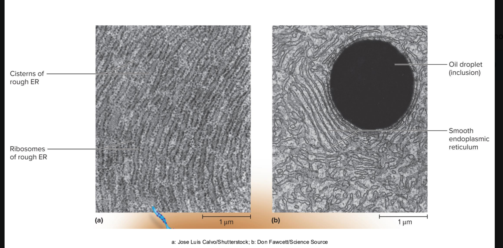
Endoplasmic reticulum (ER)—
network of interconnected parralel
membranous channels called cisterns
rough ER- covered in ribosomes.
continuous with outer membrane of nuclear envelope.
Rough endoplasmic reticulum—
parallel, flattened sacs
covered with ribosomes
• Continuous with outer membrane of nuclear envelope
• Produces phospholipids and proteins of nearly all cell membranes
• Synthesizes proteins that are packaged in other organelles or
secreted from cell
Smooth endoplasmic reticulum—
tubular ER lacking
ribosomes
• Synthesizes steroids and other lipids
• Detoxifies alcohol and other drugs
• Calcium storage

ribosomes
small granules of protein and RNA found in the nucleoli cytosol, mitochondira, rough ER< and nuclear envelope.
REads coded genetic messages (mRNA) and assemble into proteins.
free ribosomes make proteins that stay in the cell
what is this?
Golgi Complex
a system of membranous cisterns that
synthesizes carbohydrates and modifies newly synthesized
proteins
• Receives newly synthesized proteins from rough ER
• Sorts proteins, splices some, adds carbohydrate moieties
to some, and packages them into membrane-bound Golgi
vesicles
• Some vesicles become lysosomes
• Some vesicles migrate to plasma membrane and fuse to it
• Some become secretory vesicles that store a protein product for
later release

Lysosomes—
package of enzymes bound by a membrane
• Functions: autophagy and auto lysis
• Intracellular hydrolytic digestion of proteins, nucleic acids, complex
carbohydrates, phospholipids, and other substances
•
Autophagy—
digestion of cell’s surplus organelles