Special Senses
 Call Kai
 Learn
Learn Practice Test
 Spaced Repetition
 Match
Match Flashcards
 Knowt Play
1/46

There's no tags or description
Looks like no tags are added yet.
Name | Mastery | Learn | Test | Matching | Spaced | Call with Kai | Chat |
|---|
No analytics yet
Send a link to your students to track their progress
47 Terms
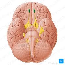
Cranial Nerve 1
Stripe on the bottom of the frontal lobe
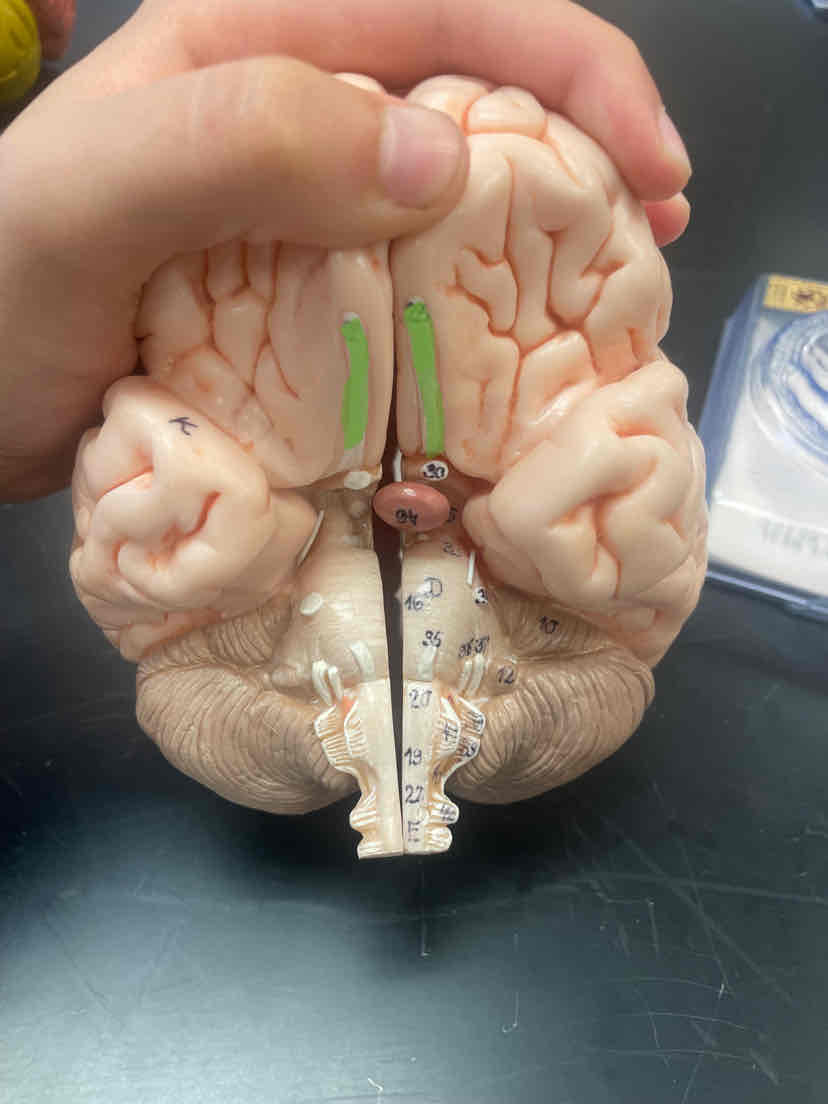
Olfactory bulb
Anterior bulb on the cranial nerve
Cribriform Plate
Things layer of bone that separates the nasal cavity and the Cranial Cavity
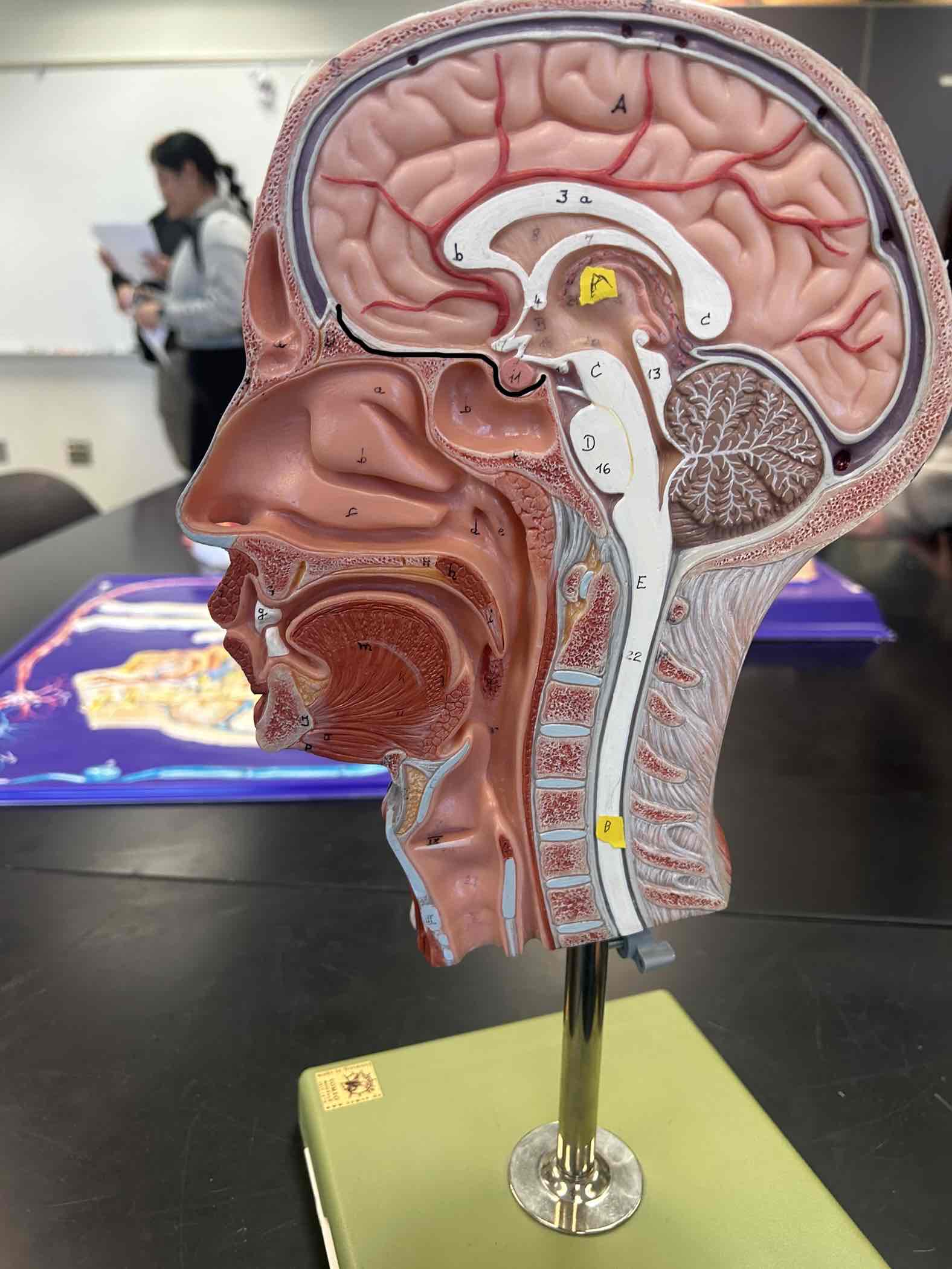
Nasal Cavities
Spaces inside of your nose
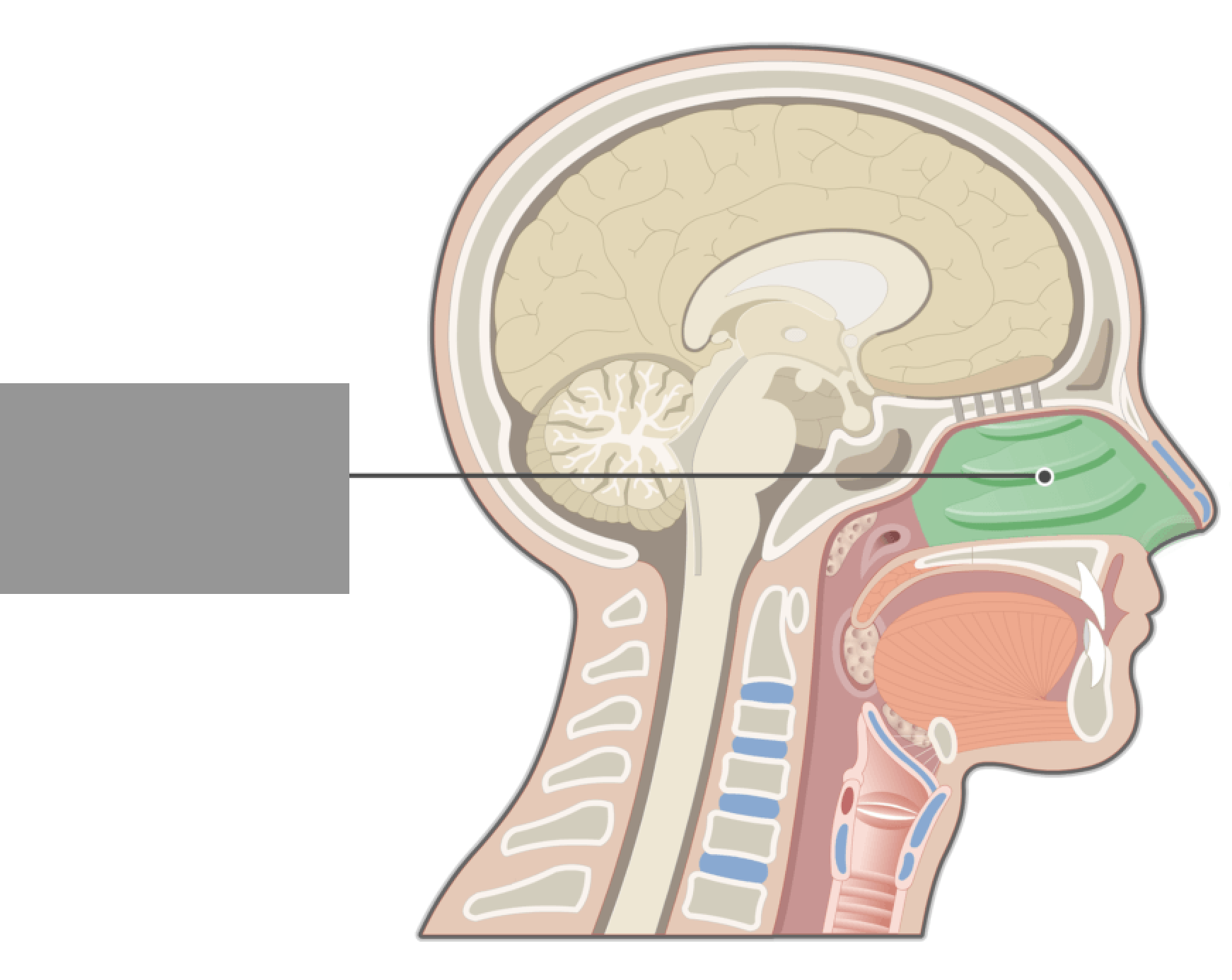
External Nares
Nostrils/ nose holes
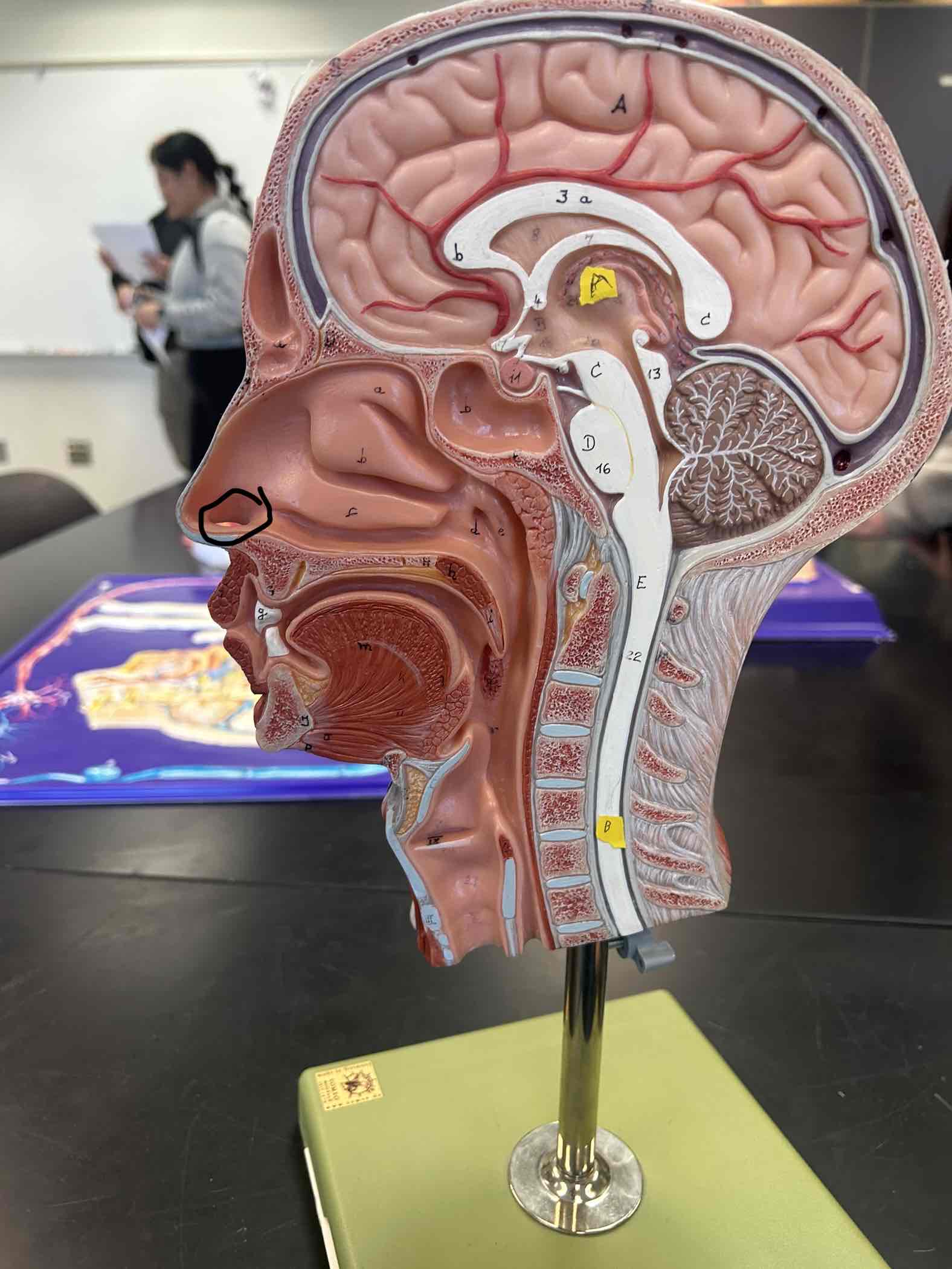
External Ear
has the funnel of cartilage and the ear canal
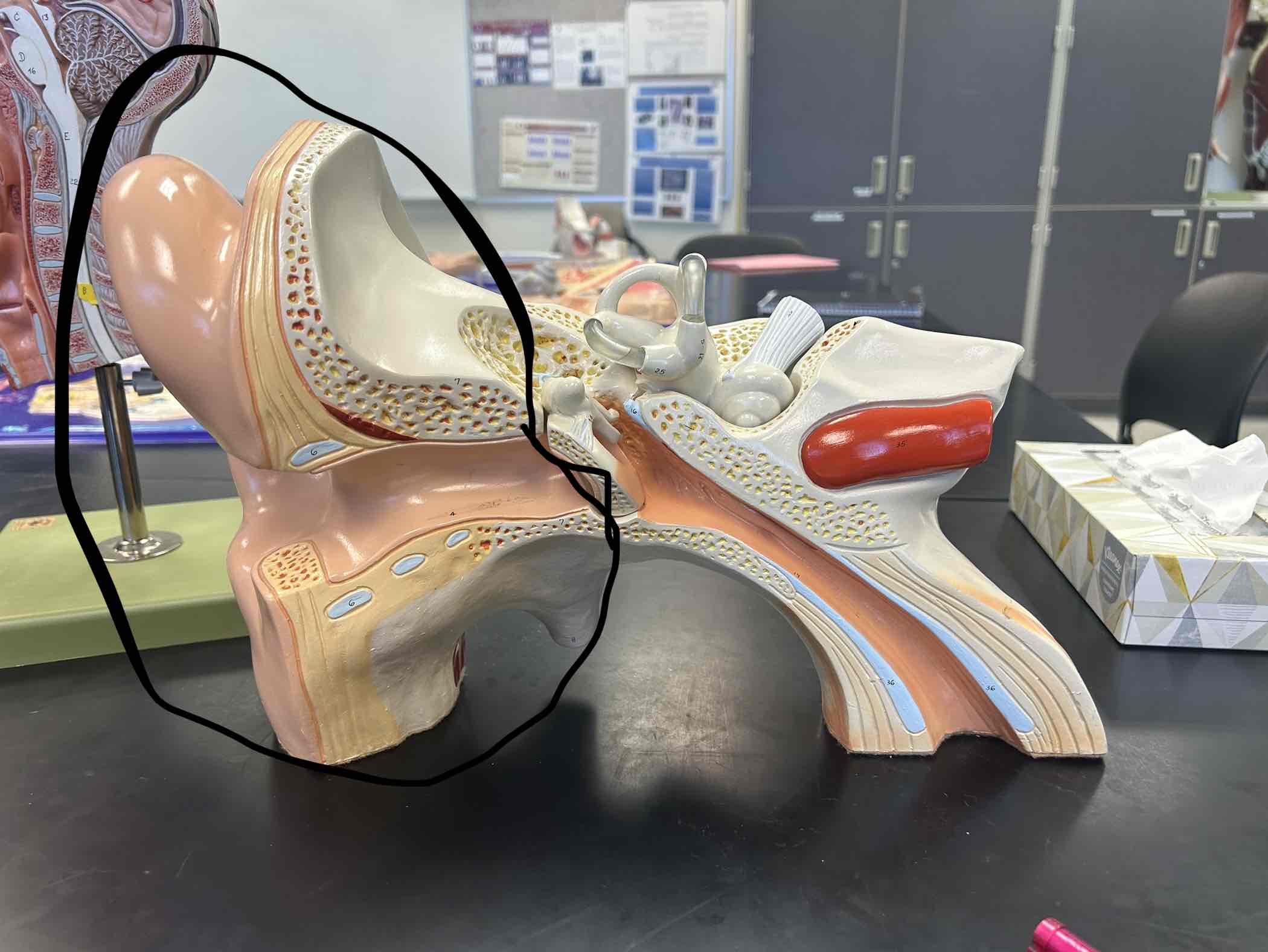
Middle Ear
See through Pieces and the 3 bones
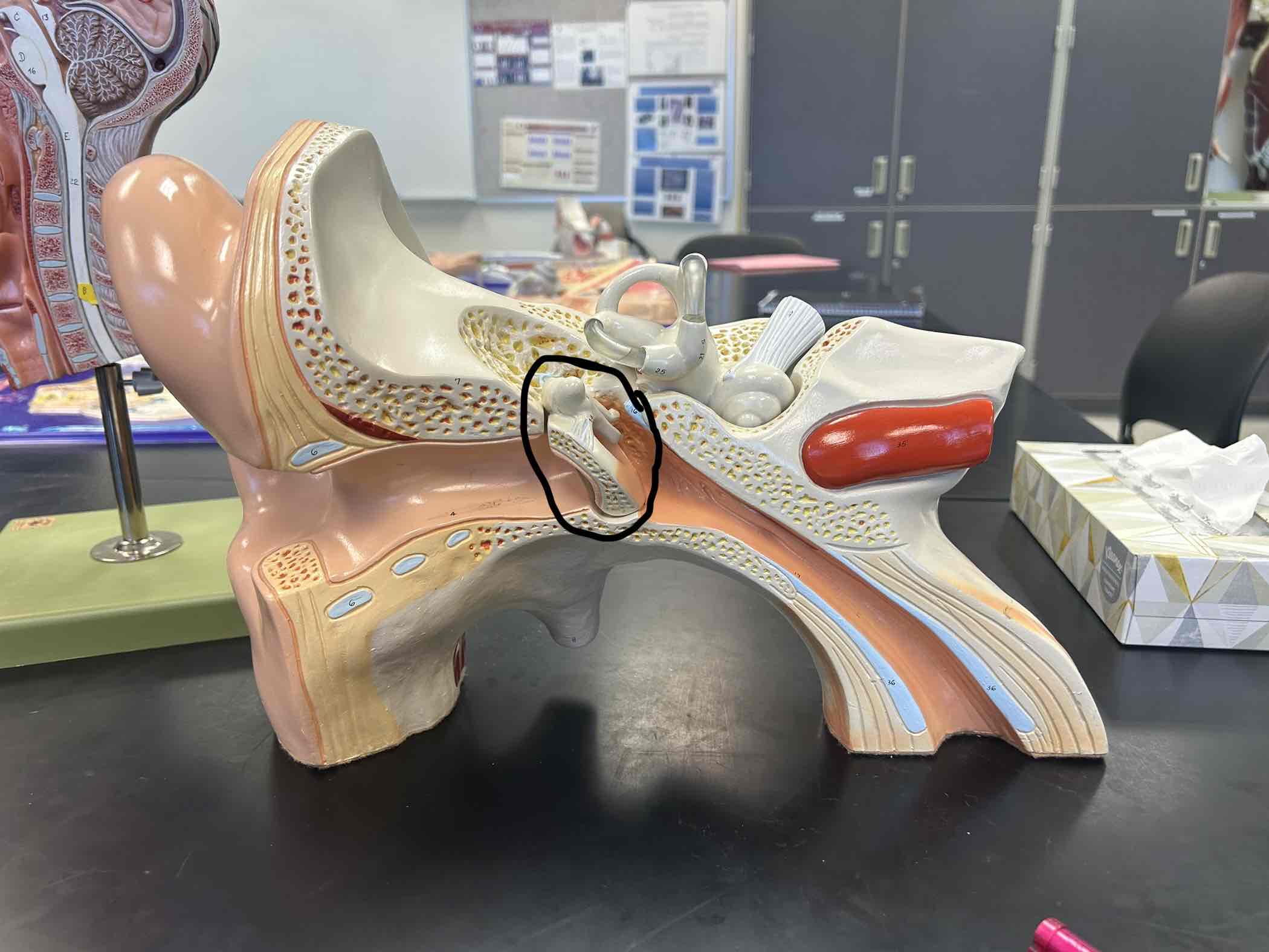
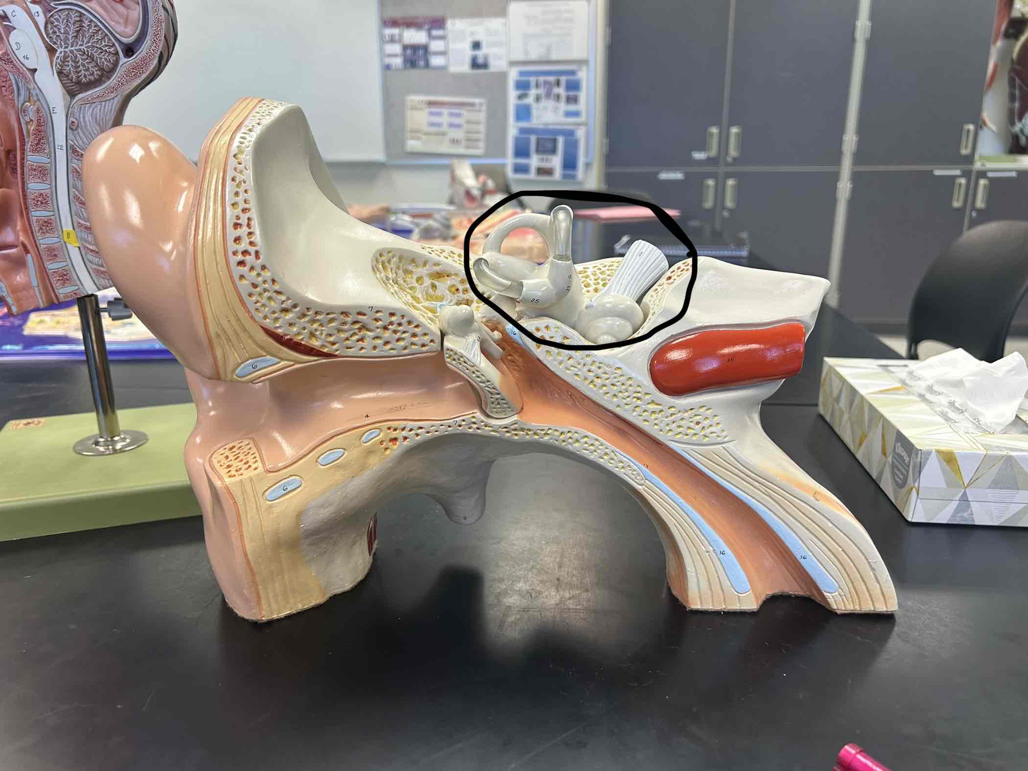
Inner Ear
Has 3 loops, snail shell, and nerve
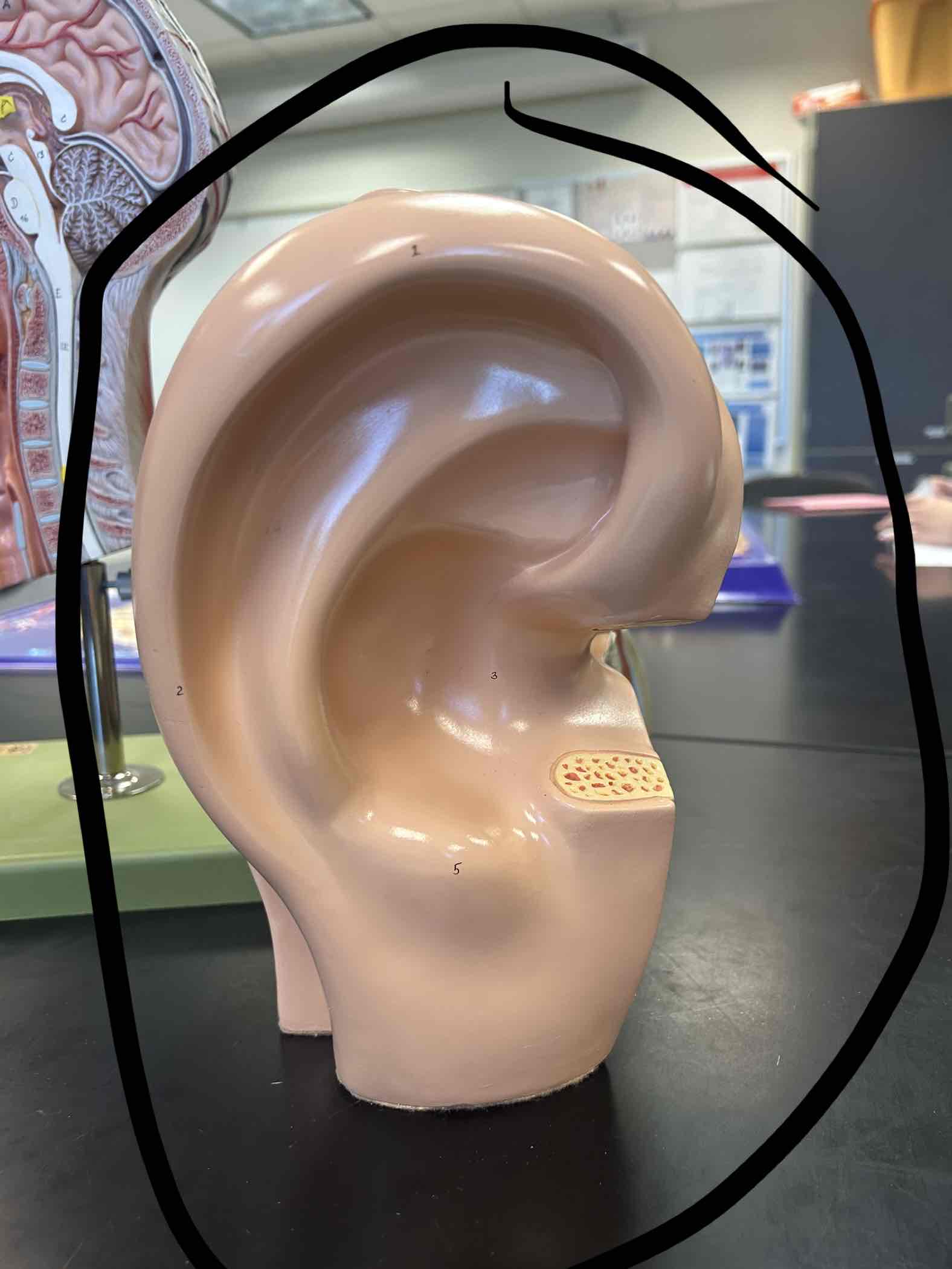
Auricle
External Funnel made of Cartilage
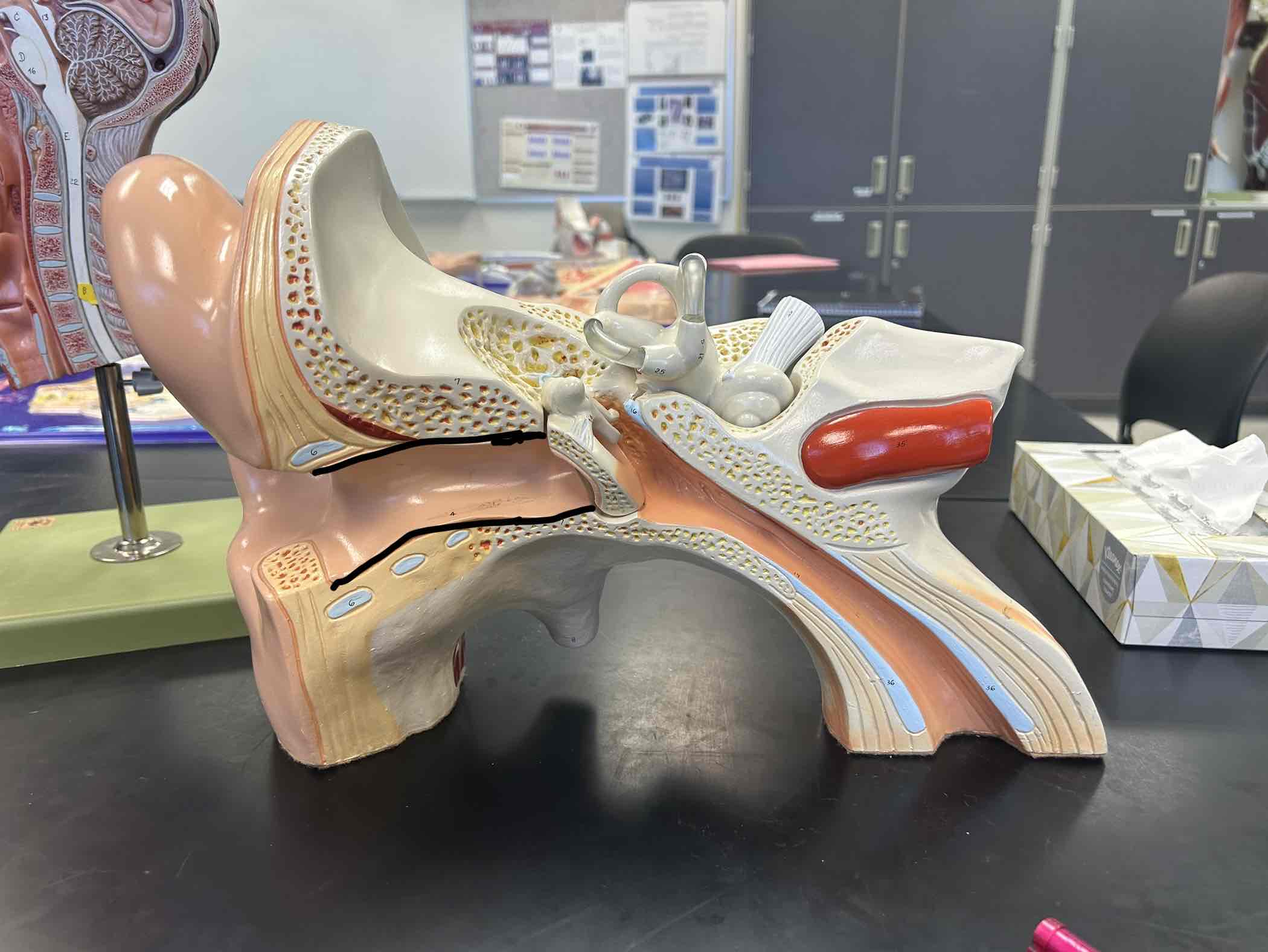
External Acoustic Meatus
hole in temporal bone for the ear canal
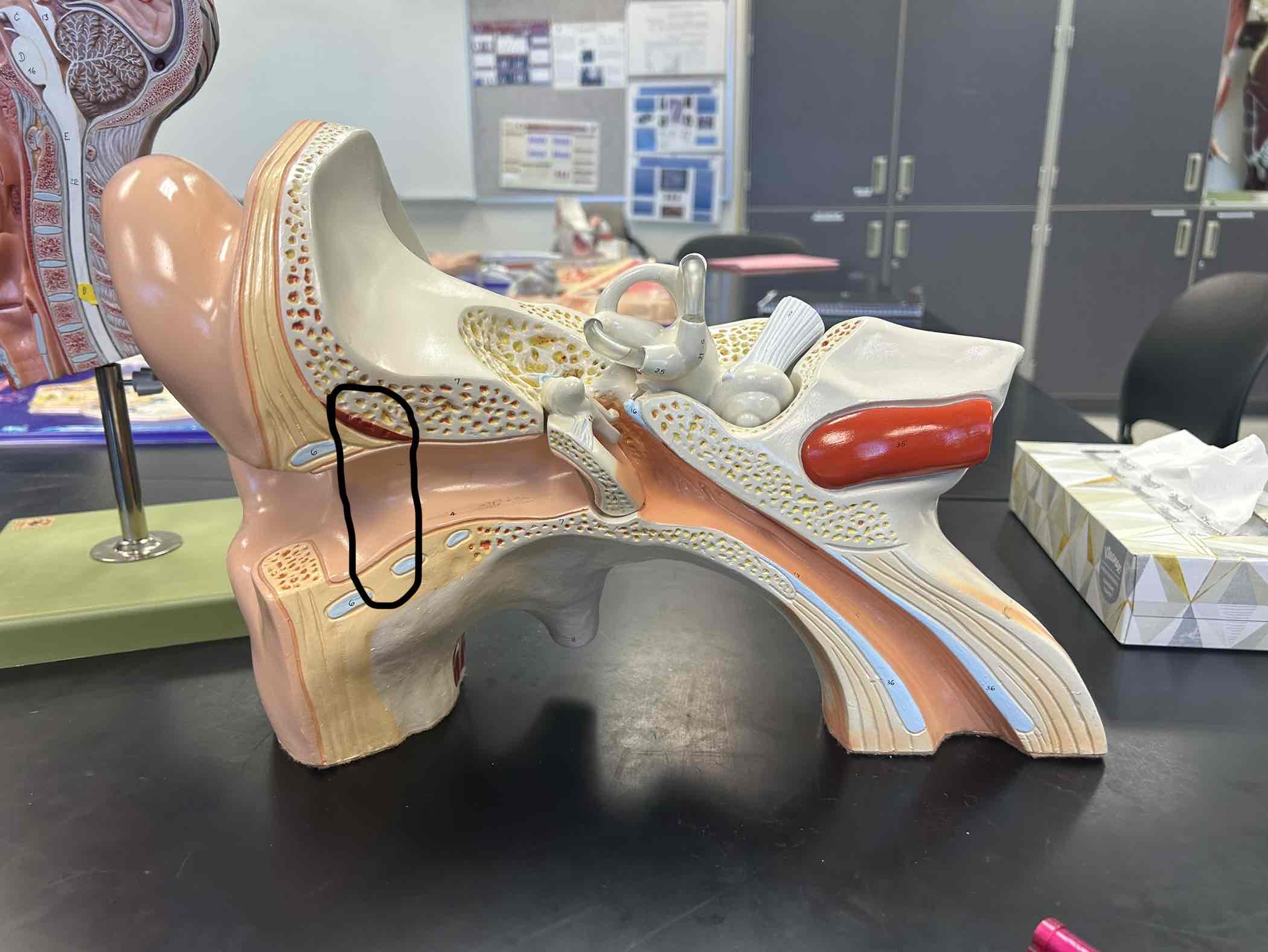
Auditory Canal
ear canal
Tympanic membrane
see through piece/ ear drum
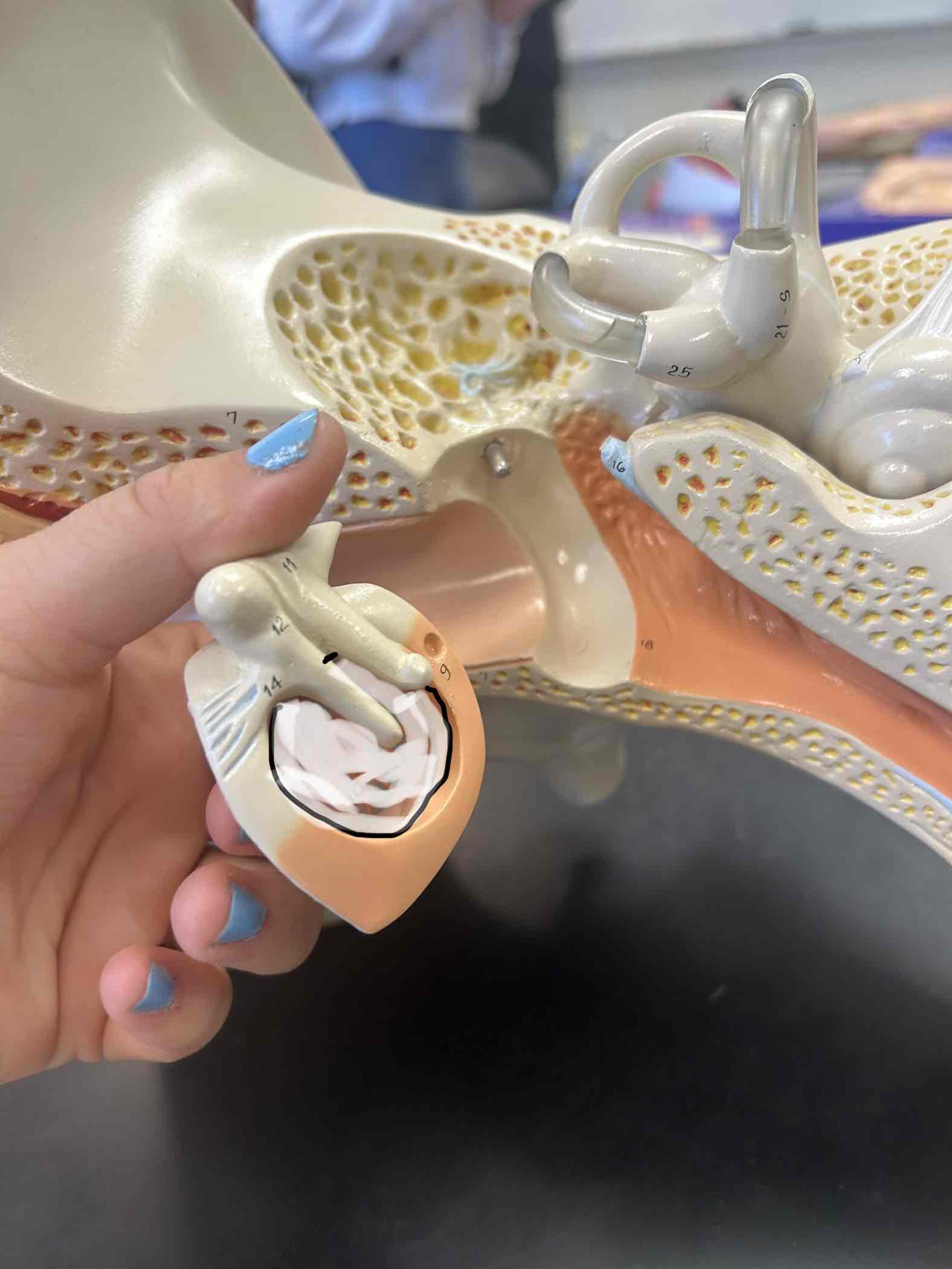
Auditory ossicles
3 tiny bones inside your ear
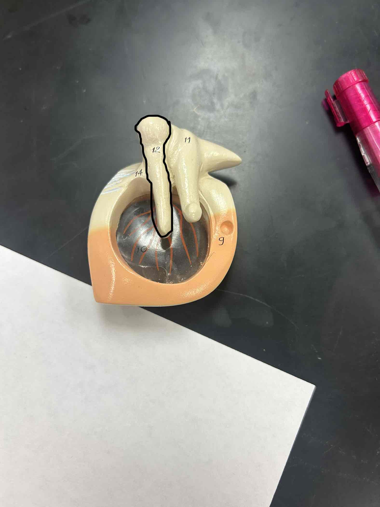
Malleus
connected directly to the Tympanic Membrane
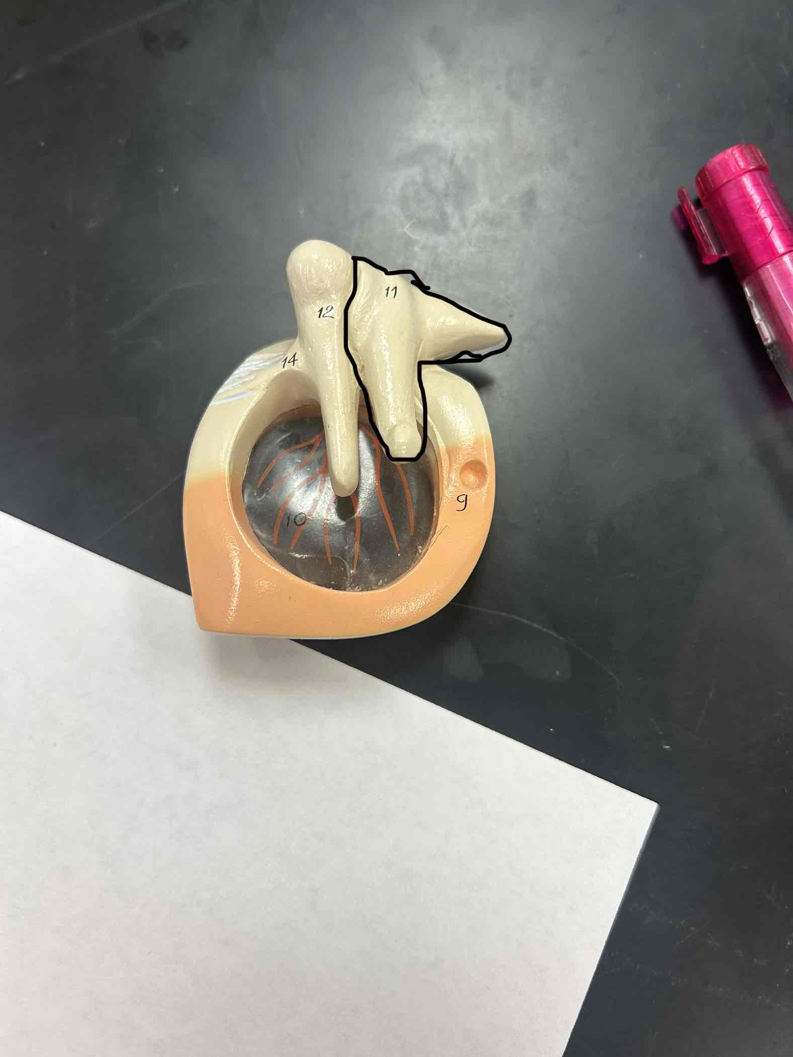
Incus
has the triangular or < coming off of it
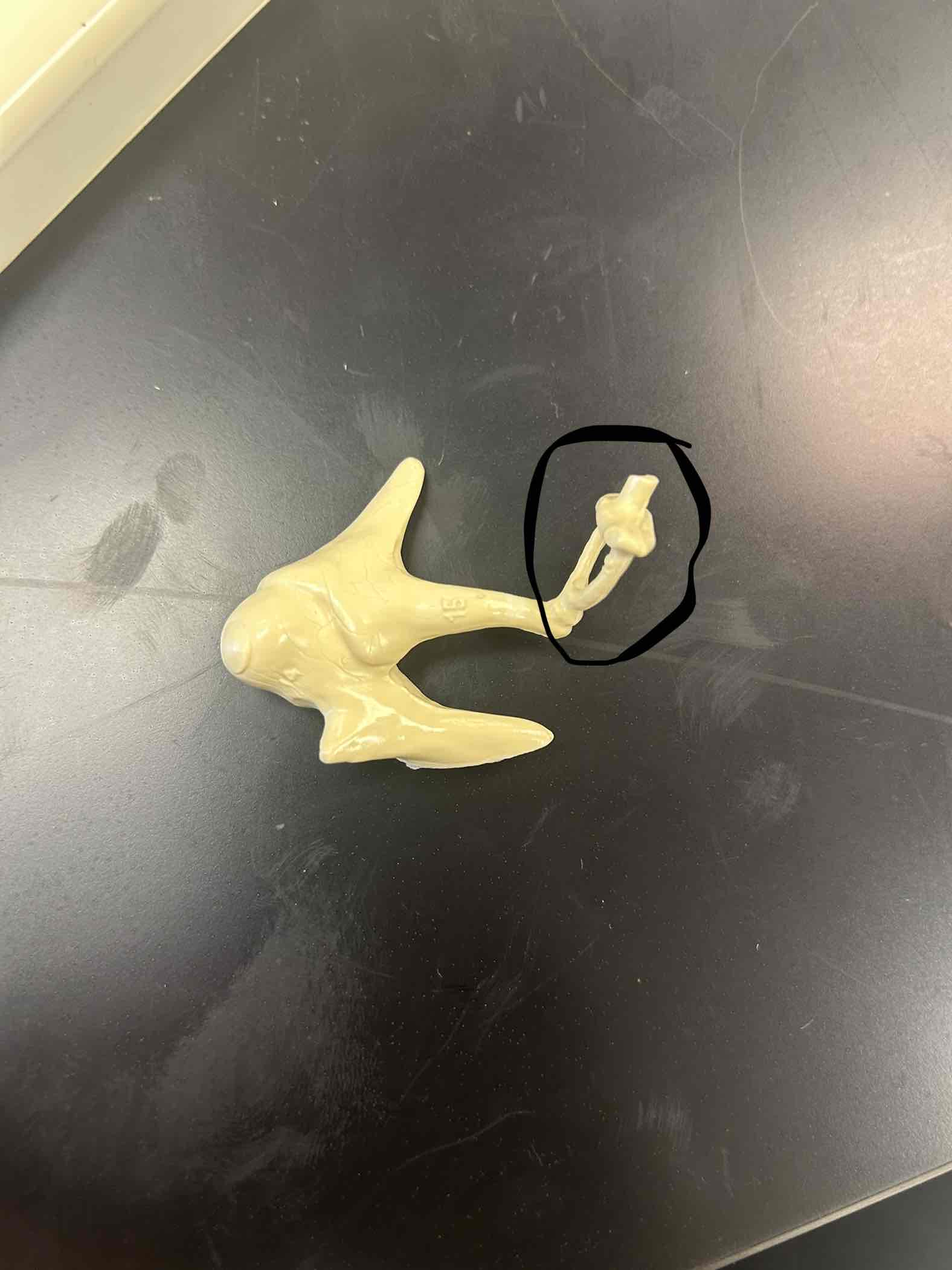
Stapes
Has the hole in the middle
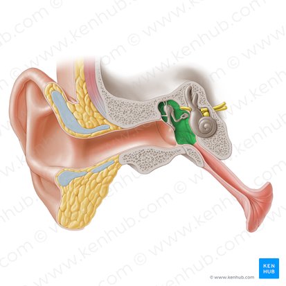
Tympanic Cavity
cavity behind the tympanic membrane
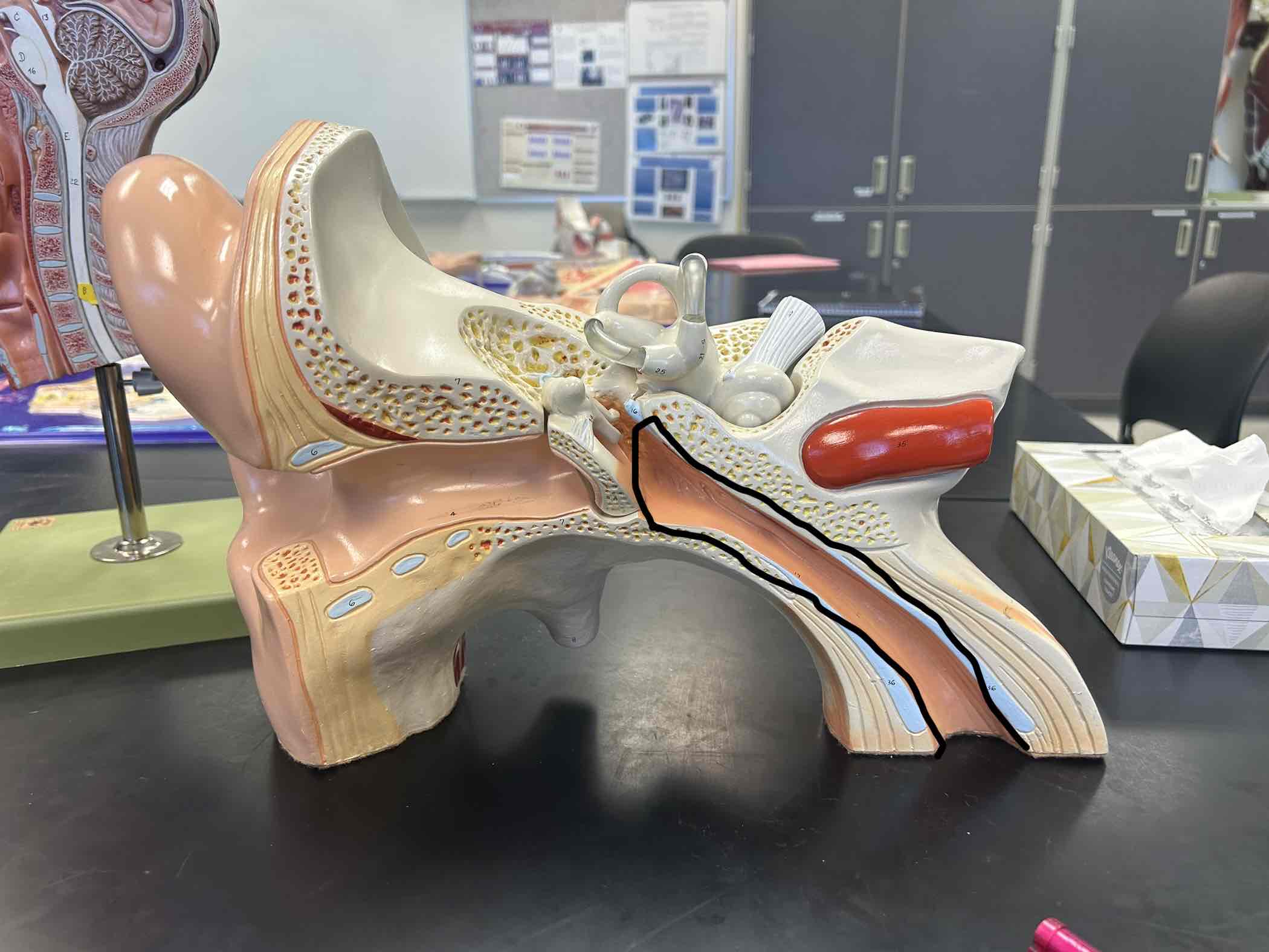
Auditory tube
Medial to the tympanic cavity
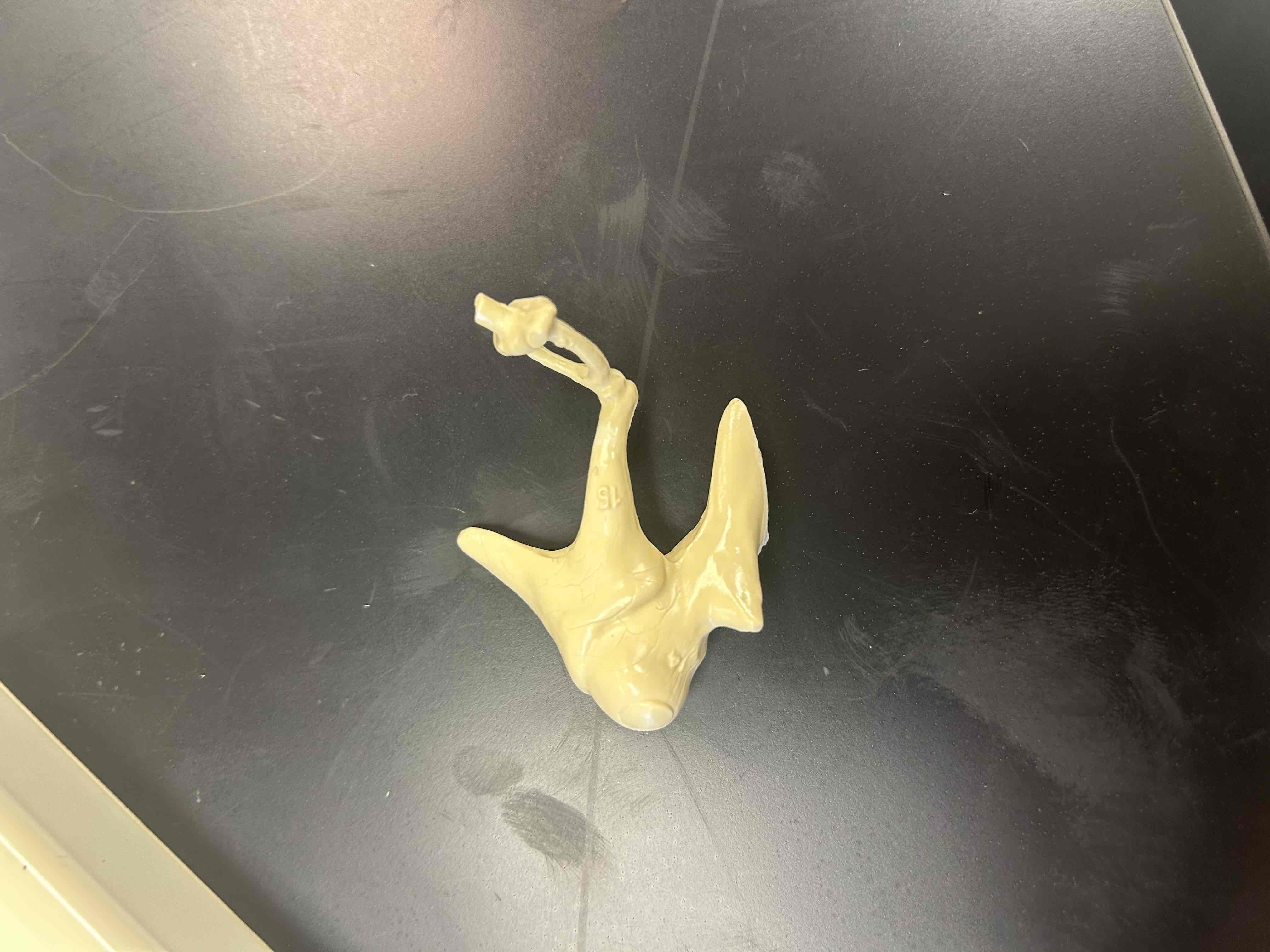
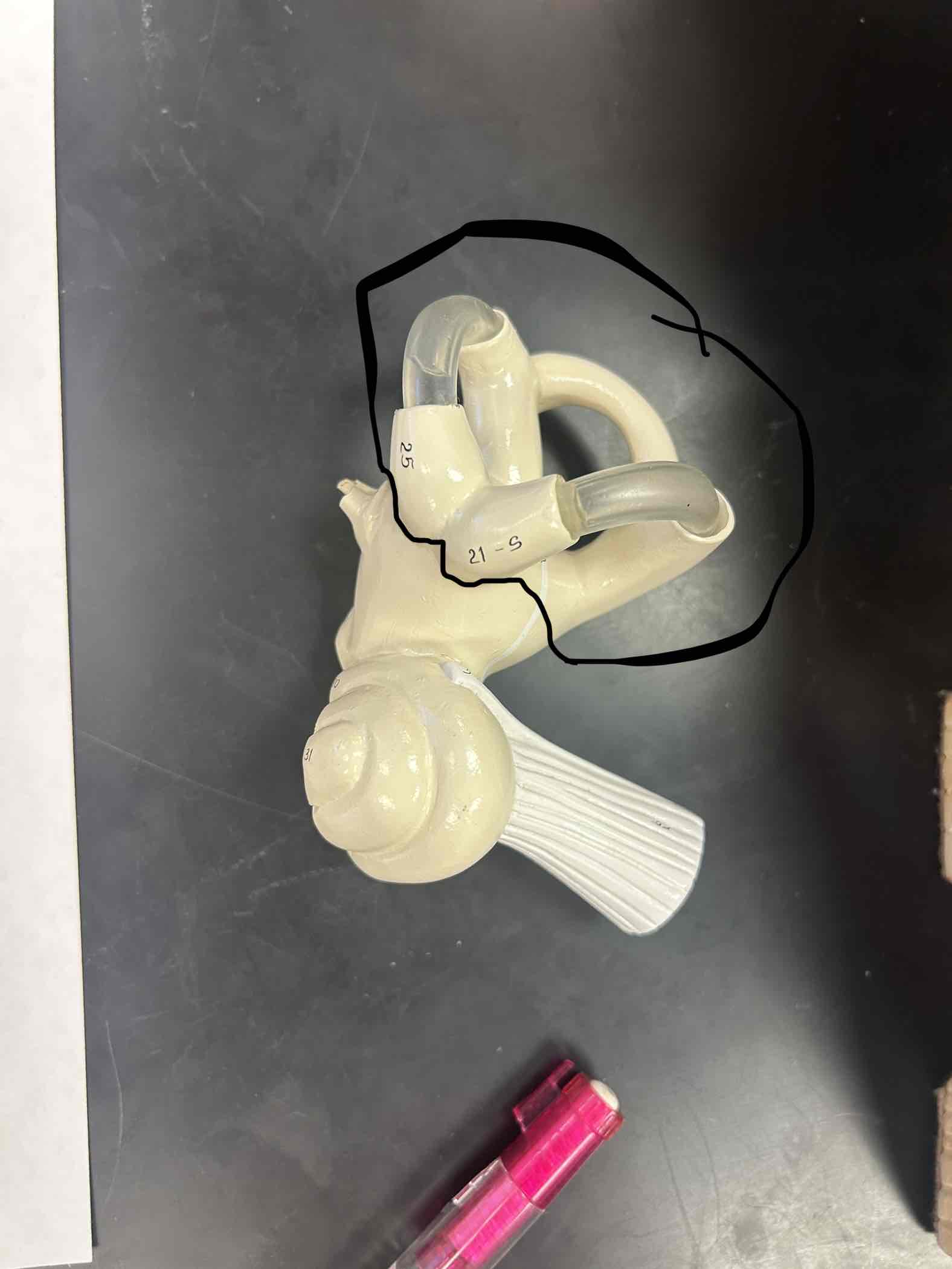
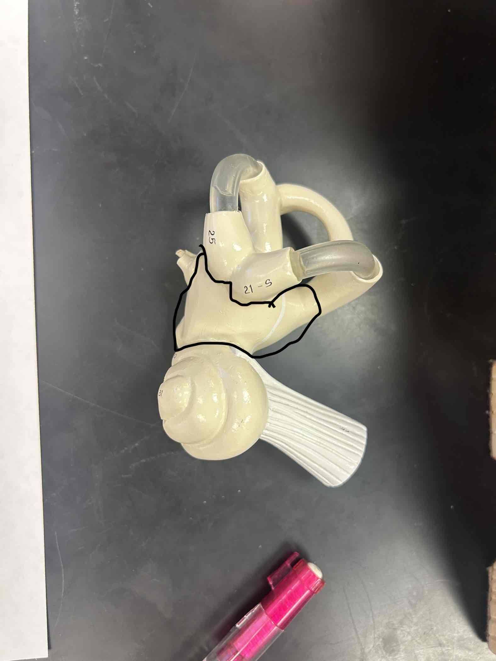
Semicircular canals
3 lopes of the inner ear
Vestibule
it is the object between the 3 lopes and the snail shell
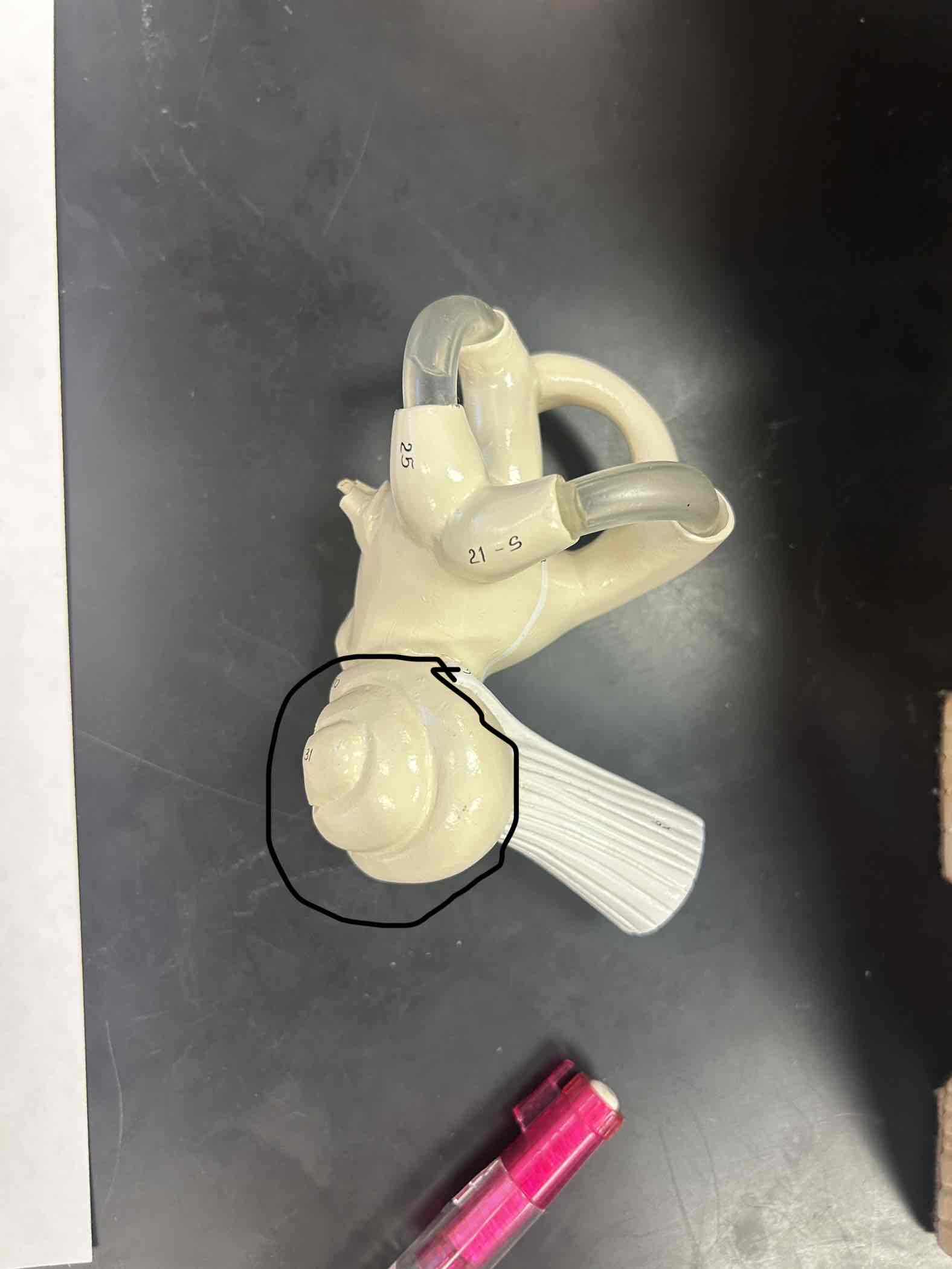
Cochlea
Snail shell looking thing in the inner ear
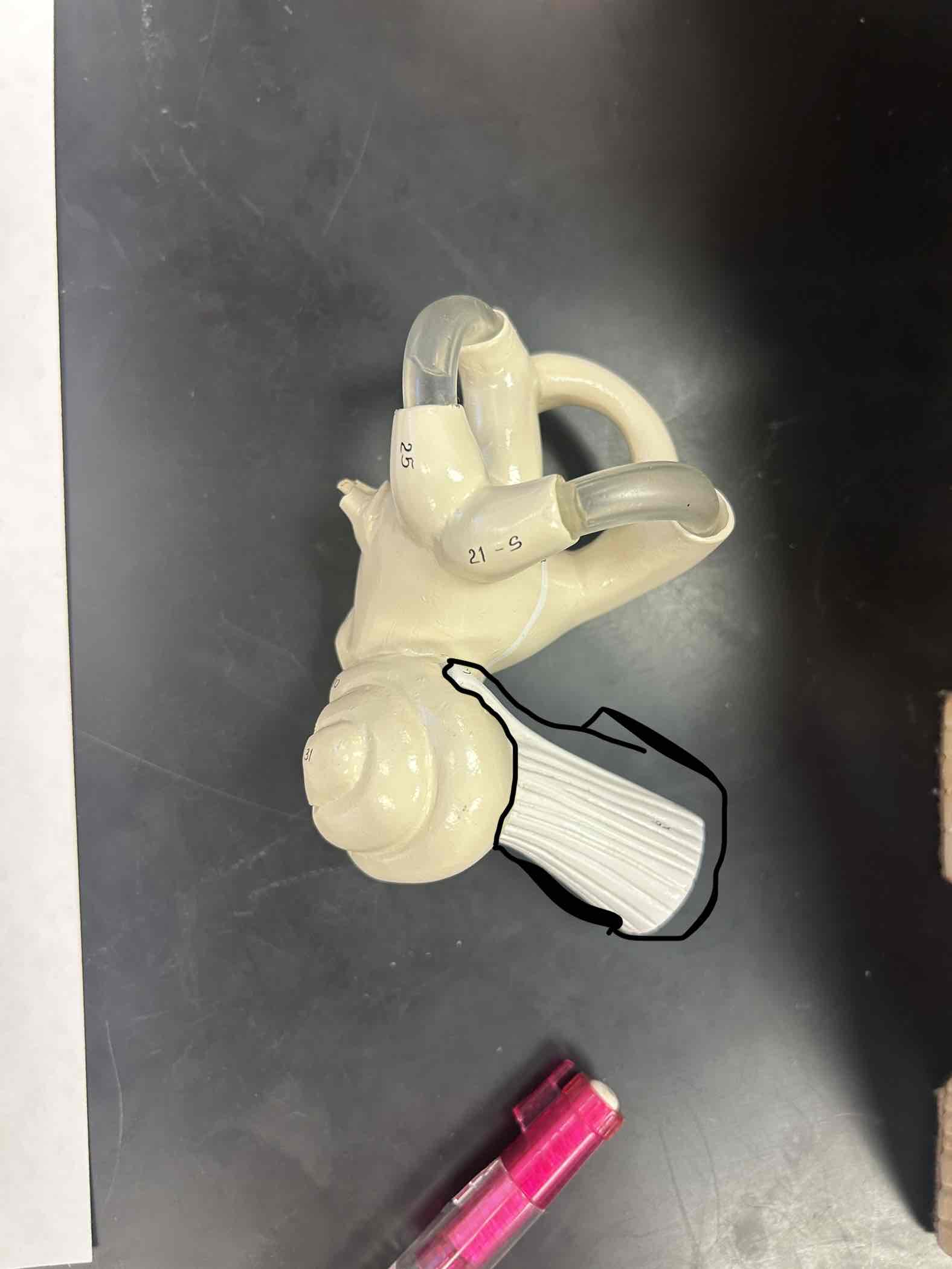
Cranial nerve 8
Connected to the snail shell
Extrinsic eye muscles
Muscles that are connected to the eye, there are 6 of them
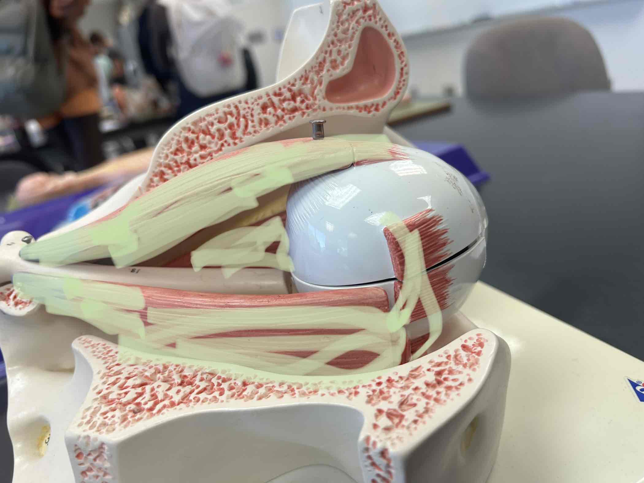
Medial Rectus
Connects from the posterior orbit to the Medial eye
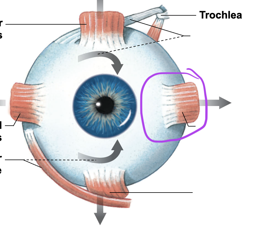
Lateral Rectus
Connects from the posterior orbit to the lateral eye
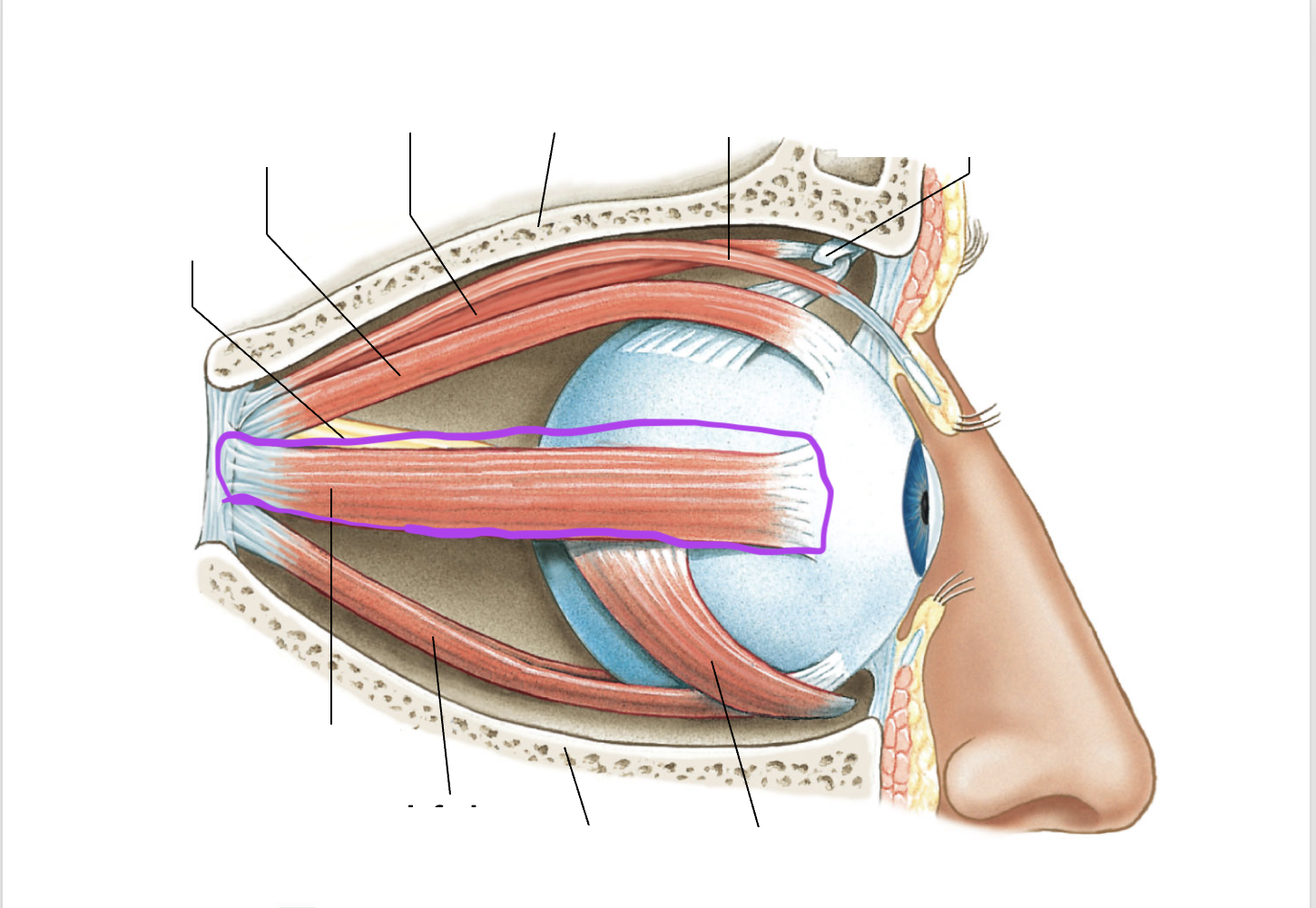
superior rectus
Connects from the posterior orbit to the superior eye
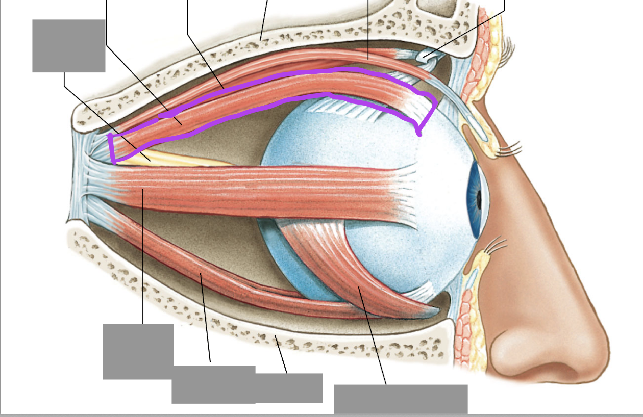
inferior rectus
Connects from the posterior orbit to the inferior eye
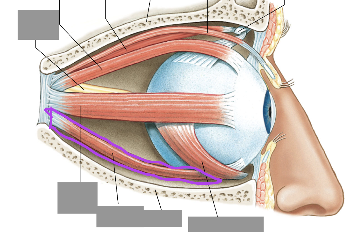
Superior oblique
Connects from the posterior orbit to the lateral posterior eye
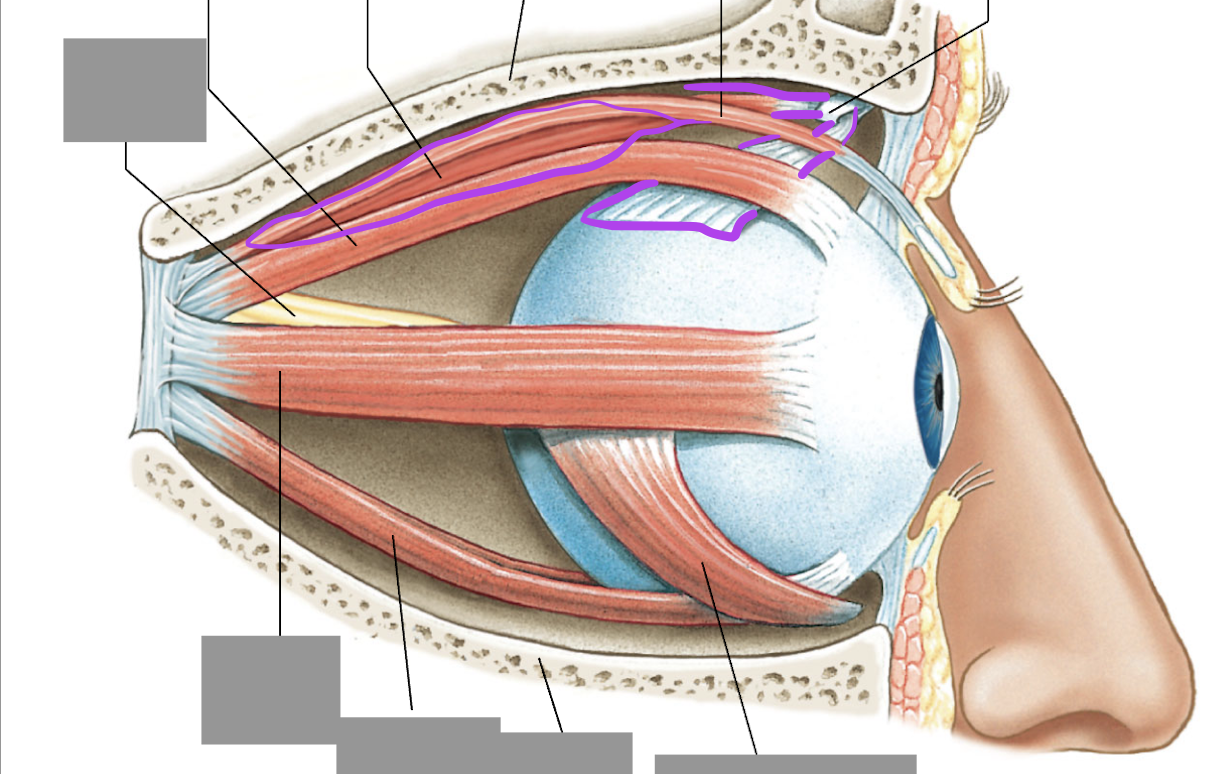
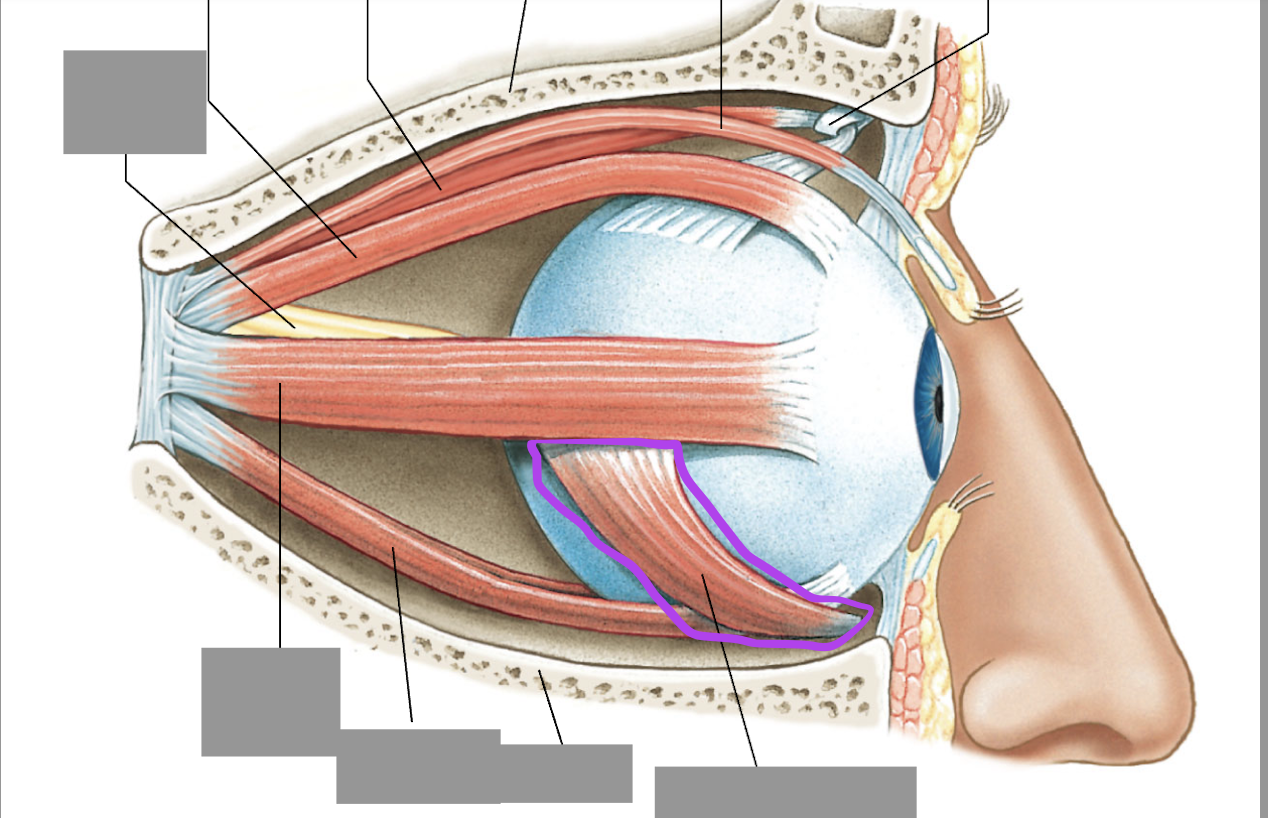
Inferior Oblique
connects from the medial inferior orbit to the lateral posterior eye
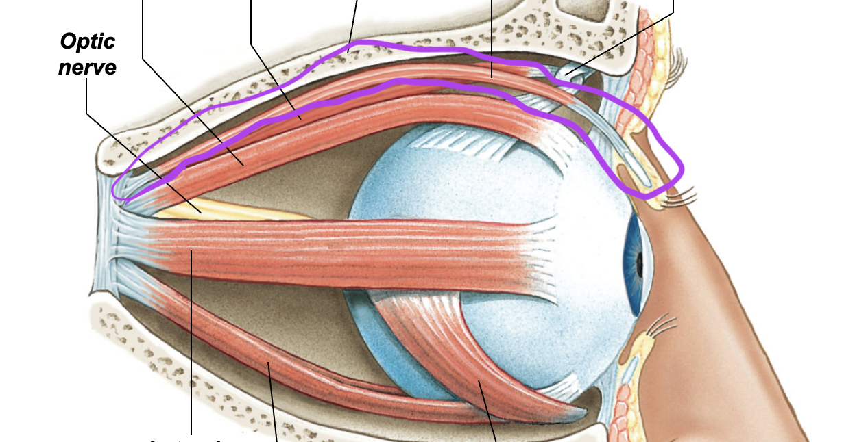
Levator palpebrae superorius
Superior to the superior rectus
fibrous tunic
outermost layer of eye tissue
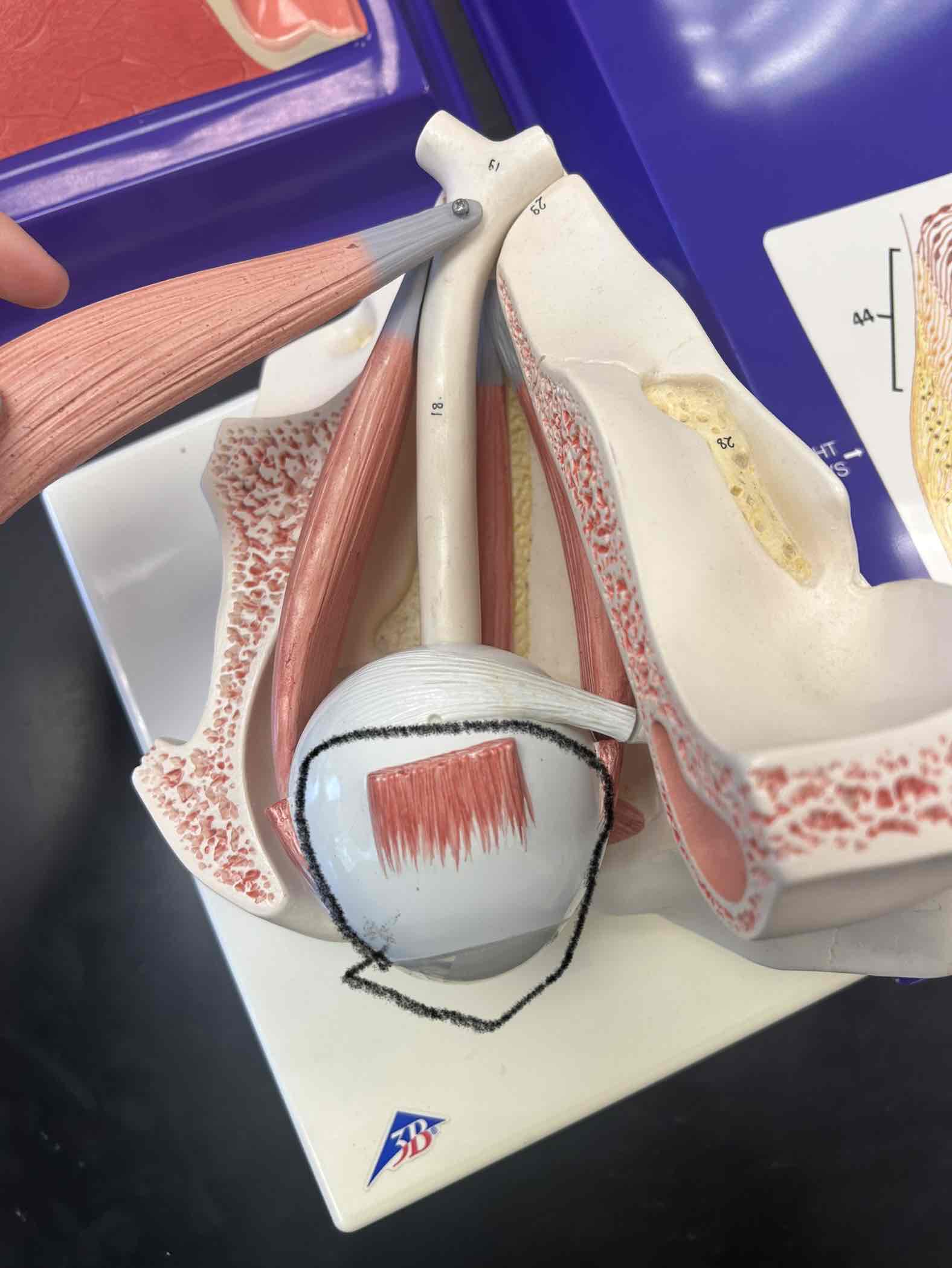
Sclera
white material of eye
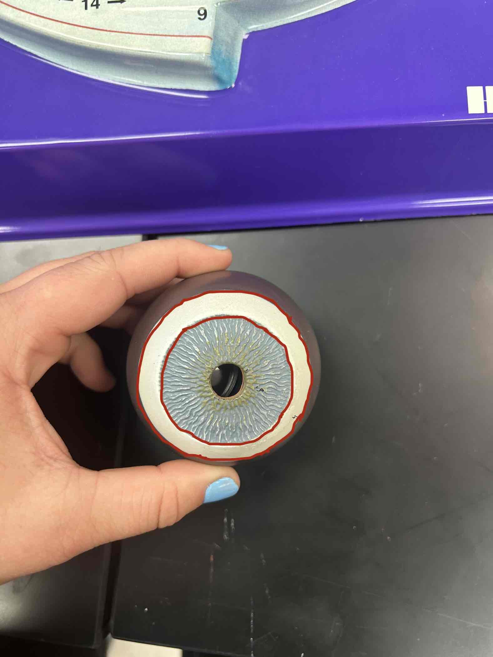
Cornea
Clear thing that covers the pupil
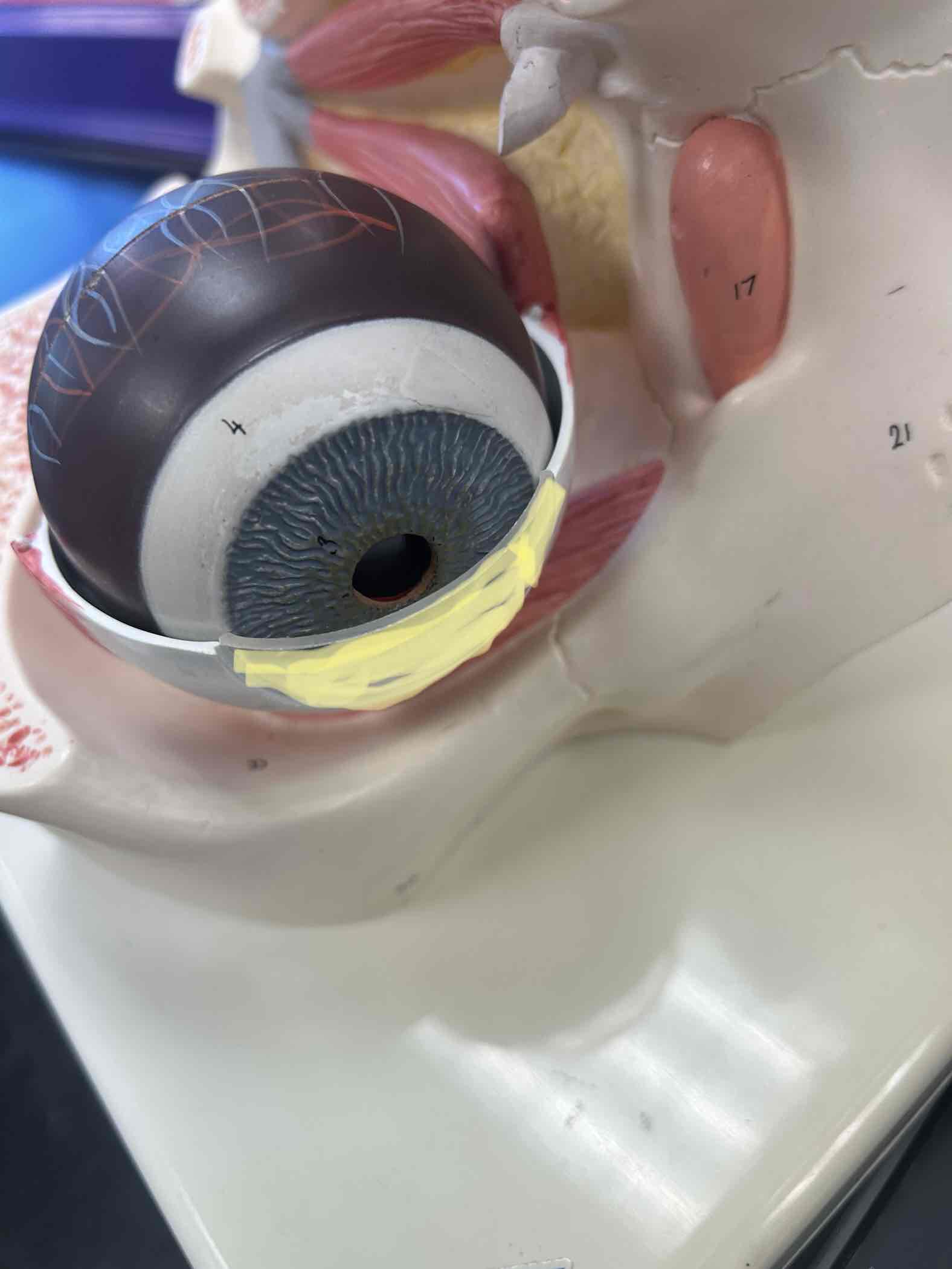
Vascular tunic
Middle layer of eye tissue
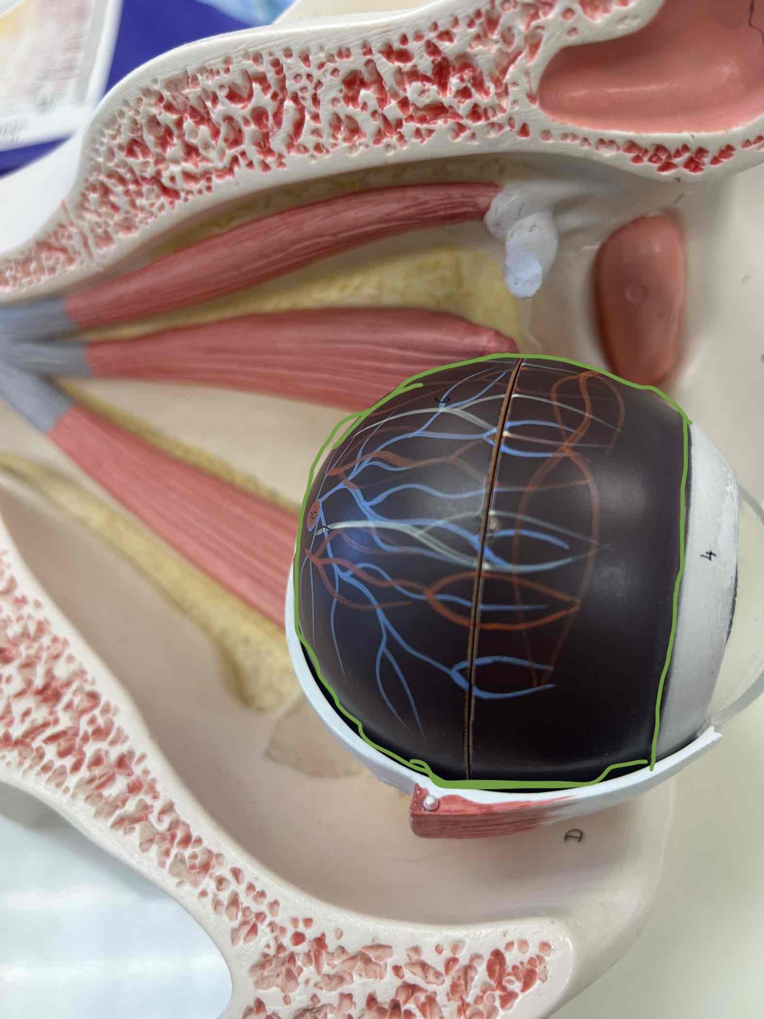
Choroid
has nerve & blood vessels on it
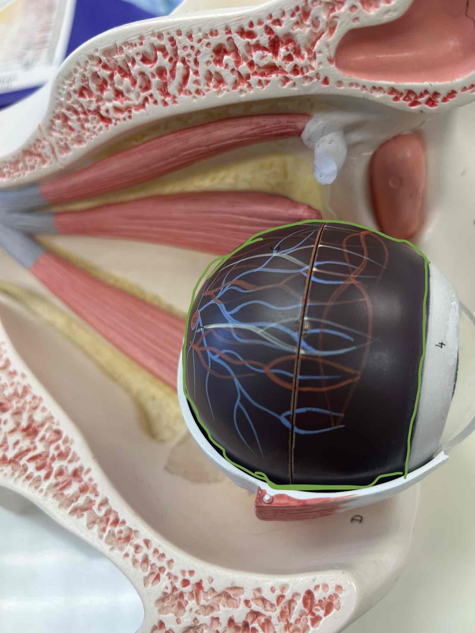
Iris
Colored part of the eye
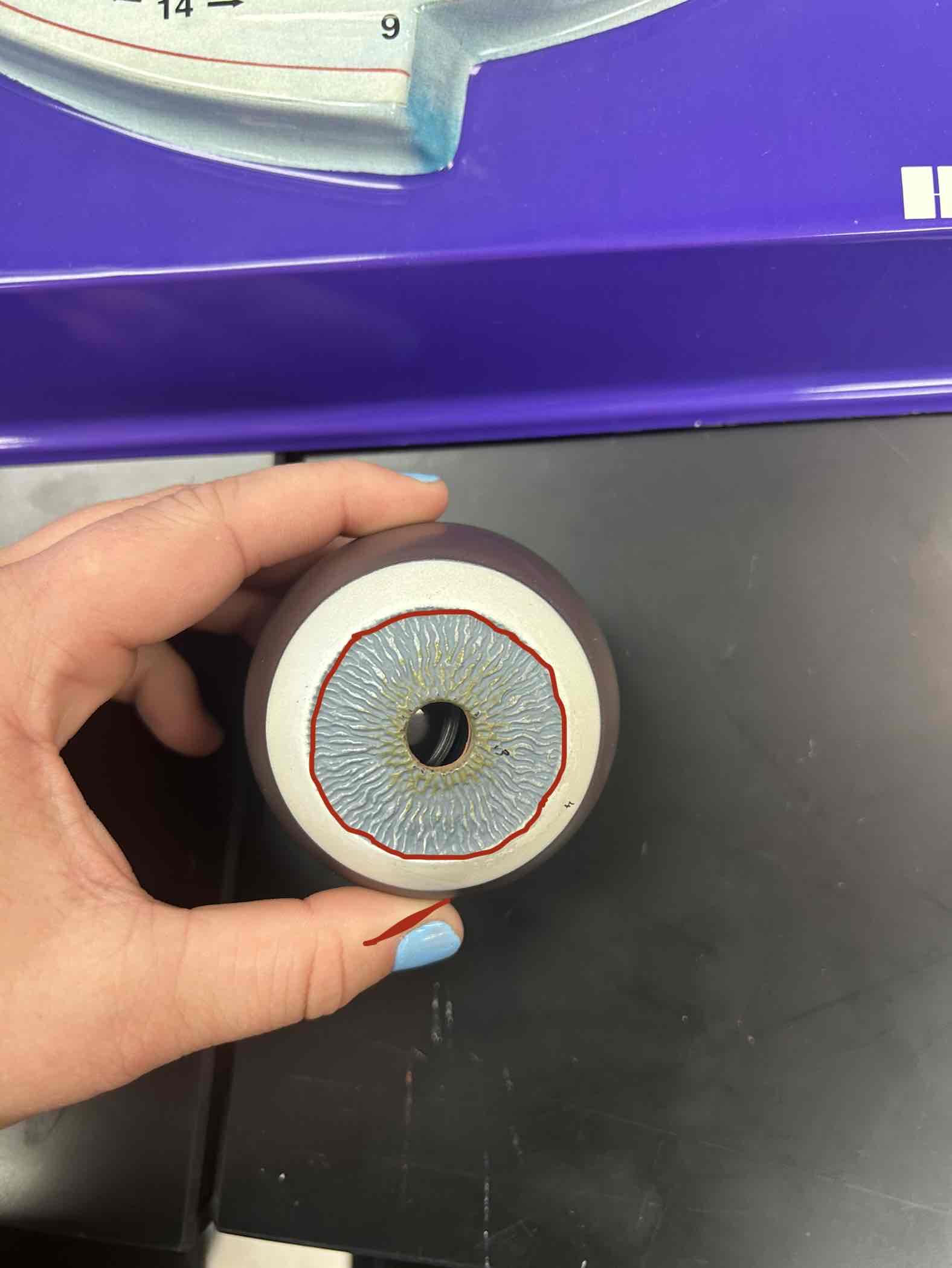
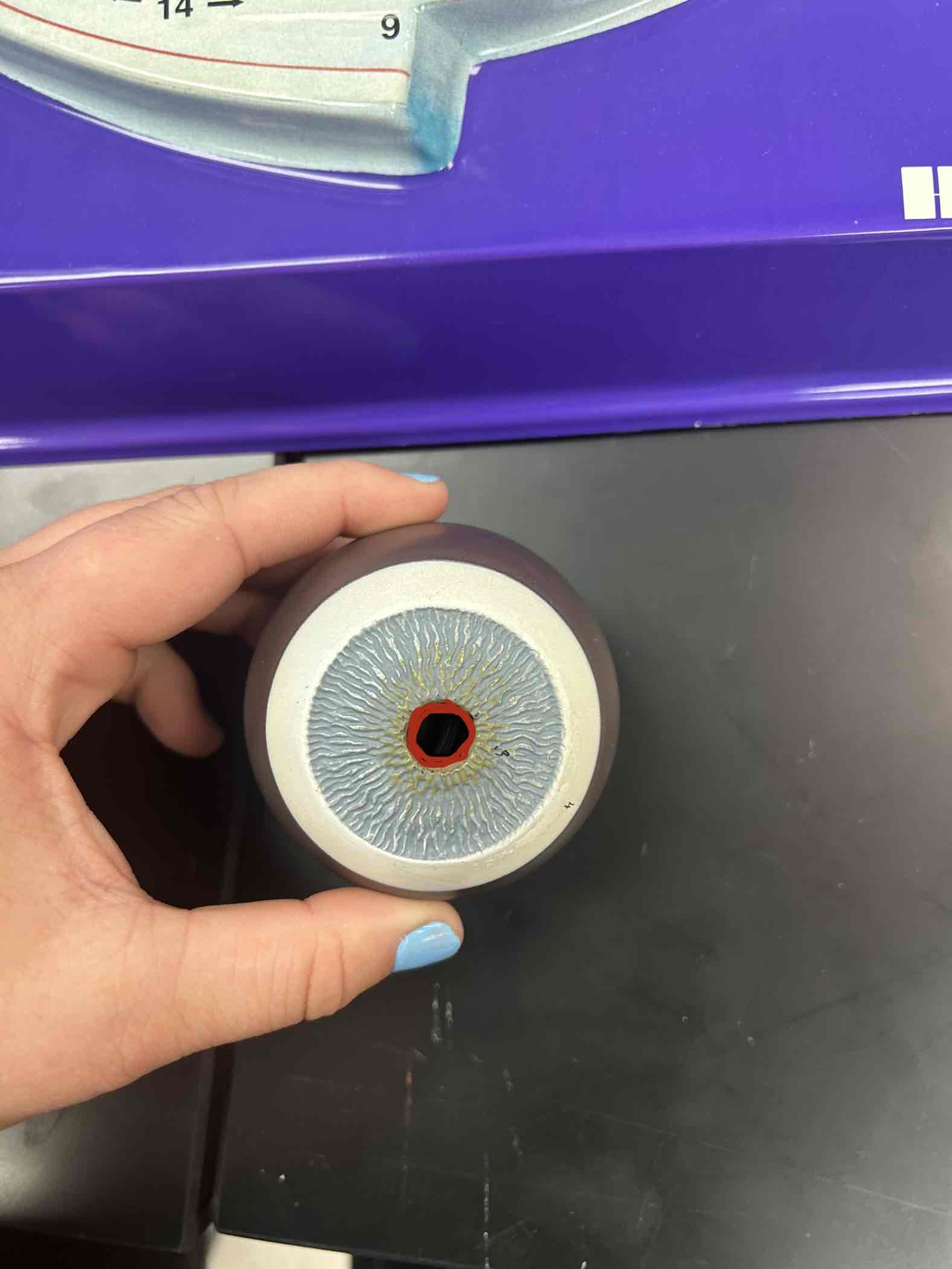
Pupil
Black hole in the middle of the eye
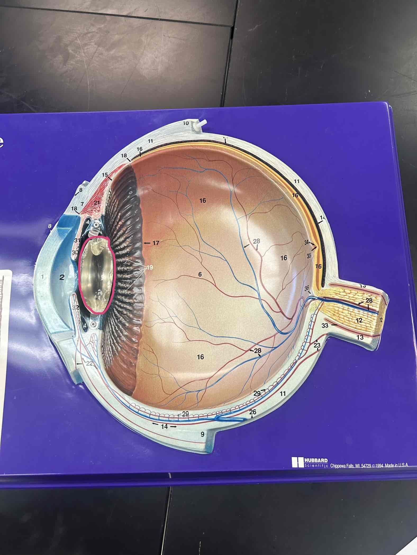
Lens
Clear colorless part inside of the pupil
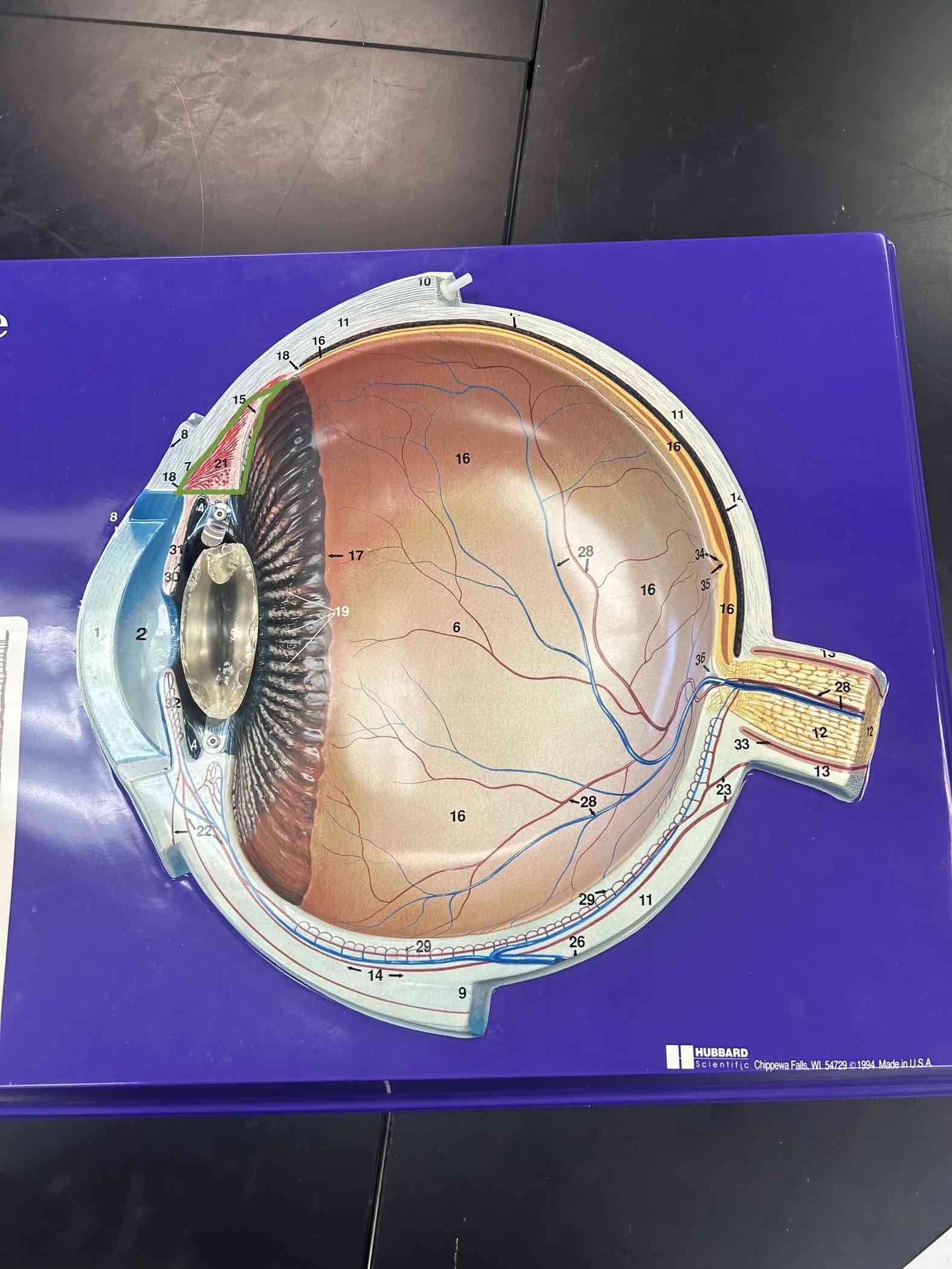
Ciliary body
ring of muscle around lens
Suspensory ligaments
attach ciliary body to lens
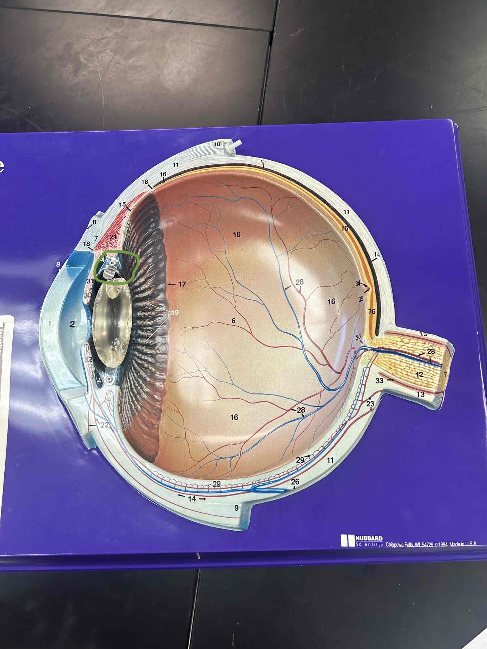
Nervous tunic
Deepest layer of tissue
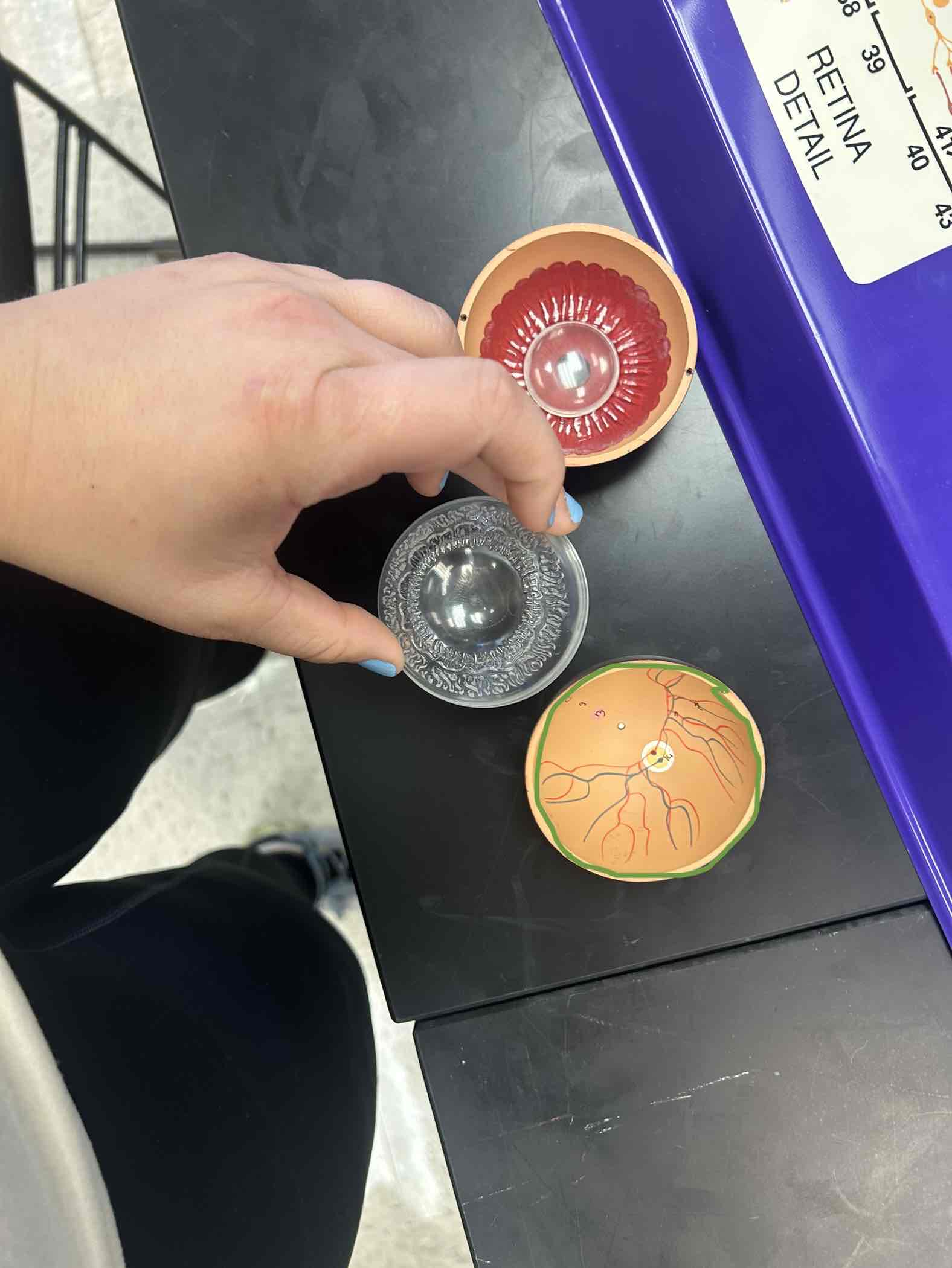
Retina
inner part of the eye, has cells that can detect radiation
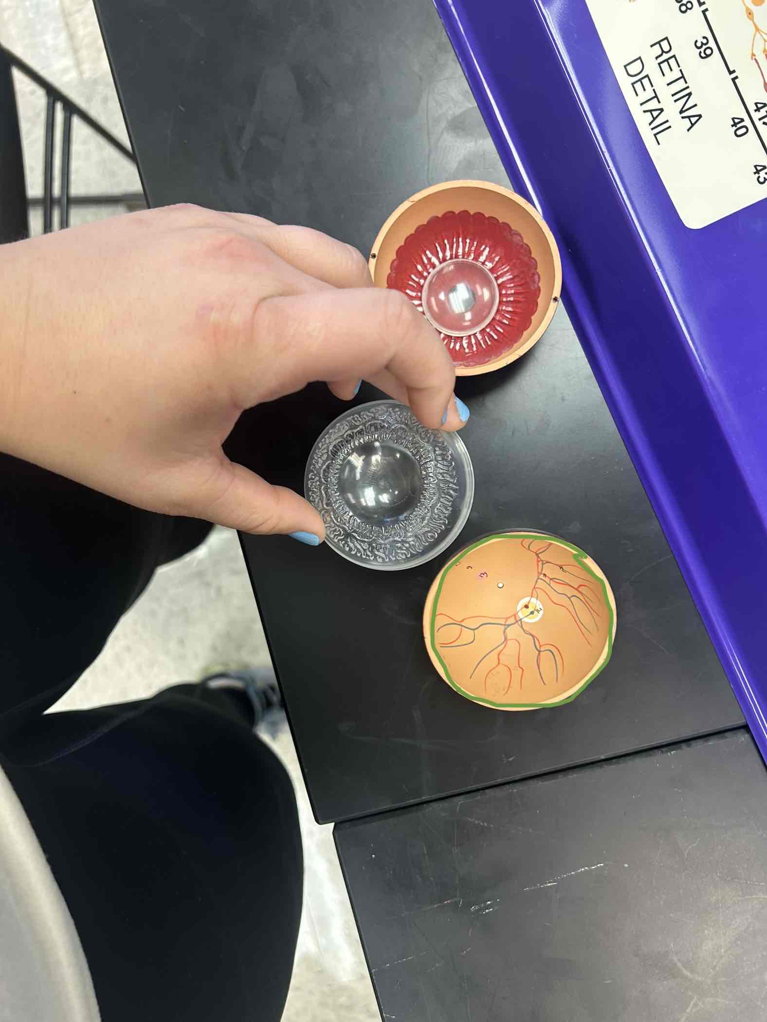
Fovea centralis
Indentation on the back of the eye, but superior to the Optic Disk
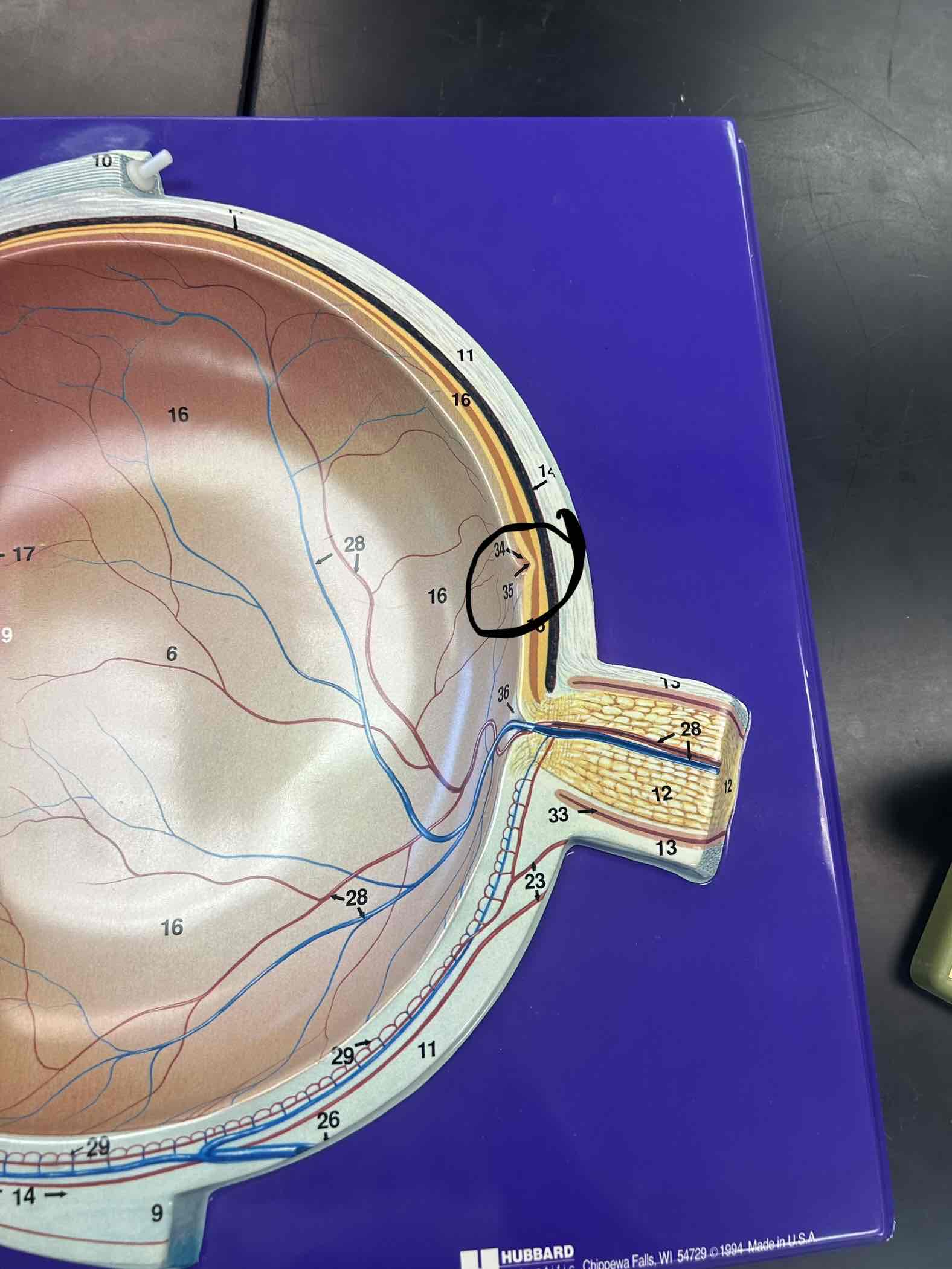
Optic Disk
Blind spot in the back, indentations
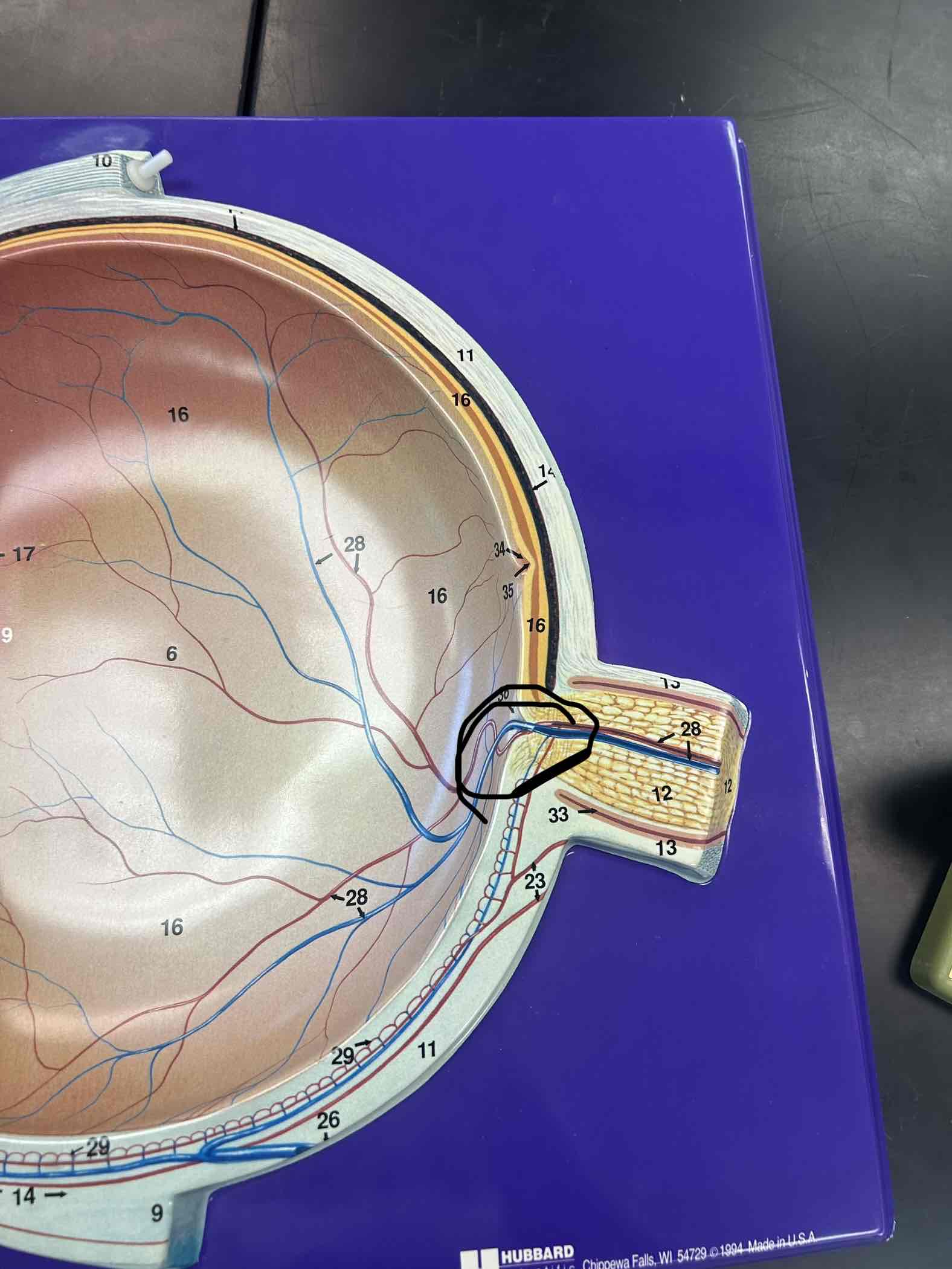
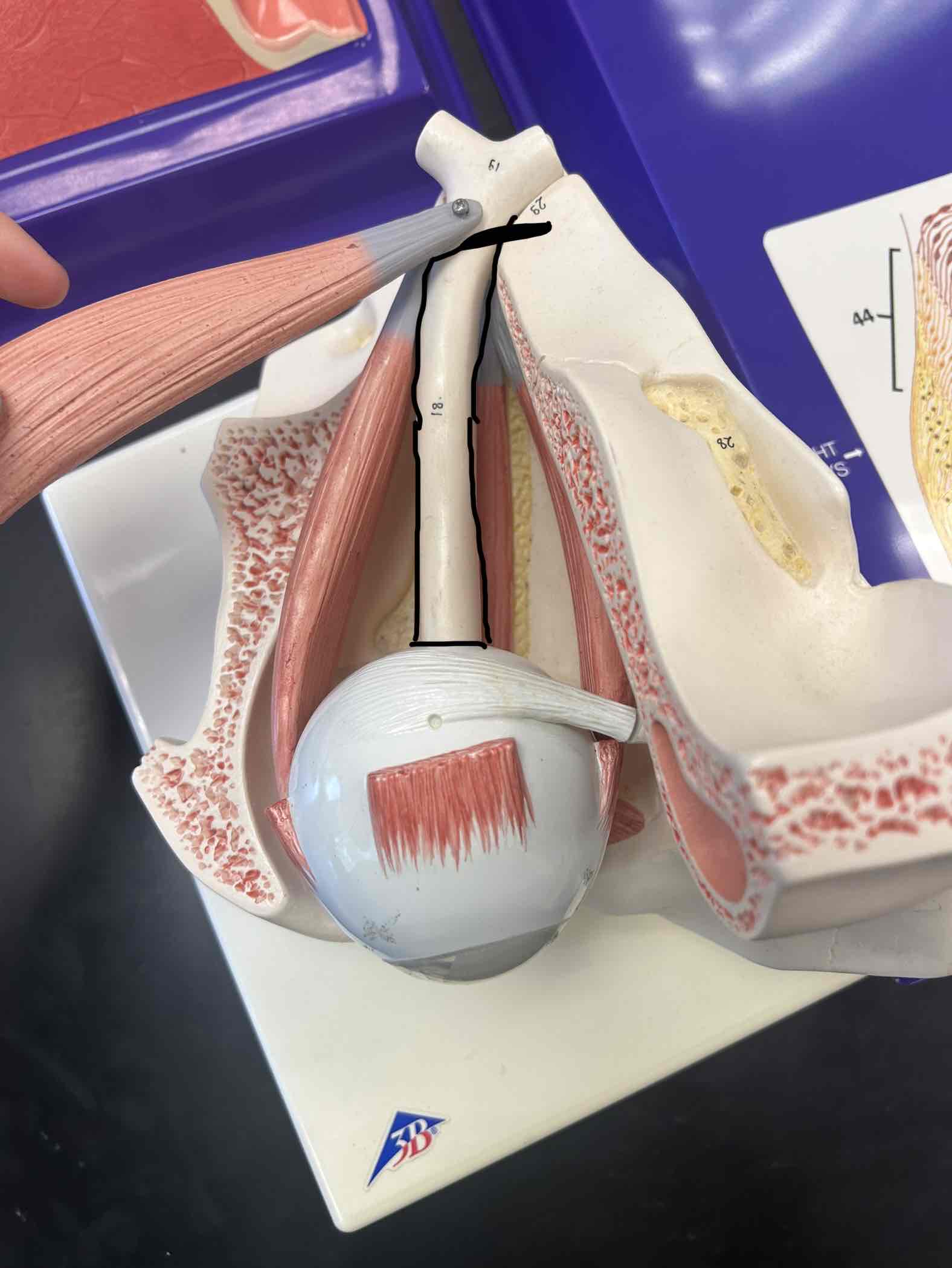
Cranial Nerve 2
Stem that connects to the optic disk on the posterior portion of the eye
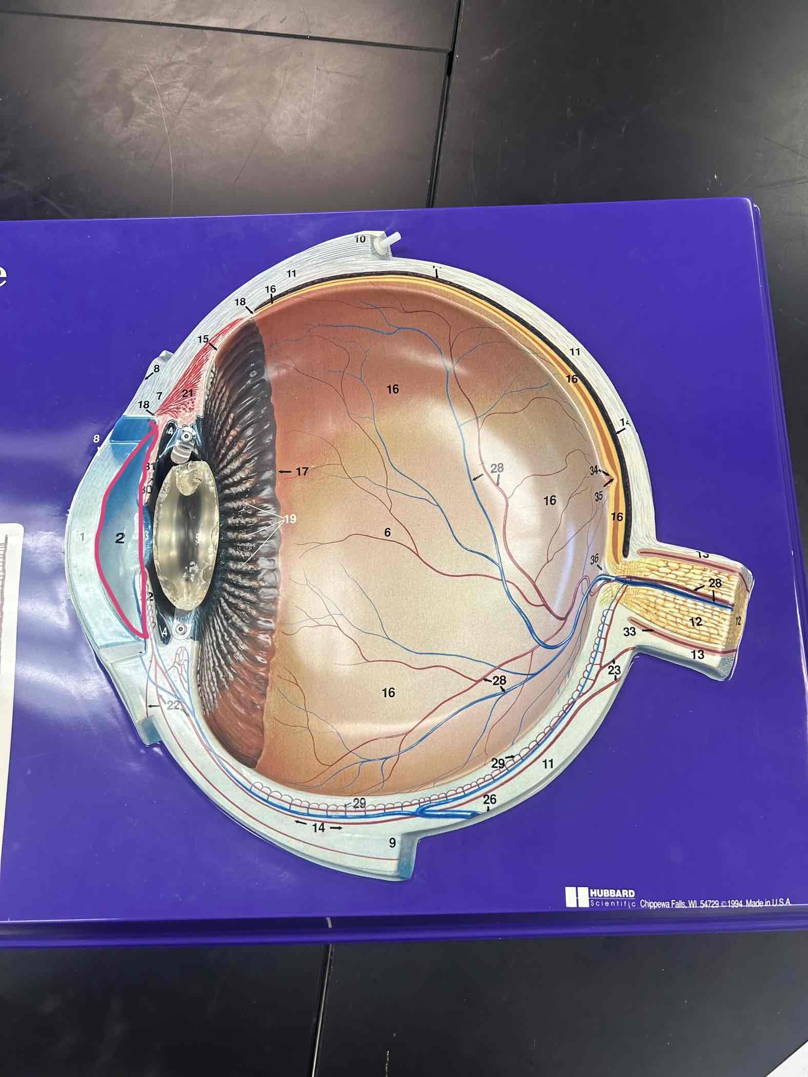
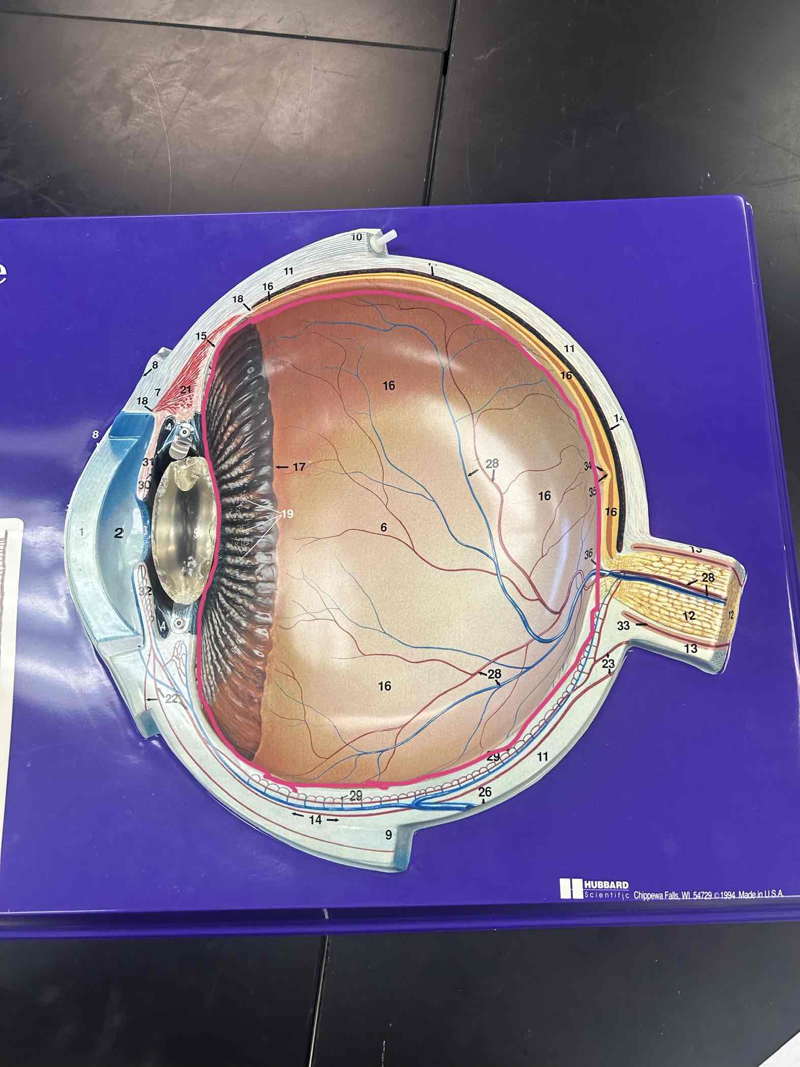
Vitreous humor
Posterior cavity to the lens
Aqueous humor
Anterior cavity to the lens