Unit 16: Cardiovascular system
 Call Kai
 Learn
 Practice Test
 Spaced Repetition
 Match
 Flashcards
 Knowt Play
1/33

Earn XP
Description and Tags
HTHS 2111
34 Terms
What structures are enclosed in the mediastinum? Mark all that apply.
Aortic arch
Esophagus
heart
inferior vena cava
superior vena cava
thymus
trachea
Systemic circulation structures
renal artery and vein, hepatic artery and vein, inferior vena cava, superior vena cava, hepatic portal vein, aorta
Pulmonary circulation structures
pulmonary arteries and veins, right ventricle, left atrium, pulmonary capillaries capillary bed of lungs
Blood enters the right atrium from the inferior and superior venae cavae. Starting with #1 = right atrium, identify the structures that the blood passes through, in the correct order.
1 & 17: right atrium
2: tricuspid valve
3: right ventricle
4: pulmonary valve
5: pulmonary arteries
6: capillary bed of the lungs
7: pulmonary veins
8: left atrium
9: mitral (bicuspid valve)
10: left ventricle
11: aortic valve
12: aorta
13: systemic arteries
14: capillary beds of body tissues
15: systemic veins
16: superior and inferior venae cavae
P wave
atrial systole/depolarization
T wave
ventricular repolarization
QRS Complex
ventricular systole/depolarization
SA node
natural pacemaker
contractile cells
myocardiocytes
conduction cells
SA node, AV node, purkinje fibers
QT interval
time of depolarization and repolarization of ventricles
These vessels have a thick tunica media of smooth muscle to allow for dilation and constriction for movement of blood
arteries
These vessels have a very thick tunica externa and in general do not control their own flow
veins
These are found in veins to regulate blood flow and prevent backflow
valves
These are generally larger arteries that must expand and contract as pulse pressure changes, e.g. aorta
elastic arteries
These vessels have larger smooth muscle layers to control blood flow; an example is the brachial artery
muscular artery
These vessels are microscopic and have very thin walls to allow for gas and nutrient exchange between bloodstream and tissue
capillary

What type of capillary is this
continuous

What type of capillary is this
fenestrated

What type of capillary is this
sinusoid

Using this image as a guide, what would the net movement of fluid be if capillary hydrostatic pressure was 40 mm Hg and blood colloidal osmotic pressure was 40 mm Hg?
no net movement

Using this image as a guide, what would the net movement of fluid be if capillary hydrostatic pressure was 35 mm Hg and blood colloidal osmotic pressure was 25 mm Hg?
fluid would move into tissues
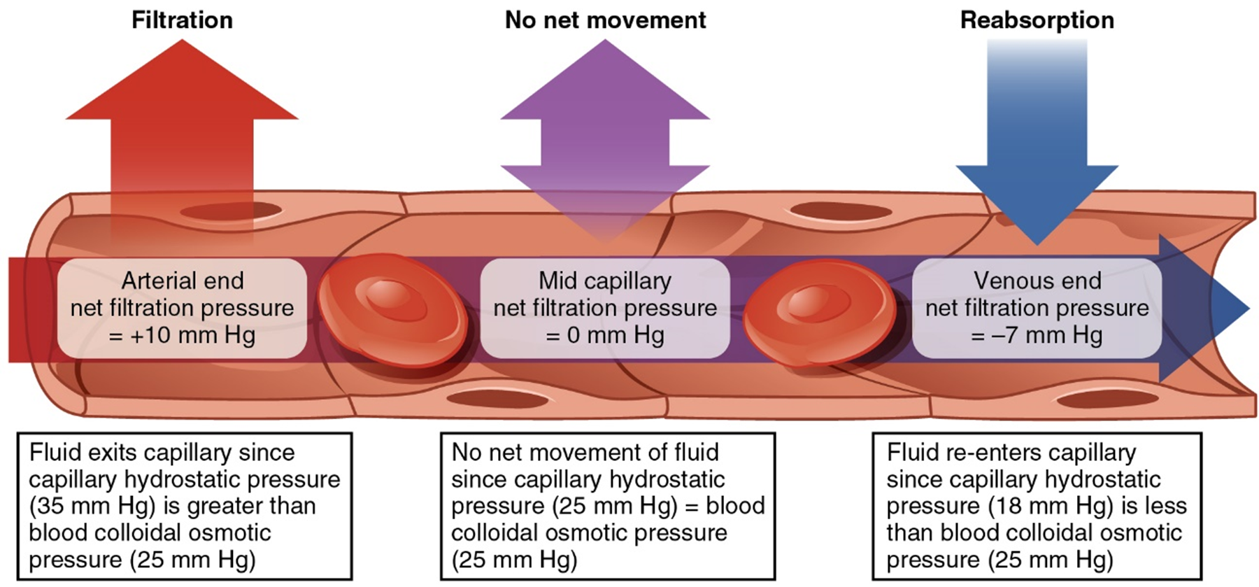
Using this image as a guide, what would the net movement of fluid be if capillary hydrostatic pressure was 25 mm Hg and blood colloidal osmotic pressure was 32 mm Hg?
fluid would reenter capillary
Right atrium and Left atrium
Right atrium receives blood from the body, Left atrium receives blood from the lungs
Right ventricle and Left ventricle
Right ventricle pumps blood to the lungs, Left ventricle pumps blood to the body
Right ventricle filled with what type of blood
deoxygenated
pericardium
encloses the heart
muscle (myocardium)
cardiac tamponade
pressure of the pericardial
fluid exceeds the pressure of the
atria
A special kind of Na+ channel (Na+ “funny” channels) is exclusive to the
pacemaker cells
preganglionic parasympathetic neurons
Cell bodies in vagal nucleus, axons travel to vagus nerve to end in the cardiac plexus to release acetylcholine (ACh)
postganglionic parasympathetic neurons
cell bodies in cardiac plexus, short axons release acetylcholine onto SA node to act on muscarinic ACh receptors
preganglionic sympathetic neurons
cell bodies in intermediolateral cell column (lateral horn) of spinal cord from T1-L2, axons end in sympathetic chain ganglia and release acetylcholine
postganglionic sympathetic neurons
Cell bodies in cervical and upper thoracic chain ganglia, axons end on heart muscle and release norepinephrine