(1) Radiographic Principles
 Call Kai
 Learn
 Practice Test
 Spaced Repetition
 Match
 Flashcards
 Knowt Play
1/15

There's no tags or description
Looks like no tags are added yet.
16 Terms
⭐ 1. Q: What does ALARA mean, and what is its purpose in radiography?
ALARA means “As Low As Reasonably Achievable,” and its purpose is to minimize radiation exposure while still obtaining the necessary diagnostic or therapeutic result.
The three major ALARA principles are:
Time
Distance
Shielding
💡 Think: Less time + more distance + shielding = less radiation exposure.
⭐ 2. Q: What is the Bucky tray?
The Bucky tray is a drawer-like component of an X-ray table or wall stand that holds the image receptor in place.
🔎 What this means:
The receptor may be a film cassette or digital detector.
The Bucky tray often contains a grid.
The grid reduces scatter radiation.
Reducing scatter improves image contrast because more primary X-rays, rather than scattered X-rays, reach the detector.

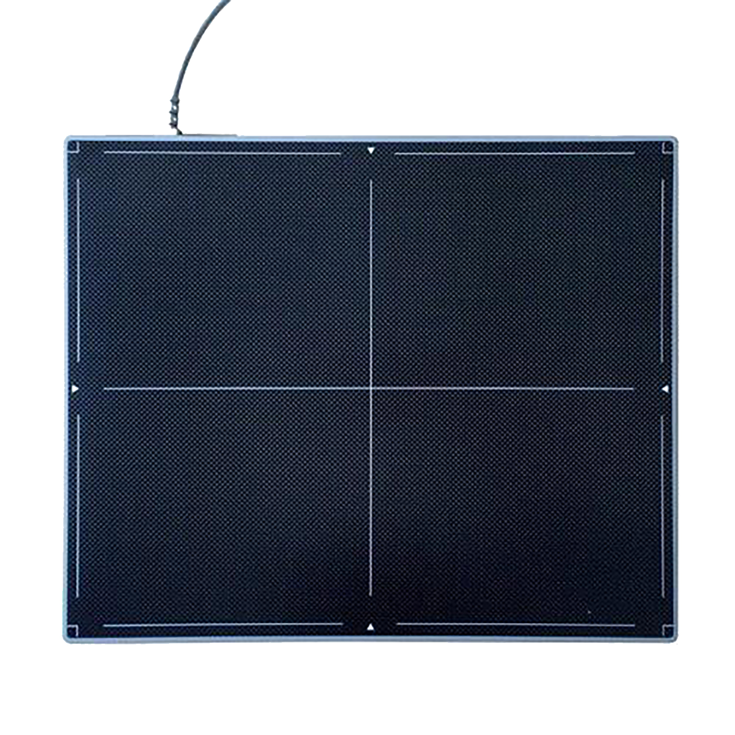
⭐ 3. Q: What is a detector plate?
A detector plate is a device in digital radiography that captures X-rays passing through the patient and converts them into a digital image.
⭐ 4. Q: How are X-rays produced in the X-ray tube?
The X-ray generator produces high-voltage electricity that accelerates electrons toward a target, usually tungsten, and X-rays are emitted when those high-energy electrons collide with the target.
🔎 What this means:
Think of it as:
Electricity → accelerate electrons → electrons hit tungsten → X-rays produced
This directly addresses the LO asking you to understand the X-ray production process.
⭐ 5. Q: What is kVp?
kVp, or kilovoltage peak, is the peak voltage applied to the X-ray tube and controls the energy and penetration power of the X-ray beam.
🔎 What this means:
kVp primarily affects how energetic/penetrating the X-ray beam is.
Increasing kVp means the beam has greater penetrating ability.
💡 Think: kVp = power/penetration
⭐ 6. Q: What is mAs?
mAs, or milliampere-seconds, is the product of X-ray tube current in mA and exposure time in seconds and determines the total quantity of X-rays produced.
🔎 What this means:
mAs = mA × seconds
It controls the quantity of X-rays generated.
💡 Think:
kVp = energy/penetration
mAs = quantity
⭐ 7. Q: What is the key difference between kVp and mAs?
kVp controls the energy and penetration of the X-ray beam, whereas mAs controls the total quantity of X-rays produced.
🔎 What this means:
Setting | Main role |
|---|---|
kVp | X-ray energy and penetration |
mAs | Number/quantity of X-rays produced |
This distinction is directly identified in your learning objectives.
⭐ 8. Q: What does “technique” mean in radiography?
Radiographic technique is the combination of exposure factors, especially kVp and mAs, selected to produce an optimal image.
🔎 What this means:
Technique selection depends on:
the body part being imaged
patient size
patient condition
the radiographic machine
The glossary states that, in general, low kVp with higher mAs techniques are preferred to optimize image contrast
⭐ 9. Q: What is radiographic exposure?
Exposure is the amount of radiation received by the imaging detector or film during an X-ray examination.
Exposure directly influences image brightness and contrast.
10. Q: What is underexposure?
Underexposure occurs when insufficient radiation reaches the detector or film.
🔎 What this means:
The image may become:
grainy
overall whiter than desired
higher in noise
harder to evaluate for fine details
💡 Think: Not enough radiation → noisy, grainy image.
11. Q: What is overexposure?
Overexposure occurs when the detector receives more radiation than necessary and becomes saturated.
🔎 What this means:
It unnecessarily increases patient radiation dose.
The glossary notes that digital imaging can show paradoxical black margins in thin body regions.
Excessive exposure can reduce the diagnostic value of the study.
⭐ 12. Q: What is radiographic contrast?
Contrast is the difference in brightness between different areas of a radiographic image that allows tissues and structures to be distinguished from one another.
🔎 What this means:
High contrast = more blacks and whites.
Low contrast = more shades of gray.
The glossary states that high-contrast radiographs are preferred over low-contrast radiographs.

⭐ 13. Q: What is radiographic detail?
Detail, also called spatial resolution or image sharpness, is the clarity with which small structures can be visualized on a radiograph.
🔎 What this means:
High detail allows you to see fine anatomical structures clearly.
Major factors specifically identified in the glossary include:
patient motion
quality of the X-ray machine
14. Q: How does motion affect radiographic image quality?
Motion can cause image blurring or distortion and reduce diagnostic quality.
🔎 What this means:
Motion can come from:
the patient
respiration
imaging equipment
both patient and equipment
If structures move while the image is being acquired, their borders become less sharp
⭐ 15. Q: What is collimation?
Collimation is the process of narrowing the X-ray beam using adjustable shutters to focus on the area of interest. It is also called the field of view.
🔎 What this means:
Collimation:
decreases irradiation of surrounding tissues
decreases scatter radiation
improves image quality
improves patient safety
controls the size of the X-ray field
💡 Think: Collimation = restrict the beam
