Neurology
 Call Kai
 Learn
Learn Practice Test
 Spaced Repetition
 Match
Match Flashcards
 Knowt Play
1/150

There's no tags or description
Looks like no tags are added yet.
Name | Mastery | Learn | Test | Matching | Spaced | Call with Kai | Chat |
|---|
No analytics yet
Send a link to your students to track their progress
151 Terms
The
Cerebrum
• Largest portion of the brain
• 2 hemispheres - each controls
the opposite side of the body
• Each hemisphere has 4 lobe
• Frontal Lobe
• Parietal Lobe
• Temporal Lobe
• Occipital Lobe

Left Hemisphere (Typically Dominant) Cerebrum
• Controls right side of the body
• Language production and comprehension
• Broca’s area → speech production
• Wernicke’s area → language comprehension
Right Hemisphere (Typically Non-Dominant)
• Controls left side of the body
• Spatial and temporal awareness
• Non-verbal analysis (e.g., visual patterns, facial recognition)
• Emotion communication (interpreting tone, body language)
Gyri & Sulci
•Gyri
• Folds of the brain
• Increase cortical surface area
• To ensure enough braintissue for all it’s functions
•Sulci
• Valleys between the gyro
Cerebrum and how sulcus divides lobes
• The central sulcus and the lateral sulcus divide
each hemisphere into the four lobes
• Frontal & Parietal are separated by the central sulcus
• Temporal & Parietal separated by the lateral sulcus

The cerebral hemispheres communicate with each
other via the
corpus callosum
Communication within
the Brain (3 fibres)
•Commissural Fibres
• Link matching areas of the two hemispheres
• e.g., corpus callosum – largest of the commissures
•Association Fibres
• Pass from one part of a single hemisphere to another
• e.g., arcuate fasciculus
•Projection Fibres
• Run from Cortex to:
• Subcortical nuclei (e.g., Basal Ganglia) in the cerebral hemispheres
• Brain stem (e.g., cranial nerves)
• Spinal cord (e.g., spinal tracts

Lobe and summarized function

Different areas of the brain image

The Frontal Lobe
• Located at the front of the brain
separated from the Parietal Lobe by
the Central Sulcus
• Functions:
• Planning, Organising, Problem Solving
• Motor Control
• Selective Attention
• Personality and Higher Cognitive
Functions: Behaviour, Emotions
Prefrontal Cortex (Frontal lobe)
• Higher cognitive function
• Controls cognitive process so appropriate movements are selected at the correct time and place
• Influences personality
The Motor Area (Frontal lobe)
• Primary Motor Cortex – direct control of
muscles
• Premotor & Supplementary Motor Areas - plan and modifies movements, selection and direction of motor sequences
Broca’s Area (FL)
• Located in the dominant frontal lobe - usually left
hemisphere
• Functions:
• Controls speech production
• Plans and sequences voluntary movements
• Integrates with motor cortex for fine motor coordination of mouth, tongue, and facial muscle
Damage to Broca’s area
• Broca’s aphasia
• Difficulty expressing language/forming words
• Comprehension intact
Impairments of the
Frontal Lobe (4 main impairments)
Motor
• Loss of simple movement of various body parts
(paralysis)
• Inability to plan a sequence of complex movements
(apraxia)
Cognition
• Loss of flexibility in thinking
• Perseveration - Persistence of a single thought
• Inattention - inability to focus on tasks
• Impaired executive function - difficulty with problem
solving
Behavioural
• Emotional lability - Mood changes suddenly
• Changes in social behaviour - disinhibition or
inappropriate actions
• Changes in personality – may be subtle or noticeable
Speech Deficits
• Broca’s Aphasia - non-fluent, effortful speech.
Difficulty expressing language and coordinating speech
movements
Primary somatosensory cortex (PL)
• Located on the postcentral gyrus
• Processes sensory information from
the body, including:
• Touch
• Pressure
• Proprioception
Parietal association areas
• Integrate sensory information to
help us understand:
-Where our body is
-Where things are around us
-How our body relates to the environment
• Help use sensory information to
guide movement
Somatosensory Cortex -
The Somatosensory Homunculus (image)

Parietal Lobe Lesion (potential impairments) don’t memorize everything get the gist of it
• Sensory discrimination
• Proprioception and body awareness
• Spatial awareness
• Ability to use sensory information to guide movement
-Tactile Sensation
-Stereognosis (astereognosis) is unable to recognize an object by touch
-Sensory neglect
The Temporal Lobe (3 main areas)
-Auditory Processing
-Memory and learning
-Language comprehension
Auditory processing (TL)
• Contains the primary auditory cortex
• Processes information from the ears
• Important for recognising and interpreting
sounds
Memory & learning (TL)
• Contributes to memory formation and
retrieval
• Helps link sensory information with stored
knowledge
• Language comprehension (TL)
• The dominant temporal lobe contains
areas important for understanding language
• Wernicke's area is associated with language
Damage to temporal lobe may cause
• Difficulties processing sounds
• Memory problems
• Difficulty understanding language
Language Loop
• Network connecting Wernicke’s area, Broca’s
area, arcuate fasciculus (how they connect)
• Supports key language functions
• Comprehension
• Speech production
• Repetition
• Semantic Processing
• This region processes language in the person’s primary modality, whether spoken or signed
• Lesions anywhere in this loop - various types
of aphasia

The Occipital Lobe
• Back of the brain
• Primary lobe for vision - processes visual information
• Contains association areas that help in visual recognition of shapes and colours
Occipital Lobe Lesions (understand the get don’t need to memorize everything)
• Defects in vision (Visual Field Cuts)
• Difficulty with locating objects in
environment
• Difficulty with identifying colours (Colour
Agnosia).
• Production of hallucinations.
• Visual illusions - inaccurately seeing
objects.
• Word blindness - inability to recognize
words.
• Difficulty in recognizing drawn objects.
• Inability to recognise the movement of
object (Movement Agnosia).
• Difficulties with reading and writing
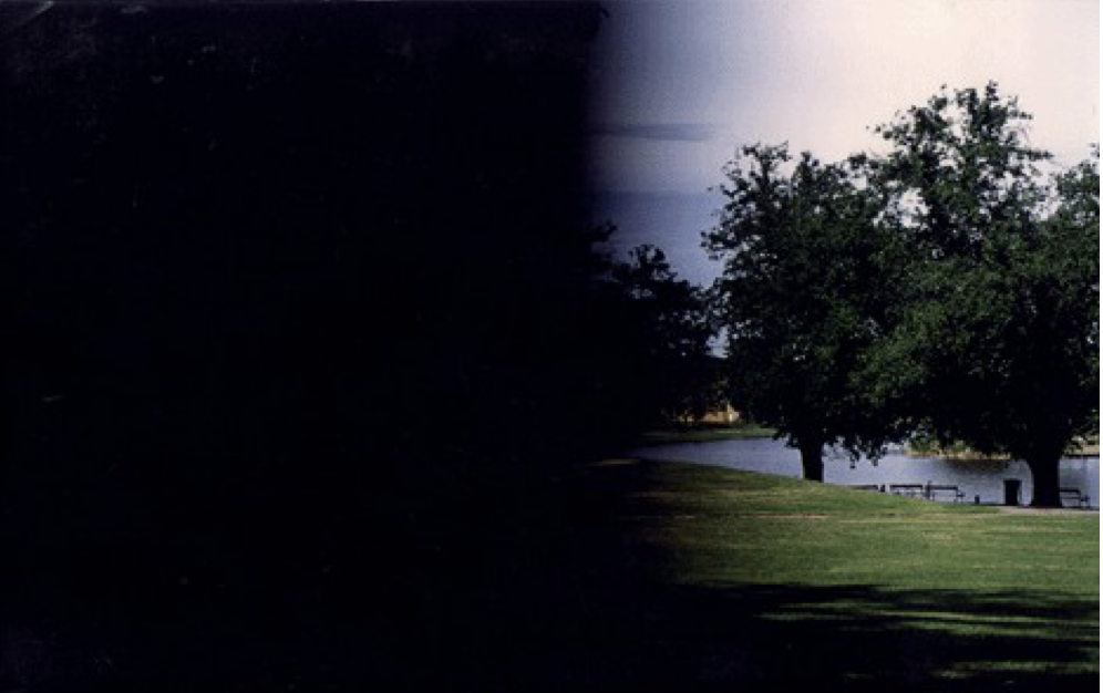
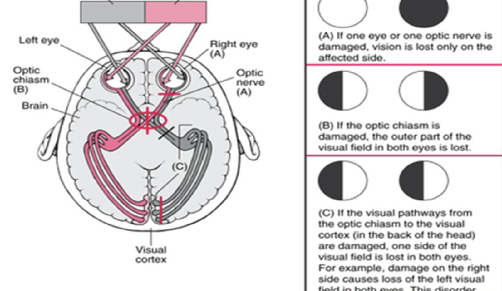
Left Homonymous Hemianopia
the loss of the left half of your field of vision in both eyes, caused by damage to the right side of your brain
Overview of Subcortical Structures (5)
• Located beneath the cerebral cortex
• They include:
• Basal ganglia – involved in motor control,
habit formation
• Thalamus – sensory and motor relay
• Internal capsule – major white matter
“highway” connecting cortex with subcortical
structures
• Cerebellum – coordination, balance, motor
learning
• Brainstem – vital autonomic functions and
cranial nerve contro
Subcortical Structures

The Basal Ganglia (5 main parts)
• Deep nuclei within the white matter of each
cerebral hemisphere
• Components:
1. Caudate
2. Putamen (together with Caudate = Striatum),
3. Globus Pallidus (GPe, GPi),
4. Subthalamic Nucleus
5. Substantia Nigra (pars compacta & reticulata)
Basal Ganglia Connections
• No direct connections with the spinal cord

Input, Intrinsic and Output Nuclei for basal ganglia components

Functions of Basal Ganglia (4)
1. Modulate motor activity
• Facilitates intended movements (direct pathway)
• Supresses unwanted or ineffective movement (indirect pathway)
• Ensures smooth, coordinated motion rather than initiating movement directly.
2. Regulate muscle tone
3. Planning, sequencing, timing of movements
4. Cognitive and emotional roles
• Through the basal ganglia-thalamocortical circuits, play a role in habit formation, decision making, and emotion.
Note:
• Dopamine is a key neurotransmitter for motor control, motivation, and cognition produced in SNpc. It helps the striatum and BG to regulate smooth voluntary
moveme
Basal Ganglia Lesions
Bradykinesia
Slowness of movement
Hypokinesia
Reduced amplitude or range of movement
Akinesia
Difficulty initiating movement (freezing)

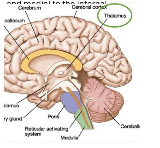
The Thalamus location and general function
• Situated under the corpus callosum, medial to the
internal capsule
• Composed of distinct sensory and motor areas
• The thalamus is a major relay/integration point for
sensory information and also participates in motor
circuits
The thalamus Sensory and motor functions
• Sensory Function:
• Relays sensory input to the primary sensory areas of
the cortex
• Filters and prioritises sensory signals before they
reach the cortex
• Motor Functions:
• Relays motor information from the Basal Ganglia and
cerebellum to the motor areas of the cortex
The Internal Capsule Location and Motor/sensory tracts
• Location: Between the head of caudate & thalamus
medially & the lenticular nucleus laterally
• Area of white matter, containing sensory & motor
tracts
• Motor tracts: corticospinal and corticobulbar
descend from the cerebral cortex through the
internal capsule to the spinal cord and brainstem
• Sensory tracts: thalamocortical fibres ascend from
the thalamus to the cortex
Internal Capsule damage
contralateral hemiparesis or hemiplegia,
+/- spasticity, abnormal reflexes, impaired motor
contro
Anatomy of the Cerebellum (3 lobes and 3 major fissures)
• Location: inferior to the occipital lobe
• Two hemispheres and central vermis
• Gray matter outside, white matter inside
• Three lobes:
1. Anterior
2. Posterior
3. Flocculonodular
• Major fissures:
1. Primary
2. Posterior
3. Horizonta

Cerebellar Peduncles 3
• Superior Peduncles: connect the cerebellum to the midbrain and thalamus (output)
• Middle Peduncles: connect the pons to the cerebellum (input)
• Inferior Peduncle: connects the brainstem and spinal cord to the cerebellum (sensory input)
Vestibulocerebellum
• Receives visual input from visual cortex
• Receives vestibular input from semicircular canals &
vestibular nuclei
• Regulates balance and eye movements with the vermis

Spinocerebellum
• Regulates body and limb movements
• Receives proprioceptive input from dorsal columns of the spinal cord
• Receives input from visual and auditory systems
• Sends fibres to deep cerebellar nuclei
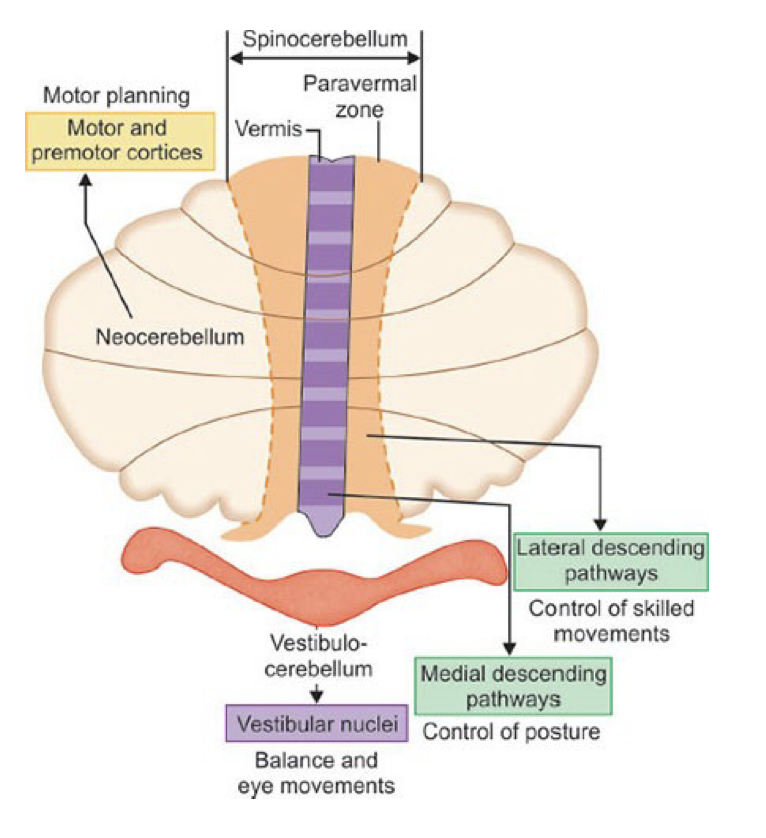
Neocerebellum or Cerebrocerebellum
• Involved in planning movement that is about to occur
• Evaluating sensory information
• Receives inputs from the cerebral cortex
• Sends nerve fibres mainly to the thalamus (connected to motor
areas)

Function of the Cerebellum (4)
1. Coordinates movement and postural control by comparing actual
motor output with the intended movement
2. Corrects motor activities to ensure smooth, accurate motion
3. Receives sensory information from
• Muscle spindles, Golgi tendon organs (proprioception)
• Cutaneous mechanoreceptors (skin and joint sensation)
• Travels to the cerebellum via the spinocerebellar tracts.
4. Responsible for timing, rhythm and precision of movement
Clinical Relevance of cerebellum
Essential for rehabilitation of motor control,
balance, and coordination after neurological injury
Cerebellar Lesions (
• Common Causes: Lack of blood supply (ischaemia or infarct), trauma, tumours, or degeneration
• Clinical Features:
• Ataxia – lack of muscle control – jerky movements
• Dysmetria – impaired coordination – past pointing on the finger/nose or heel/shin tests (dysmetria)
- Hypermetria – over shooting
- Hypometria – under shooting
• Broad based gait - increased stance for
balance
• Dyssynergia – poor proximal stability - uncoordinated movement
• Intention tremor: tremor occurs during voluntary movement
• Hypotonia: decreased muscle tone in affected limbs
• Nystagmus or impaired eye movements (if vestibulocerebellum involved
The Brain Stem
The Brain Stem
• Connects cerebrum, cerebellum, and spinal cord
• Conduit for ascending sensory and descending motor tracts
• Houses cranial nerve nuclei and autonomic centres
• Maintains consciousness via reticular formation

Midbrain 3 segments
-Anterior midbrain
-Tegmentum
-Tectum (dorsal midbrain)
Anterior midbrain (2)
• Cerebral peduncles (crus cerebri) – contain major
descending motor fibres from the cerebral cortex,
including corticospinal, corticobulbar and
corticopontine fibres
• Substantia nigra – involved in basal ganglia circuits
and movement regulation

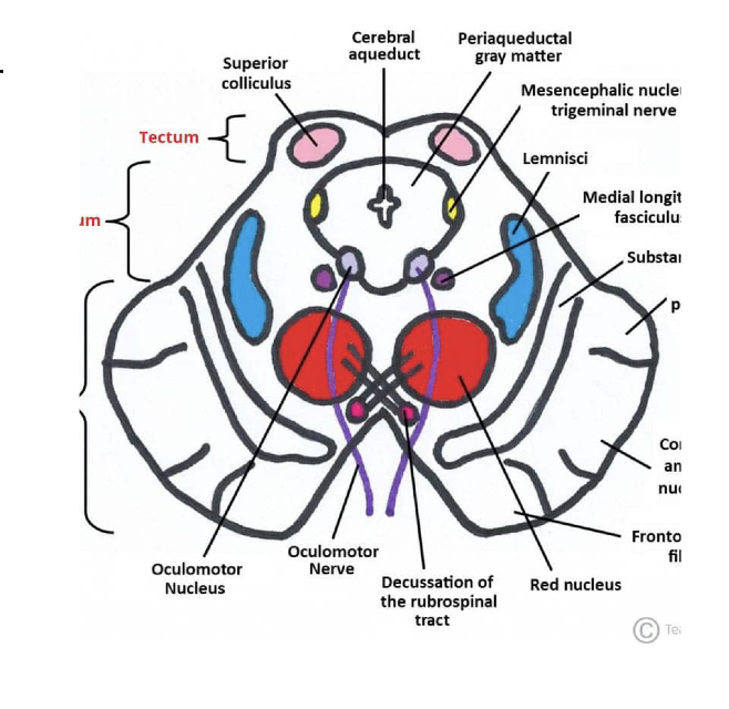
Tegmentum 2 things
• Red nucleus – involved in motor control; origin of
the rubrospinal tract
• Cranial nerve nuclei III and IV – involved in eye
movement control

Tectum (dorsal midbrain)
• Superior colliculi – visual orienting/reflexes
• Inferior colliculi – auditory pathway/reflex
processing
The Pons contains (5)
• Corticospinal fibres – descending motor pathways passing through the pons
• Pontocerebellar fibres – carry information from the cerebral cortex, via pontine nuclei, to the cerebellum; important for motor planning and coordination
• Pontine respiratory centres – contribute to regulation of breathing
• Tegmentum – contains ascending sensory pathways, including fibres carrying information towards the thalamus
• Cranial nerve nuclei – V, VI, VII and VIII (with some VIII structures extending into the pontomedullary junction
The pons clinical relevance
Locked-in syndrome: severe paralysis of voluntary movement, typically including quadriplegia and anarthria, with consciousness preserved and vertical eye movements and/or blinking often preserved
• Impaired facial movement/expression and other cranial nerve functions depending on the structures affected
The Medulla Oblongata key structures and pathways
Key structures & pathways
• Continuous with the spinal cord through the foramen magnum
• Corticospinal fibres decussate (cross) in the caudal medulla
• Gracile & cuneate nuclei receive fine touch, vibration and
proprioceptive information
• Cranial nerve nuclei: IX, X, XI and XII
• Vital autonomic centres: cardiovascular and respiratory regulation
• Protective reflexes: swallowing, coughing, vomiting and sneezing
The Reticular Formation (RF)
• A matrix of neurons extending the length of the brainstem.
• Consists of multiple nuclei and fibre tracts that integrate sensory,
motor, and autonomic functions
The Reticular Formation (RF) functions (brainstem)
• Modulates muscle tone and posture
• Coordinates reflexes and basic motor patterns
• Influences cardiovascular, respiratory, and digestive activity
• Reticular Activating System (RAS) - Maintains alertness, attention,
and wakefulness
The Reticular Formation (RF) clinical relevance
• Lesions may cause coma, reduced consciousness, impaired
arousal and attention, abnormal tone or postural control
Brainstem Lesions affect
• Motor control: corticospinal tract, decerebrate posturing
• Cranial nerves: eye movement, facial
expression, swallowing
• Autonomic function: respiration, heart rate
• Consciousness: reticular formation
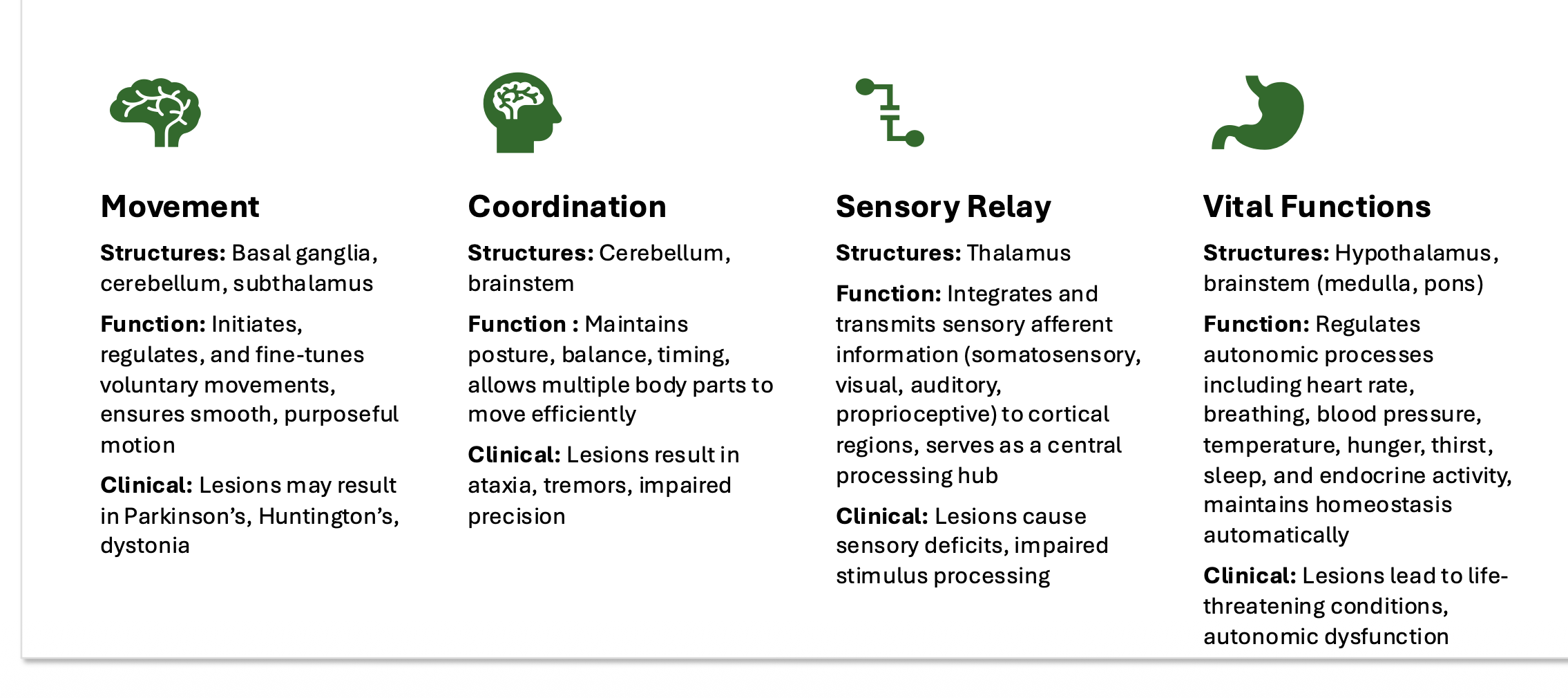
Functional Roles of Subcortical Structures (summary)
Structure of the Spinal Cord
•Central canal: CSF-filled
•Grey matter: H-shapedcentre
•White matter: outerregions

Grey Matter
H-shaped; contains neuron cell bodies & synapses
• Dorsal horns → sensory nuclei
• Ventral horns → motor neurons
• Lateral/intermediate horns → autonomic neurons
(thoracolumbar levels)
• Shape varies by level (lumbar = more grey for limb
control
Spinal Nerves
• 31 pairs: 8 cervical, 12 thoracic, 5 lumbar, 5 sacral, 1 coccygeal
• Formed from dorsal (sensory) and ventral (motor) roots
• Branch into dorsal ramus (back muscles & skin) and ventral ramus (limbs & anterior trunk)
• Dorsal root ganglia contain sensory neuron cell bodies
Spinal Reflexes
-Automatic responses mediated by the spinal cord.
-They allow our body to respond quickly to stretch, tension, or painful stimuli without needing the brain
-The brain does have a role in modulating them
-Monosynaptic - fast (1 synapse between sensory and motor neuron)
-Polysynaptic – slower (2+ synapses and 1 or more interneurons)
-Reflex testing informs treatment planning, especially for patients with neurological disorders, spasticity, or motor control deficits
Monosynaptic Stretch Reflex • Example: Knee
Jerk/Patellar Tendon Reflex
• Function: Maintains tone & posture
• Pathway: Muscle spindle
- Sensory neuron - Motor neuron - Muscle
contraction

Golgi Tendon Reflex
• Example: Lifting a very heavy weight, the muscle suddenly “gives out”
• Function: Protection - Inhibits muscle contraction in response to
excessive tension
• Pathway:
• Golgi tendon organ senses tension
- Sensory neuron - Inhibitory interneuron - α-motor neuron inhibited - Muscle relaxes
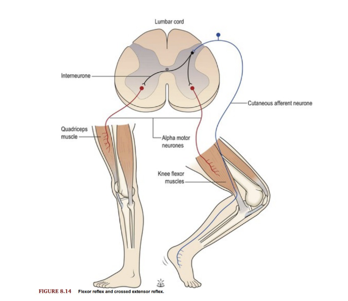
Crossed Extensor Reflex
• Example: Stepping on a Sharp Object
• Function: Supports weight on opposite limb (extension) when one limb withdraws (flexes)
• Pathway: Interneurons cross to contralateral side
- Extensor muscles contract
Two Main Types of spinal chord tracts
• Ascending – carries sensory information up
the spinal cord to the brain
• Descending – carries motor impulses from the brain down the spinal cord to the periphery
• Ascending pathways typically involve three neurons.
• Descending pathways typically involve two,
an UMN and LMN.

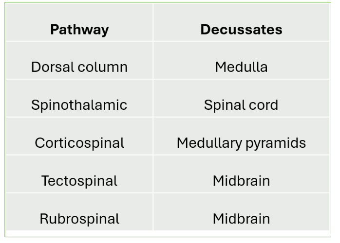
Decussation table
Sensory (Ascending) Pathways 3 neuron pathway
1st order Neuron (Afferent)
• Origin: Sensory receptors
• Cell body: Dorsal or cranial root ganglion
• Axon travels to the SC or brainstem.
• Synapses with the second-order neuron in the dorsal horn or brainstem nuclei.
2nd order Neuron
• Cell body: SC or brainstem
• Axon often decussates
• Ascends to the thalamus
3rd order Neuron
• Cell body: Thalamus.
• Axon projects to the primary sensory cortex
• Responsible for conscious perception of the stimulus

3 sensory pathways
• Dorsal Column / Posterior Column
• Spinothalamic tract
• Spinocerebellar tracts
Dorsal Column
Function: Transmit fine touch, vibration,
proprioception, deep & discriminative touch, and
some visceral pain.
Location: Posterior (dorsal) columns of the spinal
cord.
Two major fasciculi
• Gracile fasciculus (LB T6)
• Cuneate fasciculus. (UB T6)
Decussation - In the medulla, fibres from both gracile
and cuneate nuclei cross to the opposite side and
form the medial lemniscus.
Sensory Fibres → Thalamus →Primary
somatosensory cortex

Gracile vs Cuneate fasciculus (Dorsal Column)

Spinothalamic tract
Location: Anterior & lateral columns of the spinal cord
Function:
Anterior spinothalamic tract - crude touch & pressure
Lateral spinothalamic tract - pain & temperature
Pathway:
• 1st order neuron: Sensory Neuron → dorsal horn of SC
• 2nd order neuron: Decussates in spinal cord (within 1-2 segments) →ascend to thalamus
• 3rd order neuron: Thalamus → cerebral cortex

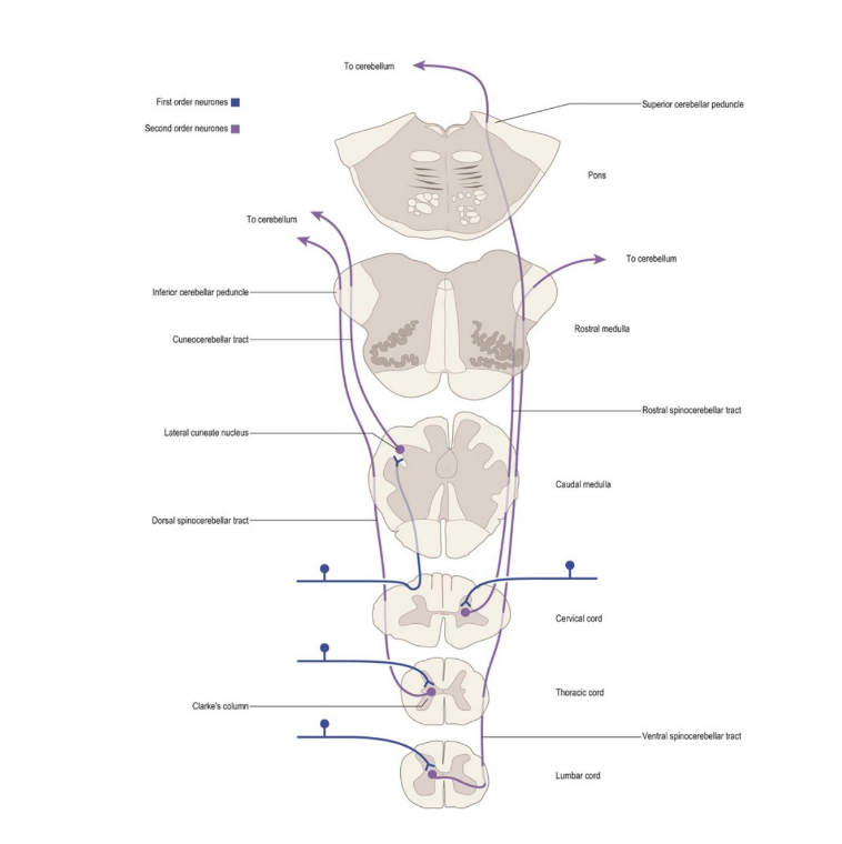
Spinocerebellar tracts (SCT
• Location: Dorsal & ventral tracts run in the lateral column of the spinal cord
• Function: Carry proprioceptive signals from the limbs and trunk to the cerebellum
• Pathway: only 2 neurons
• 1st order: proprioceptors (muscles, joints, tendons) → dorsal horn of the SC
• 2nd order : dorsal horn of the SC → cerebellum
• Dorsal SCT - stays ipsilateral, enters cerebellum via inferior cerebellar peduncle
• Ventral SCT - crosses in spinal cord, ascends contralaterally, then crosses back in cerebellum, enters cerebellum via superior cerebellar peduncle
• As the ventral SCT crosses twice, both tracts give ipsilateral cerebellar feedback
Motor (Descending) Pathways 5
Corticospinal Tract
• Lateral & anterior columns
Reticulospinal Tract
• Anterior & lateral columns
Tectospinal Tract
• Anterior column
Vestibulospinal Tract
• Anterior column
Rubrospinal Tract
• Lateral column
Corticospinal tract
• Location: Descends through internal capsule → brainstem → lateral & anterior columns of spinal cord
• Function: Voluntary fine motor control (especially distal limbs)
• Pathway:
• Origin: Primary motor cortex
• Fibres descend → pyramidal decussation in medulla (most cross → lateral CST; some uncrossed → anterior CST)
• Synapse on anterior horn motor neurons

Reticulospinal tract
• Location: Anterior & lateral columns of spinal cord
• Functions:
• Motor - Posture, locomotion, and muscle tone
• Autonomic: cardiovascular and respiratory regulation
Pathway:
• Origin: Reticular formation (pons & medulla)
• UMN fibres descends ipsilaterally & bilaterally in the SC
• Synapse on interneurons & lower motor neurons (LMNs) in spinal
cord

Tectospinal tract
Location: Descends in anterior column of spinal cord
• Function: Reflex postural movements in response to visual/auditory stimuli (head & eye orientation)
• Pathway:
• Origin: Superior colliculus (midbrain tectum)
• UMN fibres cross in midbrain, descend contralaterally in anterior column
• Synapse on LMNs in cervical spinal cord – activate neck muscles

Vestibulospinal tract
• Location: Anterior column of spinal cord
• Function:
• Balance & posture via activation of extensor muscles
• Helps maintain posture and balance, especially in response to vestibular input
• Pathway:
• Begins in vestibular nuclei (brainstem)
• UMN fibres:
• Lateral vestibulospinal tract: descend ipsilaterally
• Medial vestibulospinal tract: descend bilaterally
• Synapse on LMNs controlling axial and limb extensors

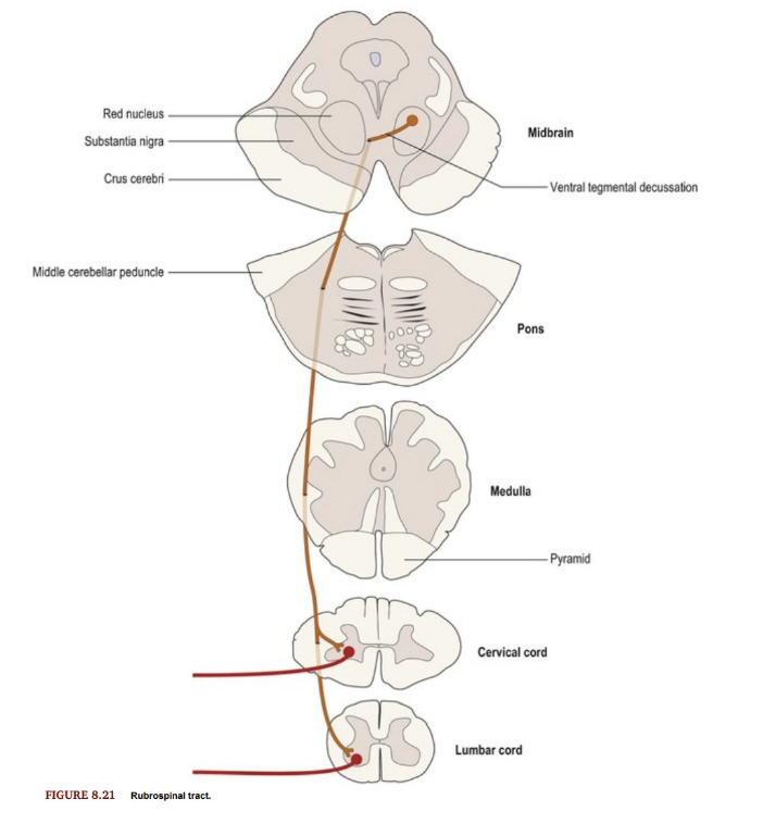
Rubrospinal tract
• Location: Lateral column of SC
• Function:
• Motor control of upper limb flexors
• Small role in regulation of tone
• Pathway:
• From red nucleus (midbrain)
• UMN fibres cross in midbrain, and descend contralaterally in lateral
column
• Synapse on lower motor neurons in cervical spinal cord to activate
upper limb flexor muscles
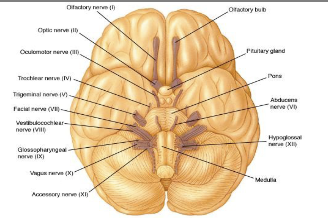
Cranial Nerves origin
• Forebrain: CN I, II
• Midbrain: CN III, IV
• Pons: CN V, VI, VII
• Ponto-medullary junction: CN VIII
• Medulla: CN IX, X, XI, XII

Inferior angle of brain showing where cranial nerves are
Function of Cranial Nerves - Sensory table

Function of Cranial Nerves –
Motor table

Function of Cranial Nerves -
Mixed table

Cranial Nerve II – Optic Nerve damage image (nerve, chasm and cortex)
CN III - Oculomotor nerve
• Supplies: all eye muscles (Medial rectus, Superior rectus, Inferior rectus, Inferior oblique) except superior oblique & lateral rectus
• Function: Eye movement, eyelid elevation, pupil constriction
(parasympathetic)
• Effects of Damage: Ptosis, dilated pupil, inability to move eye, eye tends to drift laterally at rest, inability to focus and Diplopia
• Clinical Tests: Pupil light reflex, eye trackin
CN IV –Trochlear
• Supplies: Superior oblique muscle of the eye
• Function: eye movement
• Effects of Damage: Double vision and inability to rotate eye
inferiolaterally. Patient often tilts head towards the affected side
CN VI – Abducens
• Supplies: Lateral rectus muscle
• Function: Abduction of the eye
• Effects of Damage: eye deviates medially at rest, due to action of
antagonistic muscles
• Clinical Test: Eye tracking side to side
Three Major Sensory Contributors to
Movement

The Vestibular System summary (read)
• Peripheral organs (semicircular canals, otoliths)
• Central pathways (vestibular nuclei, cerebellum, cortex).
• Function: detect head movement, maintain balance, stabilize gaze.
• Role in: balance, posture, and spatial orientation
• Works in close coordination with visual and somatosensory
systems.
• Vestibular dysfunction - dizziness, vertigo, imbalance,
risk of falls
Peripheral Components of the
Vestibular System
• Located in the inner ear within the bony labyrinth
• Sensory receptors in semicircular canals and
otoliths
• Semicircular canals (3: anterior, posterior, lateral) (Vestibular Peripheral component )
• Detect angular acceleration (rotational head
movements).
• Each has an ampulla containing crista ampullaris
with hair cells
Otolith organs (utricle & saccule) (vestibular peripheral components)
• Detect linear acceleration and gravity.
• Contain maculae with hair cells embedded in otolithic membrane (with calcium carbonate crystals).
• Hair cells (vestibular peripheral components)
• Movement of hair cells in one direction excites them (depolarisation), while movement in the opposite direction inhibits them (hyperpolarisation).
• Signals transmitted via vestibular nerve (CN VIII).
Central Component s of the Vestibular
System
-Vestibular nerve
-Vestibular nuclei
Vestibular Nerve
Vestibular Division of CN VIII
Vestibular nucle
• located in the brainstem - closely
connected with the cerebellum
• Act as relay stations, integrating
input from:
• Vestibular apparatus
• Cerebellum
• Spinal cord
• Visual system
Connections of Vestibular Nuclei (4)
• Vestibulospinal tracts -posture & balance (adjusts trunk/limb muscles).
• Medial longitudinal fasciculus - coordinates eye movements with
head movements (vestibulo-ocular reflex, VOR).
• Cerebellum (flocculonodular lobe) - fine tunes balance.
• Thalamus - conscious perception of motion & orientation
